Urinary Patho
1/69
There's no tags or description
Looks like no tags are added yet.
Name | Mastery | Learn | Test | Matching | Spaced | Call with Kai |
|---|
No analytics yet
Send a link to your students to track their progress
70 Terms
Urinary system consists of…
Upper urinary tract & lower urinary tract (bladder and urethra).
- 2 Kidneys (upper)
- 2 Ureters (upper)
- Urinary bladder (lower)
- Urethra (lower)
GU tract congenital anomalies are what?
more common than other organ systems
What can anomalies cause
obstruction or stasis, create infections, impair renal function, and/or prompt stone formation.
What kidney sits lower
right (liver vs. spleen)
How do kidneys assist in homeostasis
They detoxify the blood, maintain normal pH, iron, and salt levels in the blood. also work to regulate blood pressure by producing the enzyme renin
Urine is formed and excreted through ____ and _____ of blood
filtration and reabsorption
How much urine is made per day
1-1.5L
Nephron
Functional unit of the kidney responsible for formation of urine (filtration)
Reservoir until excretion
urinary bladder
Gerota’s Fascia
Covers kidney and adrenal gland.
Cortex
Outer Portion/Renal parenchyma
Medulla
Inner portion from base of pyramids to center of kidney
Pyramids
Anechoic triangles of collecting tubules that are between the cortex and sinus in the medulla
Sinus
Hyperechoic area that contains the calyces, fat, renal pelvis, connective tissue, vessels and lymphatics
Renal Pelvis
Funnel shaped area between the major calyces and ureter
Hilum
Area where the vein/artery/ureter enter/exit
What muscles are posterior to kidneys
Psoas, Quadratus Lumborum
urinalysis
for renal function includes but is not limited to, an evaluation for the presence of bacteria, pus, blood, and protein in the urine.
BUN (blood urea nitrogen)
measures the amount of urea nitrogen, a byproduct of protein metabolism that occurs within the liver and is excreted by the kidneys.
Creatinine
measures the amount of creatinine phosphate.
LDH (lactate dehydrogenase)
additional enzyme found within the blood that may be used to monitor renal function and other abnormalities, including some forms of cancer
GFR (glomerular filtration rate)
can be used to evaluate the overall function of the kidneys.
Clinical indication for kidneys
Flank pain, hematuria, abnormal labs/ images, abdominal trauma, pre and post transplant eval,
Normal kidney length
9-12 cm
Echogenicity for cortex
hypo or isoechoic compared to liver/spleen
Echogenicity for pyramids
anechoic
Echogenicity for renal sinus
hyperechoic
Renal Agenesis
Absence of kidney on one side (unilateral) large kidney on contralateral side
Left kidney agenesis is more common. +Men. Bilateral agenesis often presents in utero with oligohydramnios and pulmonary hypoplasia, and is incompatible with life.
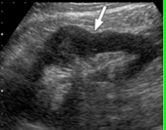
Dromedary Hump
Local/anterior bulge of anterior border of the kidney
dromedary hump image
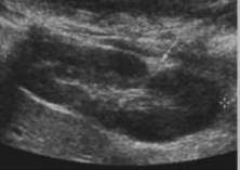
Junctional Parenchymal Defect (JPD
Wedge-shaped hyperechoic defect, anterior, usually right
JPD image
Hypertrophied Column of Bertin (HCB)
Layer of renal cortex is folded toward the center of kidney. Normal vs HCB
HCB image
Hypoplasia
Developed but small. Clinical significance depends on volume of output, hypertension generally accompanies this anomaly.
hypoplasia image
Hyperplasia
Overdeveloped kidney (LARGE). Often associated with renal agenesis or hypoplasia of contralateral kidney.
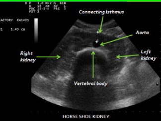
Horseshoe Kidney
Most common fusion anomaly. Lower poles of the kidneys are joined across the midline by a band of soft tissue. +Men. Generally unimpaired – but if obstruction present, surgery possible
horseshoe kidney image
Crossed Ectopia
One kidney lies across the midline and is fused to the other kidney. Second most common fusion anomaly.
crossed ectopia image
Ectopic Kidney
Having one kidney that is out of its normal position (from birth, failure to ascend).
ectopic kidney

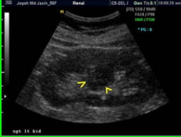
Double Collecting System (aka Duplex Kidney)
Duplication of collecting system - may be complete (2 ureters) or incomplete (1 ureter).
image of duplex kidney
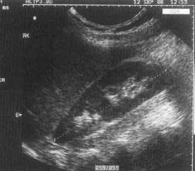
sonographically of duplex kidney
Band of cortical tissue seen throughout a mid area section of the kidney that can be seen in both long and trans.
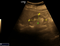
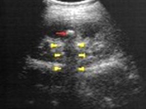
Nephrolithiasis (kidney stones)
Stones in the kidney. They are composed of calcium and salts. Kidneys are the second most common area for calculi in the body.
clinical indications for nephrolithiasis
Causes include metabolic disorders (calcium), high calcium intake, and chronic UTI’s. Common in Men >30 years old. Patients will present with acute back/flank pain that may radiate into the ipsilateral groin. Possible dysuria, hematuria, cloudy urine, fever, and/or chills.
How are kidney stones usually diagnosed
Radiography, IVU, Retrograde Pyelogram, Ultrasound, and CT
treatment options for kidney stones
Depending on size, may pass naturally (strainer), or lithotripsy, medication, or surgery.
nephrolithiasis prognosis
excellent
Kidney stone image
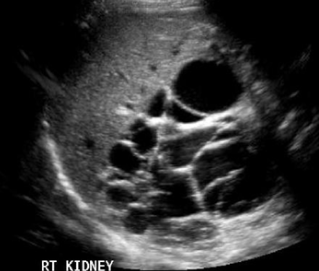
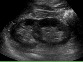
Hydronephrosis
Obstructive disease of the urinary system – dilatation of calyces and renal pelvis with urine. Long term can cause atrophy and loss of renal function.
Clinical indications for hydronephrosis
Most common cause is calculus/stone. Signs/Symptoms include flank pain and blood or pus in urine. May have abnormal labs if damage is present.
Treatment options for hydronephrosis
removal of stone
prognosis for hydronphrosis
excellent if caught prior to damage
hydronephrosis image
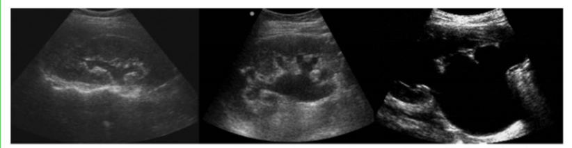
Autosomal Recessive Polycystic Kidney Disease (ARPKD)
Genetic condition, formation of innumerable tiny cysts.
Four kinds: Perinatal, neonatal, infantile, and juvenile.
clinical indications for ARPKD
Portal hypertension (in severe cases), Renal insufficiency - shown via elevated BUN/Creatinine - decreased GFR.
How is ARPKD usually diagnosed
Often during ultrasounds in pregnancy - enlarged hyperechoic kidneys, oligohydramnios, and pulmonary hypoplasia.
Treatment options for ARPKD
If prolonged survival (past birth) - kidney/liver transplant.
prognosis for ARPKD
Severity/progression vary. Often poor, due to lack of fluid and lung development. With prolonged survival kidney/liver transplant may help.
Radiological images description for ARPKD
Enlarged hyperechoic kidneys. May see hepatic fibrosis and splenomegaly in severe cases (with prolonged survival).
ARPKD images
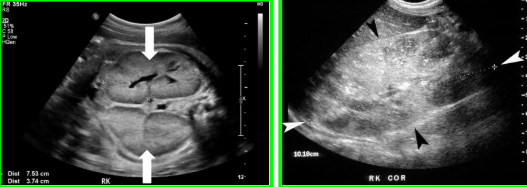
Autosomal Dominant Polycystic Kidney Disease (ADPKD)
More common than ARPKD. Bilateral renal enlargement due to formation of many cysts of various sizes. The cysts gradually enlarge as the patient ages. Over time cysts will compress and destroy normal tissue.
ADPKD indications
Chronic UTI’s, Stone Formation, Back Pain, Headache, Increasing Abdomen Size, High blood pressure, Renal Insufficiency. (50% diagnosed with renal hypertension)(50% will need dialysis or transplant due to renal failure). Increased Bun & Creatinine, Decreased GFR
How is ADPKD usually diagnosed
Typically seen sonographically around 20-30 years of age with sonography, MRI, CT, Xray.
Treatment options for ADPKD
Medications, lifestyle modifications, dialysis, transplant.
Prognosis: Worse as pt gets older due to complications relating to: HTN - Renal Insufficiency and then failure - with a need for dialysis, transplant.
Radiological Images description ADPKD
Cysts may also be present in the liver, pancreas, and/or spleen. Multiple cysts noted.
ADPKD image