mri lec 2
1/30
There's no tags or description
Looks like no tags are added yet.
Name | Mastery | Learn | Test | Matching | Spaced | Call with Kai |
|---|
No analytics yet
Send a link to your students to track their progress
31 Terms
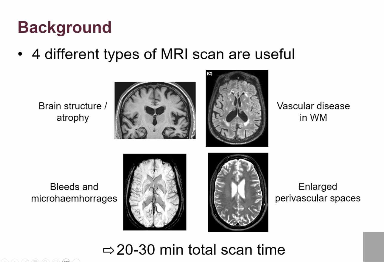
list the 4 different types of MRI scans:
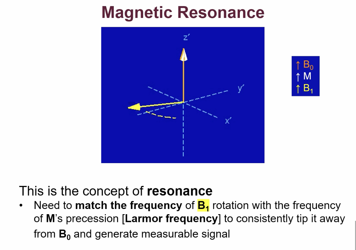
definition of resonance:
when something that moves back and forth gives a very strong response because it is being driven at the same frequency as its natural motion/frequency.
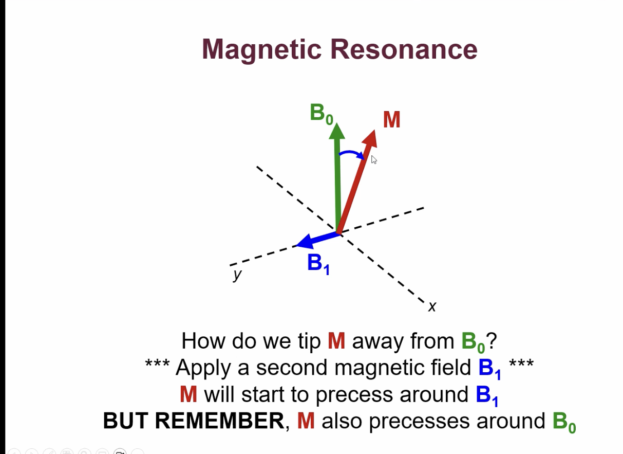
what is magnetic resonance?
when subject is in MRI scanner the bulk magnetisation(M), aligns along the B0 direction. Because M isn’t moving we can’t detect it, so we need to tip it so it can precess.
to tip M away from B0 we need to apply a second magnetic field(B1) perpendicular to M.
M starts to precess around B1.
M will soon then be on opposite side of B0 Now B1 will push M back up towards B0.
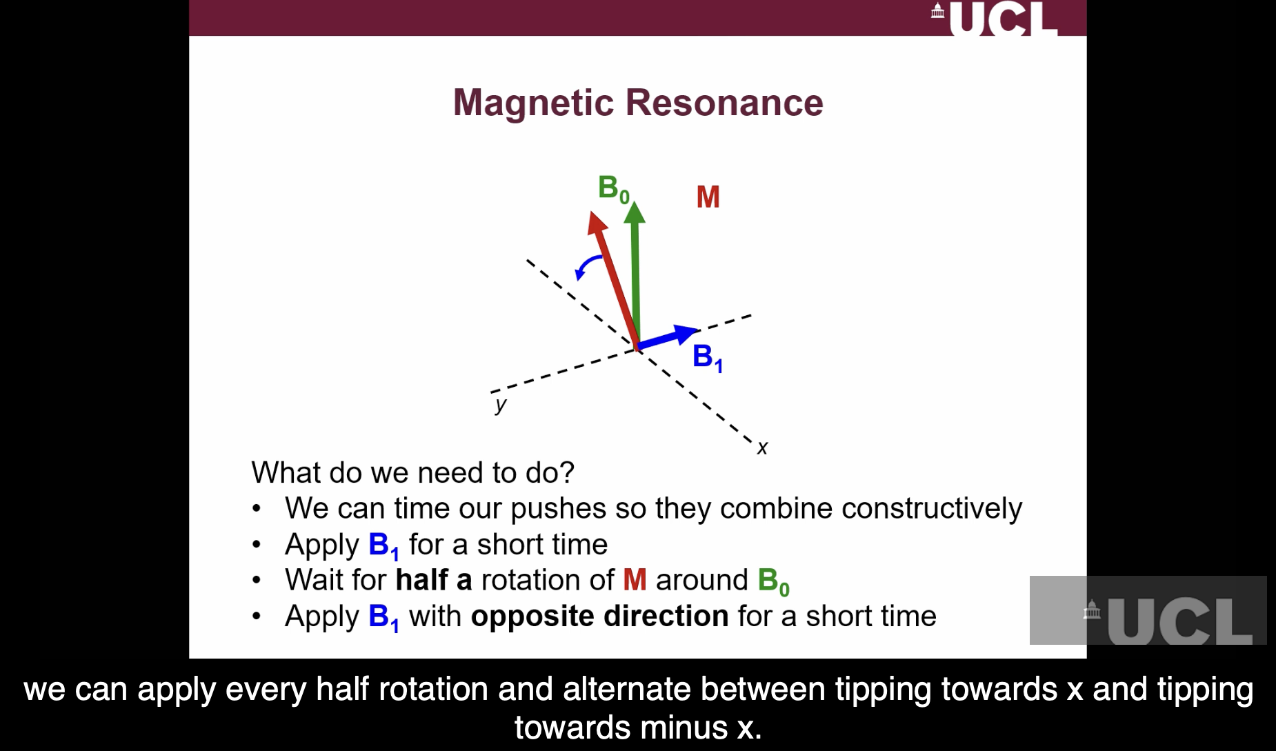
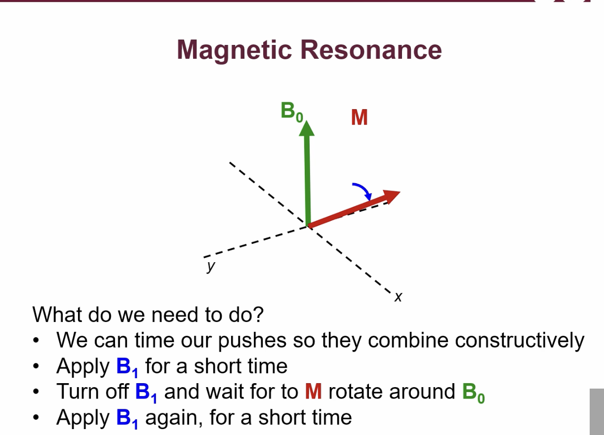
What 2 ways can we time the precessions/pushes in magnetic resonance so they combine constructively?
what is the concept of resonance?
what is a radiofrequency pulse?
the B1 field that is applied to tip M away from B0
magnetic field that is turned on + off.
rotates at larmor frequency , to stay synchronised with M as it precesses.
what is used to create the B1 field?
RF coils
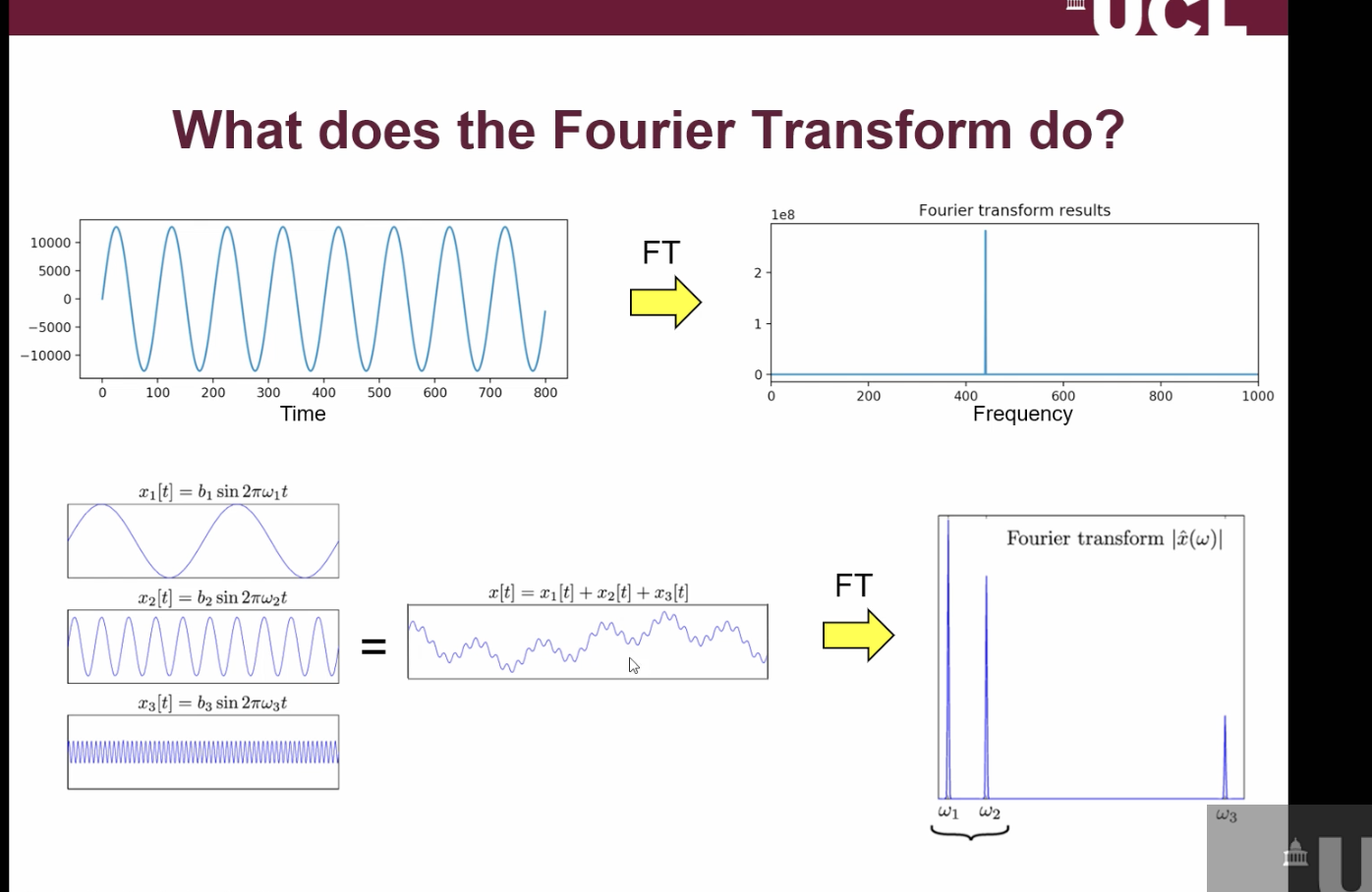
what is the Fourier transform?
equation that takes the signals that you have and tells you how much of each frequency it contains + therefore how much signal is coming from each position within the MRI scanner.
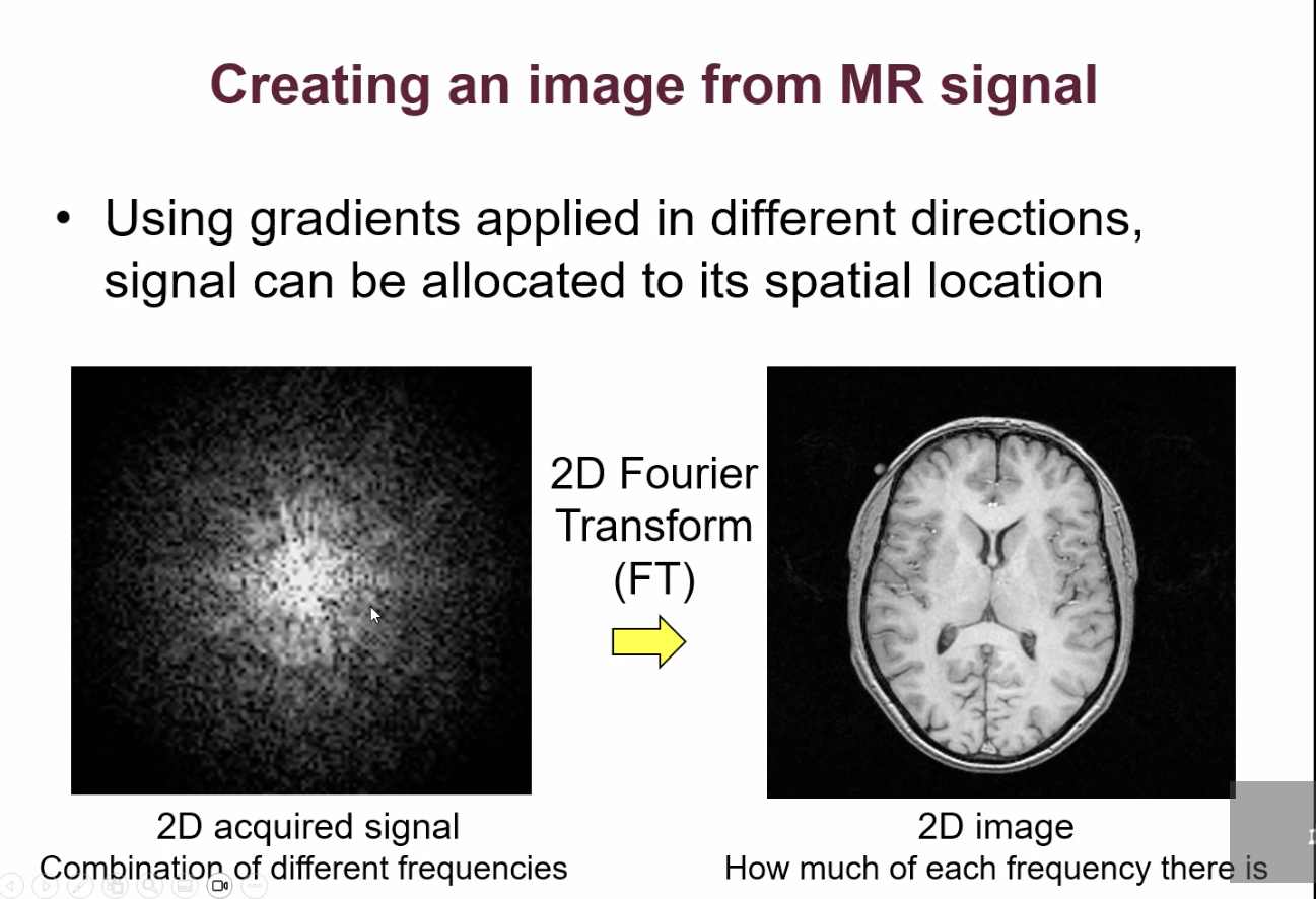
How can we create an image from MR signal?
using magnetic field gradients
what are the 2 main purposes of RF coils in MRI scanner?
to detect signal
to tip M away from B0
What is the main purpose of the main magnet in the MRI scanner?
to align spins and generate M/bulk magnetisation.hat is the mIN PURPOSE OF MAGNETIC FIELD GRANDIENTS w
What is the main purpose of magnetic field gradients in MRI scanners?
to make signal frequency depend on position
How do you get contrast in MRI images?
water concentration in tissues
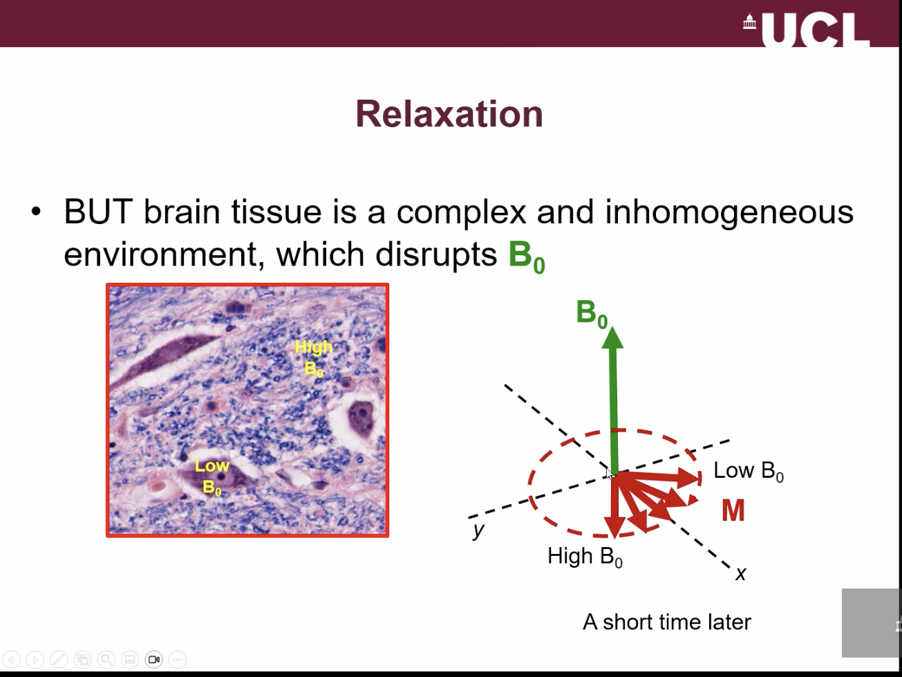
what is the concept of relaxation:
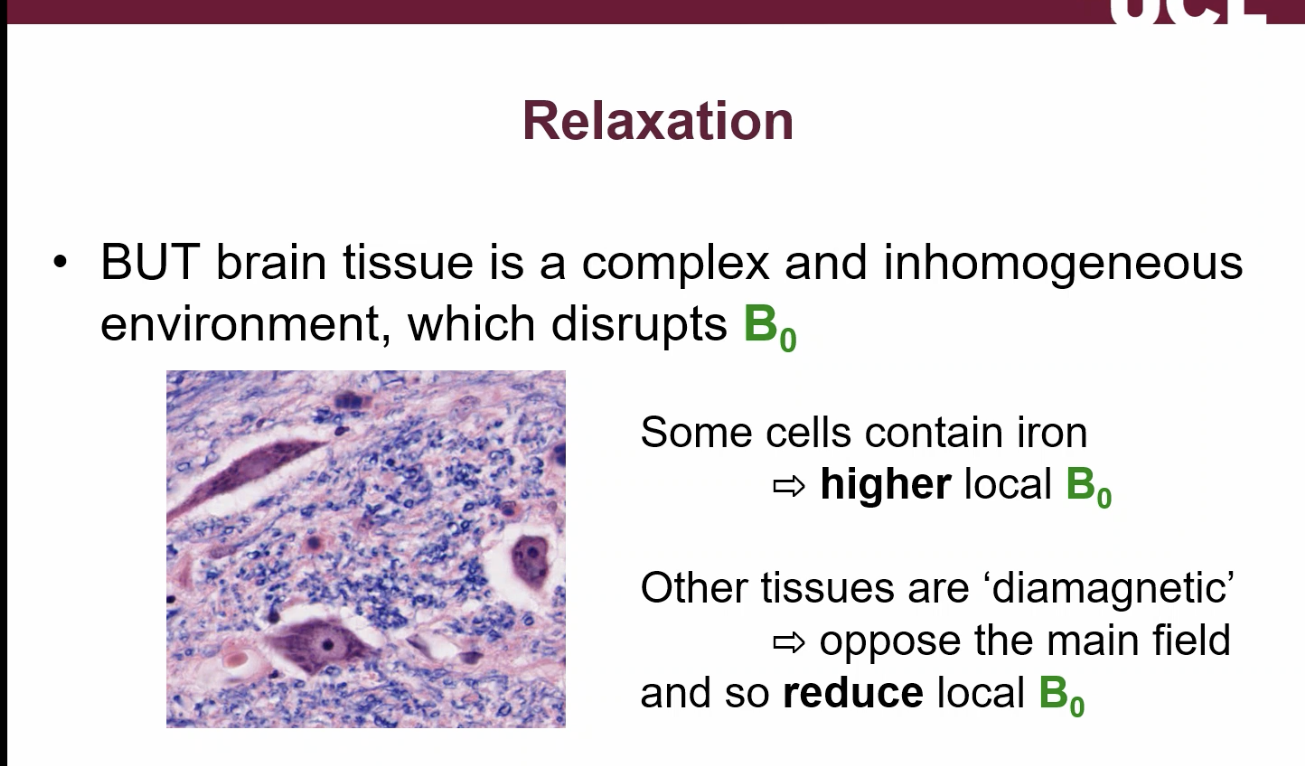
what happens after M is tipped into the XY plane in relaxation?
after M is tipped into XY plane, water which is closer to the region where there is a higher B1 will precess faster (because precession frequency is related to the B0 field).
water in the region where there is a lower B0 will precess slower.
so we now have spreading of the magnetisation.
difference in frequencies cause further spreading out of the magnetisation over time.
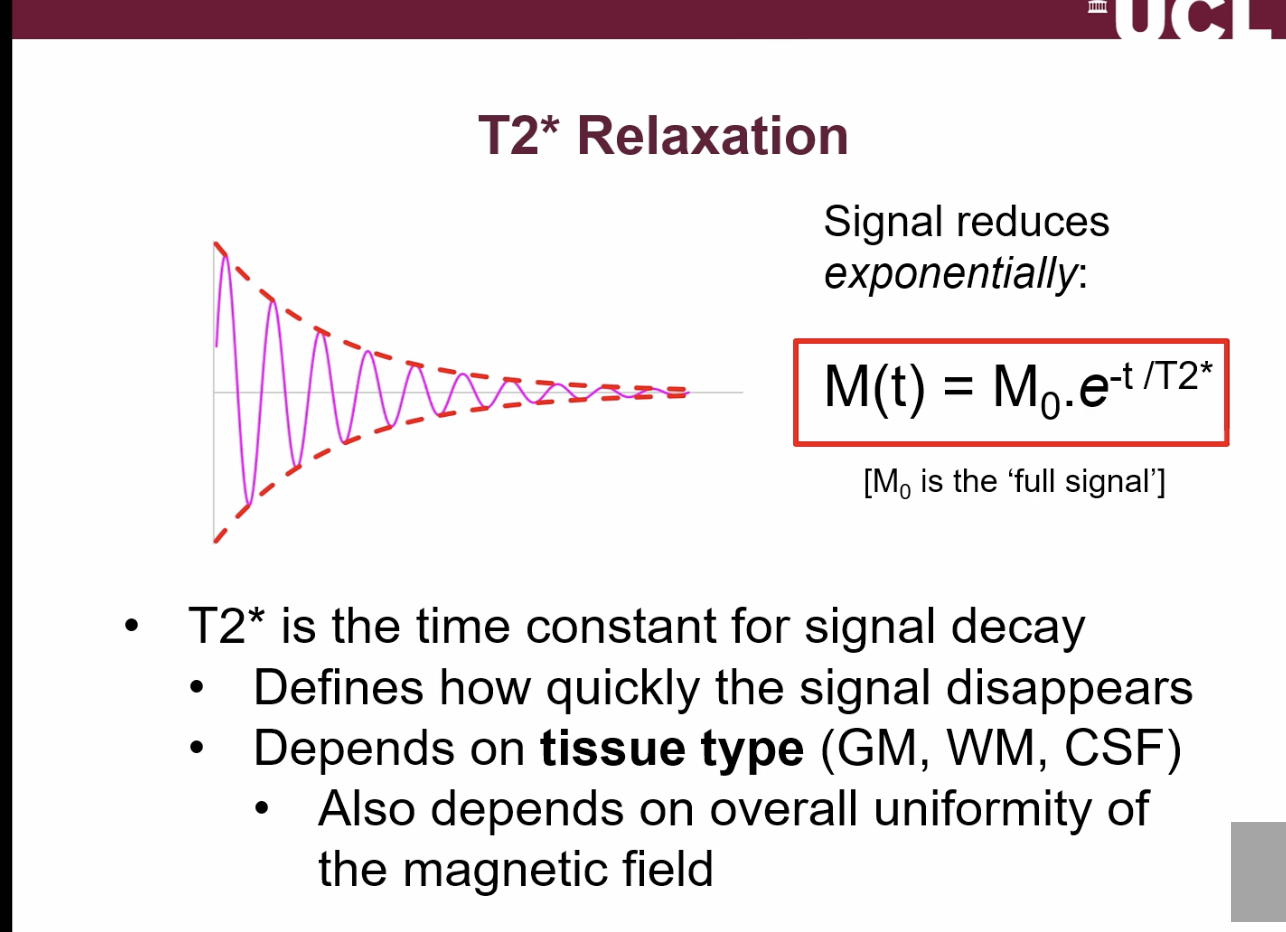
what effect does relaxation have on the signal that we measure/what is T2* relaxation?
after the excitation/90 degree pulse, the signal gradually decreases as the signal gets less coherent as the magnetisation cancels itself out.( magnetic field lines going in opposite directions tend to cancel each other out)
what does T2* stand for?
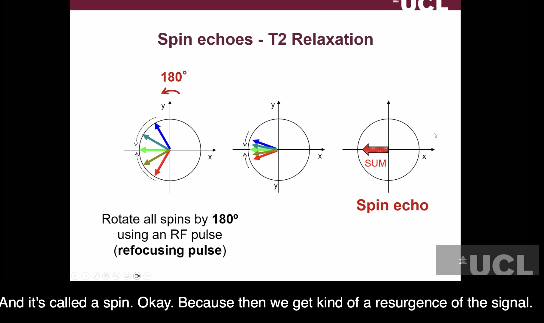
What is T2 Relaxation?
uses spin echoes to compensate for loss of signal in T2*.
to recover some of the lost signal we apply a second RF pulse/180 degree pulse.. This takes the magnetisation + flips it 180 degrees. the spins/magnetisation vectors then start to move in the same way = spin echo.
leads to signal coming back
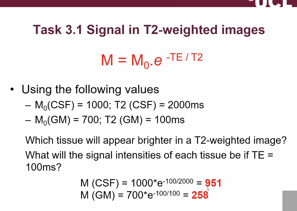
describe what the T2 values are for fluids and tissues:
fluids have long T2 and are homogenous/uniform. long T2 gives strong contrast.
tissues with complex microstructure have a shorter T2
What is the equation for exponential decay according to T2?
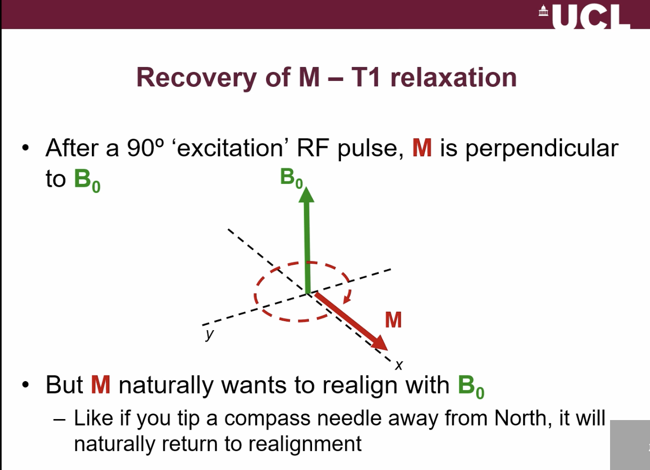
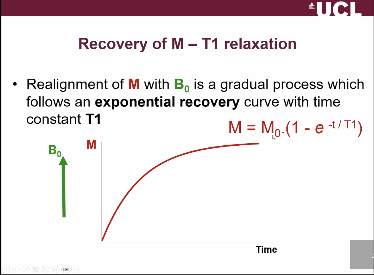
What is T1 relaxation?
T1 relaxation is the way M returns back to B0.
IT returns by an exponential recovery
Why are repeated acquisitions used in MRI?
To build a 2D or 3D image using different gradient directions.
What is repetition time (TR)?
Time between successive excitation and acquisition cycles; controls T1 contrast.
How does TR create T1 contrast?
Different tissues recover bulk magnetisation at different rates during TR → different signal.
Compare short T1 vs long T1 tissues
Short T1: fast recovery → high signal
Long T1: slow recovery → low signal
What happens if magnetisation hasn’t recovered before the next excitation?
Less magnetisation in the x-y plane → weaker signal detected.
comparing what TR and TE (echo time) are used for:
TR (repetition time) → controls T1 contrast
It depends on how quickly tissues recover bulk magnetisation between excitations.
TE (echo time) → controls T2 contrast
It depends on how quickly the signal decays after excitation.
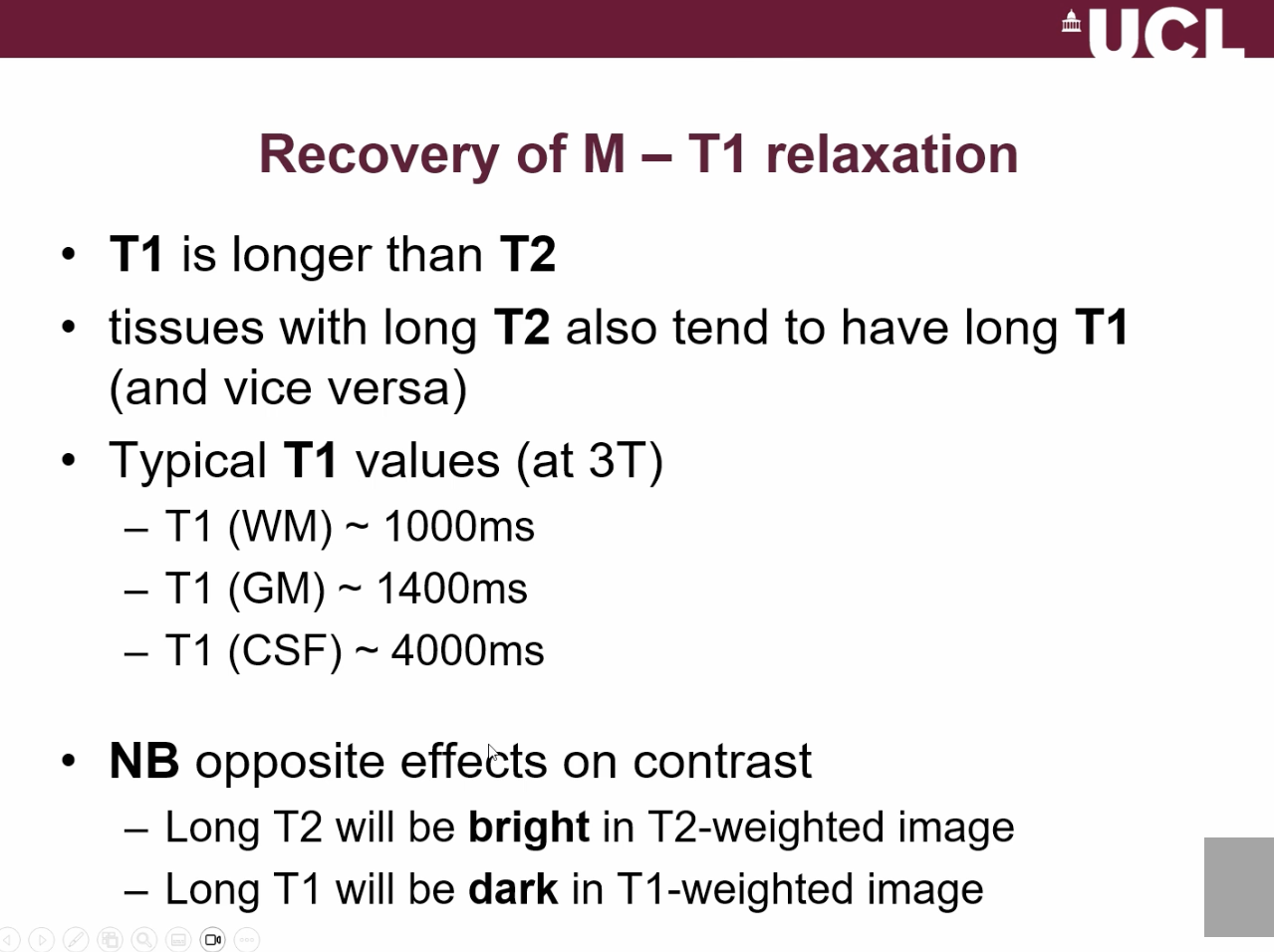
list 3 ways in which T1 + T2 are related?
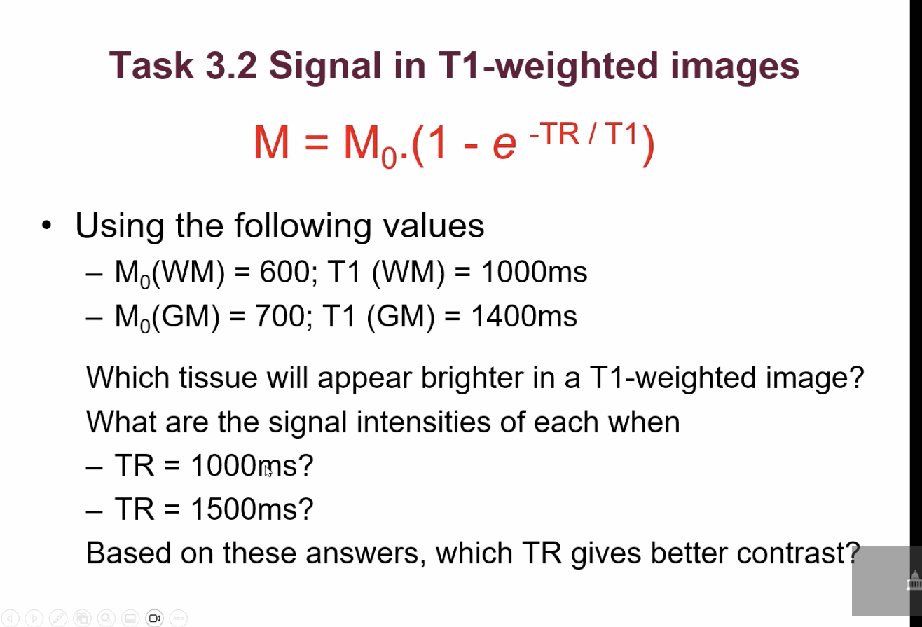
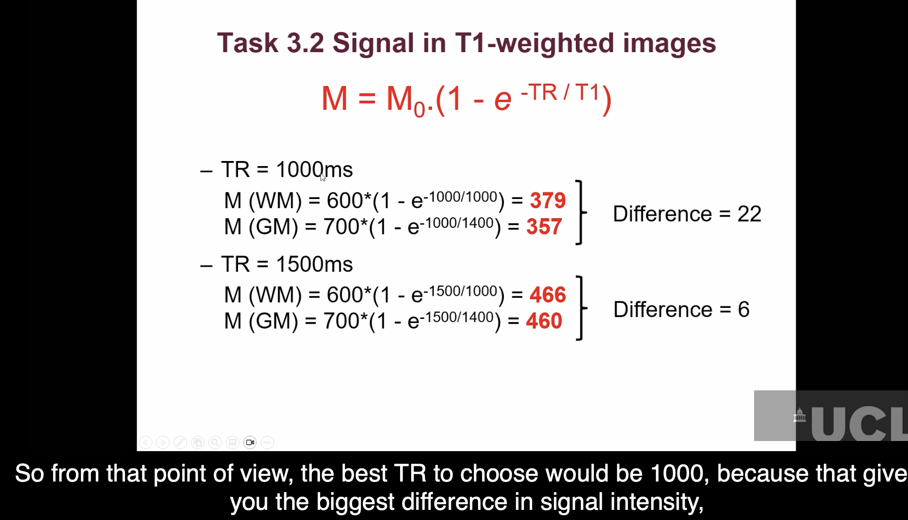
what is the equation to calculate signal in T1 weighted image?
What are T1-weighted images used for?
Structural imaging and segmentation into grey matter, white matter, and CSF; used to measure tissue volume and cortical thickness.
What are T2-weighted images used for?
Structural + clinical imaging, especially for detecting pathology (e.g., stroke, lesions, cysts) where fluid/CSF appears bright.