fundamentals of physiology
 Call Kai
 Learn
Learn Practice Test
 Spaced Repetition
 Match
Match Flashcards
 Knowt Play
1/309

There's no tags or description
Looks like no tags are added yet.
Name | Mastery | Learn | Test | Matching | Spaced | Call with Kai |
|---|
No analytics yet
Send a link to your students to track their progress
310 Terms
what is physiology used to understand
● how a healthy body works
● how to maintain health (and age well/ trying to maintain quality of life as well as expand lifespan - especially important due to ageing population)
● how living organisms cope with or adapt to different
environments e.g. temperatures, altitudes
● what goes wrong in disease (pathophysiological
conditions)
● how to treat diseases
what is the The internal environment
fluid environment around cells
why must the internal environment be maintained
● Cells need a carefully regulated
fluid environment to function.
● The internal environment must
remain stable despite changes in
the external environment.
● The relatively constant steady
state of our internal environment
is achieved by homeostasis.
homeostasis
homeostasis as an active process that keeps the body in its steady state.
homeostasis vs equilibrium
homeostasis is a steady state and not an equilibrium.
when something's in equilibrium, it's kept in balance with no energy being applied, whereas we're in a steady state, which means we're keeping everything in our bodies in balance, maintaining the appropriate internal environment, but it's costing us energy to do so.
examples of external environment
the space in the alveoli
lumen of gastrointestinal tract
examples of internal environment
cardiovascular system
epithelial lining of the lungs
epithelial lining of the GI tract
lots more
what is the internal environment divided into
extracellular fluid, which is every fluid that's outside of a cell, and we have our intracellular fluid, which is inside of the cell,
extracellular fluid, can divide into three further fluids
blood plasma
interstitial fluid (IF) - anything surrounding cells
transcellular fluid (don't need to know)
what are Vital parameters
these are the things that if they get too much out of balance, they will cause imbalance in your body and cause you to kind of move into a state of dysfunction or unhealthiness.
what is in the blood plasma (ECF)
• Oxygen
• Glucose
• Ions e.g. Ca2+, K+, Na+, H+ (pH)
what does blood plasma regulate
volume
Osmolality
what is in Interstitial fluid (ECF)
• Glucose
• Ions e.g. Ca2+, K+, Na+, H+ (pH)
what does interstitial fluid regulate
Osmolality
what is in Intracellular fluid (ICF)
● ATP
● Glucose
● Ions e.g. Ca2+, K+, Na+, H+ (pH)
what does Intracellular fluid (ICF) regulate
● Volume
● Osmolality
what must be regulated in terms of the whole body
Arterial blood pressure
core temperature
what maintains the steady state of vital parameters
homeostasis
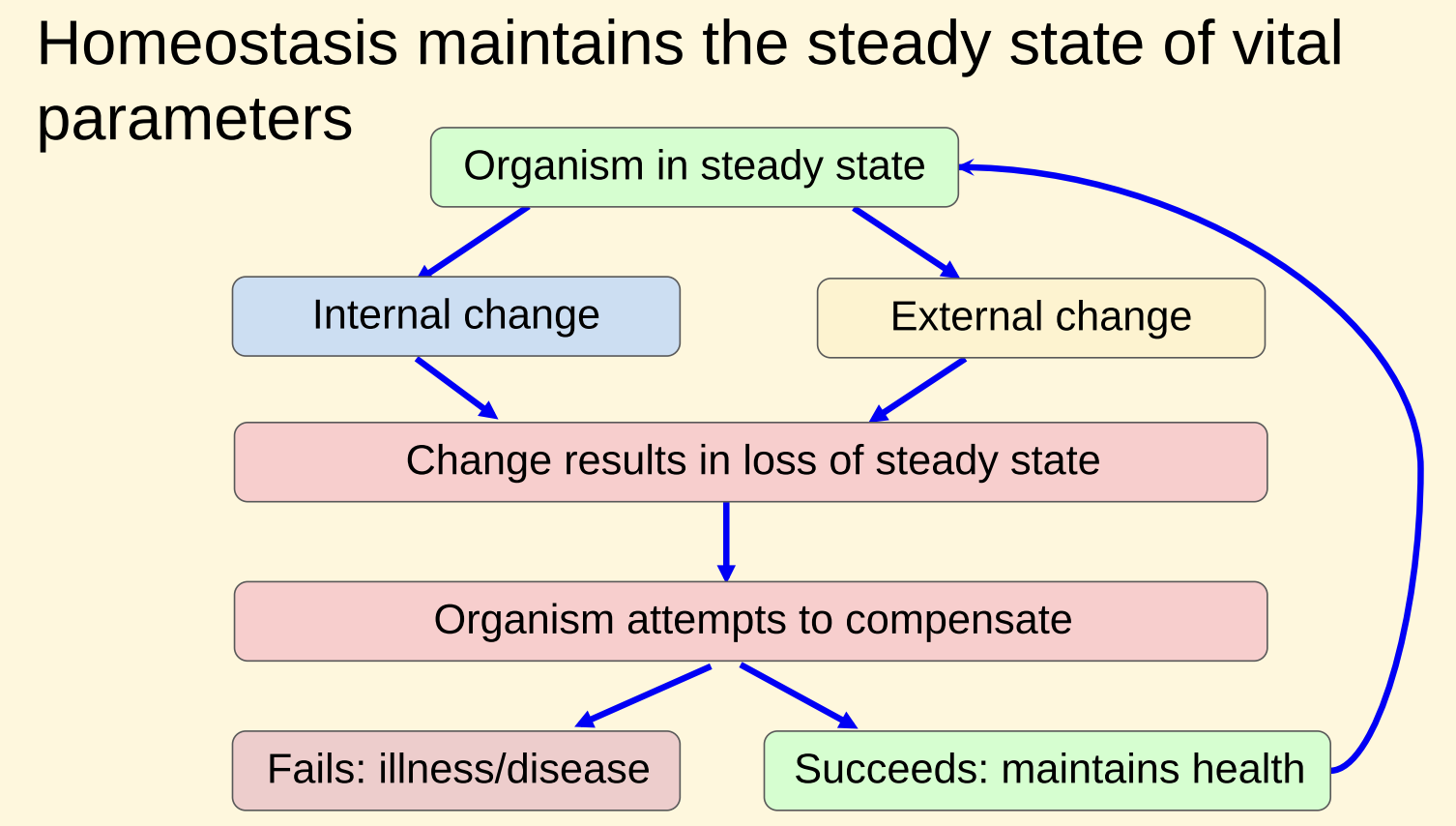
4 essential components of the negative feedback loop
Steady state disrupted
1. Receptors - Sense the vital parameter (input)
2. Control centre - Compares input against a set point
4. Effector - Enables a change to return vital parameter
3. Output signal - Signal from control centre to effector
where are receptors in the negative feedback loop normally found
usually found in the brain:
hypothalamus
medulla
pons
output signals in the negative feedback loop
nervous signal
secretion via the endocrine system
features of Thermoregulation: a negative feedback loop
● Physiological adjustment in the opposite direction
● Returns parameters back to their original level/value
● Redundancy - multiple mechanisms present for many vital parameters
order of body fluid compartments size in the internal environment
Intracellular fluid 25L
Interstitial fluid 13L
blood plasma 3L
Transcellular fluid 1L
osmolarity
the amount of water particles
Osmolality
Total concentration of all particles that are free in a solution
mOsm
milliosmoles per kg of water
is Na+ conc higher in ECF or ICF
ECF
is K+ conc higher in ECF or ICF
ICF
is Cl- conc higher in ECF or ICF
ECF
is Ca2+ conc higher in ECF or ICF
ECF but very small amounts in both
do have quite a lot of intracellular calcium, but not in the intracellular fluid
what separates blood plasma and IF
Capillary endothelium
what separates IF and ICF
cell (plasma) membrane
what is the phospholipid bilayer impermeable to
○ large molecules e.g. proteins,
nucleic acids
○ charged, water soluble
substances e.g. ions
what is the phospholipid bilayer permeable to
● hydrophobic molecules e.g. O2, CO2, steroid hormones
what is the phospholipid bilayer partially permeable to
uncharged polar molecules
what are steroid hormones derived from
derived from cholesterol, so they can move through a membrane quite easily
functions of the cell membrane
the phospholipid bilayer and specific membrane proteins:
● maintain homeostasis
● allows the movement of specific substances
● results in the development of a membrane potential
● allows the development of action potentials
tissues definition
group of similar cells and products arising from same embryonic region, working together to perform a specific physiological or structural role
what are the Four broad classifications of tissues
● Epithelial tissue - protective/barrier
● Muscle tissue - movement/heat generation
● Nervous tissue - communication/coordination
● Connective tissue - structural support/connecting
how do the Four broad classifications of tissues differ
in types + functions of cells; characteristics of the ECM; space occupied by cells/ECM, eg. muscles and epithelia ECM is scarce - much more prevalent in connective tissue
what is Epithelial Tissue
Epithelial Tissue
features of Epithelial Tissue
● First line of protection/defense - physical (stratified), chemical, biological
● Control permeability - selective physical barrier
● Secretes - mucus/enzymes onto external/internal surface, hormones into
blood, ions, acid/alkali
● Absorbs - water, macromolecules, ions
● Diffuses - gases through capillaries: tissues and lungs (squamous)
epithelia tissue common properties
● Highly cellular - tightly packed continuous cell layer → sheets
● Polarised - distinct apical + basolateral domains determine function
● Basement membrane (even simple squamous)
● Avascular + innervated - no blood vessels running through but do have nerves
● Regenerative
● Intercellular adhesion - connected by tight junctions → properties
what are the 3 types of muscle tissue
● Skeletal: Voluntary movement - bone + soft tissues e.g. tongue/
upper oesophagus, eyes, diaphragm, face, sphincters anus/urethra
● Cardiac: Heart only not vessels, contracts involuntarily to pump blood
● Smooth: Organ walls (e.g. GI tract) to move substances, involuntary
shared properties of muscle tissues
Myocytes; excitability; contractility; extensibility
skeletal muscle tissue properties
long bundles striated multinucleated fibres (cells fused in development) surrounded by connective tissue
cardiac muscle tissue properties
highly branched, striated, 1-2 nuclei, intercalated discs + gap junctions→ functional syncytium
smooth muscle tissue properties
no striations, overlapping sheets spindle-shaped cells, single nucleus, gap junctions, dense bodies
functions of neurones
carry electrical signals (action potentials) to each other to control + coordinate bodily function in CNS,PNS + ENS
structure of nerve cells
● Cell body - cellular functions
● Dendrites - cytoplasmic processes carry
impulses to cell body
● Axon - carries impulses from cell body, forms
synaptic connections
how many types of cells in nervous tissue
two
what is the ENS
the enteric nervous system. A separate nervous system, although it's in the periphery that sits within the gastrointestinal tract.
the GI tract is such a large organ system and requires such a lot of control and coordination so has its own immune system
role of Neuroglia (glial cells)
Support, protect, insulate, provide nutrition to neurones
depending on where you are, whether you are in central nervous system or the peripheral nervous system, some of the supporting cells will be given different names. PNS - Schwann cells, CNS - might have oligodendrocytes.
role of Nervous Tissue ECM
ECM supports cells - soft, porous dynamic + biologically active tissue network. Helps direct brain development, maturation + aging, synaptic function.
ECM remodeling heavily linked to brain aging + disease pathology
e.g. neurodegeneration, cancer, epilepsy
components of the nervous tissue ECM
● Hyaluronic acid (HA)
● Proteoglycans
● Interstitial matrix
● Perineuronal nets
CNS ECM - 10-20% total brain volume:
Microglia (Brain Scouts) function
brain's resident immune cells, constantly moving their branches to "survey" the dense, interconnected web of neurones and astrocytes
Enteric glia present in ENS
examples of connective tissue
● Specialised cells in CT defend body from microorganisms that enter
● Transport of fluid, nutrients, waste, chemical messengers by blood/lymph - specialised fluid CT
● Sheath surrounding muscle cells
● Tendons attach muscles - bones
● Skeleton
● Fibrous capsules/bones around organs
● Triple layer in brain meninges
● Adipose cells store surplus energy (fat) and thermally insulate the body.
role of connective tissue
Performs many functions in the body, supporting/connecting other tissues
how is the connective tissue formed
Derived from embryonic mesodermal layer→mesenchyme: highly varied, static → dynamic
Three main structural components of connective tissue
1. Specialised cells: (fibroblasts, adipocytes, macrophages, mast cells
and leukocytes)
2. Protein fibres:
○ Collagen: tensile strength - resists pulling forces (± reticular)
○ Elastic: flexibility + recoil
3. Ground substance (Interfibrillar matrix):
○ Fluid - transport/communication e.g. blood
○ Gelatinous - support/shock absorption e.g. cartilage
○ Calcified - rigid support/protection e.g. bone
what are reticular forces fibres
a specialist type of collagen fibre.
classification of connective tissue
connective tissue → proper + specialist
proper → loose (areolar) + dense (regular, irregular, elastic)
specialist → (adipose, cartilage, bone) + (blood, lymph)
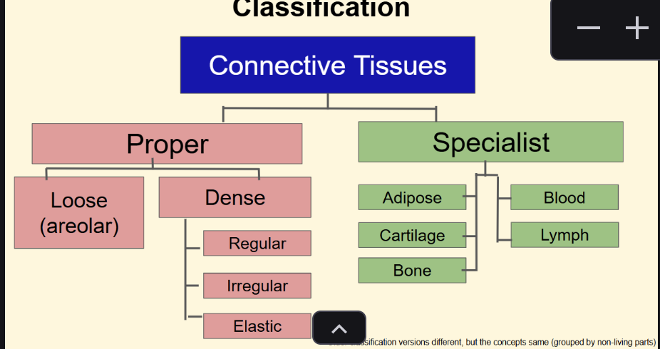
functions and features of CT proper - loose
Holds organs, structures, tissues in place:
❖ beneath epithelia
❖ surrounds blood vessels/nerves/oesophagus/trachea
❖ cell ‘mesh’ lymph nodes/spleen/bone marrow/liver
❖ fascia between muscles (lubricating HA between)
❖ mesenteries, visceral pericardium, lung pleura, meninges(2)
● Highly vascular (esp. skin)
● Variable spaces between fibres, more compact under skin
● Cells < gelatinous ECM. Collagen + elastic fibres embedded
(± reticular), permitting ECF diffusion
● Fibroblasts > adipocytes, macrophages, mast cells, leukocytes
functions and features of CT proper - dense
High tensile strength, densely packed collagen > ground substance + cells
Arrangements:
● Regular (parallel fibres of tendons/ligaments: strength/shock absorption)
● Irregular (multidirectional fibres e.g. dermis)
● Elastic (embedded elastin/fibrin e.g. arteries: concentric rings)
examples of Connective tissue migrating cells
Interstitial leukocytes - ‘immuno-wanderers’:
○ Dendritic cells - peripheral tissues → lymph nodes
○ Plasma cells - loose connective tissue < lymphoid tissue → antibodies
○ Monocytes - when stimulated → macrophages
○ Macrophages - organ specific, e.g. move over alveolar surface → phagocytosis
○ Eosinphils - WBC/leukocyte migrate through capillary walls to connective tissue
○ Mast cells - connective tissue (NB skin/gut) → release histamine
types of Specialised CT - Solid
adipose tissue - White/brown/beige/pink - determined by organelles
Lipid storage, secretory, insulating, shock absorbing
cartilage -
types of Specialised CT - Fluid
blood - Connects all bodily systems, RBC, WBC, platelets. Nutrients, salts, waste dissolved in ground substance
lymph - Collects from interstitial fluid, returns to blood to maintain fluid levels, immune response - liquid matrix + WBC
function and developmental origin of epithelia
- barriers to separate compartments
● Functional interface between two environments, e.g.:
○ Skin separates the body from the external environment
○ External environment ‘enters’ the body via respiratory system and GI tract
● Most abundant and functionally diverse tissue - all organs
● Develops from all germ layers
examples of function of epithelia
dynamic barriers that cover surfaces, line cavities and form glands:
Protection - skin
Selective barrier - BBB
Contractile - myoepithelia
Diffusion/waste - lung
Absorption - SI
Secretion - glands
Filtration - podocytes
Lubrication/propulsion - trachea
Epithelia - common properties
● Highly cellular - tightly packed continuous cell layer → sheets
● Polarised - distinct apical + basolateral domains
● Basement membrane
● Avascular + innervated
● Regenerative
● Intercellular adhesion - connected by tight junctions → properties
● Tissue specific specialisations:
○ Isoforms of laminin/collagen IV
○ Proteoglycan + accessory protein variation
○ Assembly/arrangement
● Upper BL secreted by epithelial cells
○ Collagen IV - main structural component
○ Laminin - ‘molecular glue’
● Lower RL from connective tissue fibroblasts
(collagen III + VII )
how does the basement membrane of epithelia prevent cancer
many cancers formed from epithelia cells, basement membrane stops them metastasizing and moving elsewhere in the body.
basement membrane composition
A mesh of interconnected fibres
● Physical foundation - anchors + separates
epithelial tissue from connective tissue
● Selective filter supplying nutrients, oxygen +
removing waste
● Guides epithelial cell migration during wound
healing
● Prevents carcinomas accessing lymph
what drives passive transport?
electrochemical gradients
● Always depends on the concentration gradient of the solute
● For charged molecules also depends on any difference in voltage between the ECF and ICF (e.g. Cl- and Na+)
what is the Electrochemical gradient
the sum of the 2 forces:
concentration gradient
difference in voltage between the ECF and ICF
what is simple diffusion
Movement of an uncharged, hydrophobic solute (e.g. CO2) through the lipid bilayer
what does Jx = Px ([X]o - [X]i) calculate
● How fast the solute X moves can be described by its flux (Jx).
● Flux (Jx) depends on:
○ Permeability coefficient of X (Px), how easily X can move through the membrane (NB for drug design)
○ Difference in [X] between ECF and ICF (concentration gradient)
![<p>● How fast the solute X moves can be described by its flux (Jx).</p><p>● Flux (Jx) depends on:</p><p>○ Permeability coefficient of X (Px), how easily X can move through the membrane (NB for drug design)</p><p>○ Difference in [X] between ECF and ICF (concentration gradient)</p>](https://assets.knowt.com/user-attachments/b7b61430-e6b7-4229-87ec-a76bc9d919ae.png)
what are transmembrane proteins classed as
● integral membrane proteins
● Composed of membrane-spanning ɑ- helical domains
● Can be single pass or multi pass
● How proteins move in and out of the membrane define a protein’s membrane topology
how do hydrophilic solutes pass through the membranes
transmembrane proteins
what are the types of transmembrane proteins
pore (non-gated channel)
channel (gated pore)
carrier
pump
All have multiple transmembrane segments surrounding a solute permeation pathway
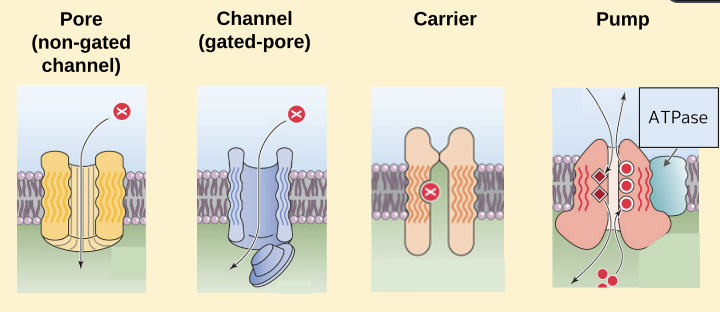
what regulates hydrophilic solutes transport rates
Membrane transport proteins and concentration
how do solutes pass through membrane transport proteins
Solutes pass through the membrane without contact with the hydrophobic membrane core (permeation pathways)
● Amphipathic helices
○ alternating hydrophobic and hydrophilic amino acids
● Hydrophobic surfaces face the lipid membrane
● Hydrophilic surfaces create a central pore
how do Pores allow facilitated diffusion
● Driving force for movement is the electrochemical gradient
● Always open
● Multiple subunits
● Example, aquaporins (AQP)
what is a non gated channel
a constantly open channel that allows the transmission of solutes from one side of the membrane, in this case the extracellular space, into the intracellular space.
how do Channels allow facilitated diffusion
● Driving force for movement is the electrochemical gradient
● Gated ion channels
● Multiple subunits
● Example, potassium channel
important in neurones
what do all channels have
1. a moveable gate
2. a sensor:
○ voltage
○ ligand
○ mechanical
3. a selectivity filter
4. an open channel pore
types of channels
voltage gated
extracellular ligand
intracellular ligand
mechanical-gated
how do Carriers allow facilitated diffusion
● Driving force for movement is the electrochemical gradient
● Never has a continuous transmembrane path
● Example, GLUT (glucose transporters)
what is Flux (Jx) limited by
○ Number of carriers in the membrane
○ Speed by which the carrier can cycle through the steps
Jmax = [X] is high enough to occupy all of the carriers
![<p>○ Number of carriers in the membrane</p><p>○ Speed by which the carrier can cycle through the steps</p><ul><li><p>Jmax = [X] is high enough to occupy all of the carriers</p></li></ul><p></p>](https://assets.knowt.com/user-attachments/67bc9b22-ead7-4262-9f1f-f0446f2e289d.png)
how can Carriers can mediate active transport
● Achieved through the use of:
○ Pumps
○ Cotransporters
○ Exchangers
what are the Two types of active transport
1. Primary active transport uses pumps
● Driving force = a chemical reaction e.g. ATP hydrolysis
2. Secondary active transport uses cotransporters and exchangers
● Driving force = coupling the downhill movement of one solute with the uphill movement of another solute.
how do Cotransporters move both solutes
in the same direction
● Requires a ‘driving’ solute whose electrochemical gradient provides the energy
○ Often the inward Na+ electrochemical gradient
● Example, Na+/glucose cotransporter
*also called symporters
how do Exchangers move solutes
in opposite directions
● Requires a ‘driving’ solute whose electrochemical gradient provides the energy
○ Often the inward Na+ electrochemical gradient
● Example, Na+/Ca2+ exchanger
*also called antiporters
what are the Two routes across an epithelial sheet
● Transcellular - crossing AM and BLM membranes (passing through cytoplasm)
● Paracellular - moving between cells through tight junctions (NB variation ‘leakiness’)
what does a Transepithelial voltage indicate
electrical resistance and permeability of TJs
if we put an electrode in the lumen and inside the cell, we can effectively measure the electrical resistance of the apical membrane.
And then we could do exactly the same on basilitral membrane.
the transepithelial resistance will be the sum of the two.
what is the the transepithelial resistance
the sum of the voltage between the lumen and cell and the international space and cell
whats the purpose of leaky epithelia
‘Leaky’ epithelial perform bulk transepithelial transport of solutes and
H2O in isosmotic conditions, e.g. :
● Small intestine (SI)
● Kidney proximal tubule (PT)
bulk transepithelial transport of solutes and therefore water, because water will eventually follow in isosmotic conditions.
whats the purpose of tight epithelia
‘Tight’ epithelia generate/maintain large transepithelial ion concentration or osmotic gradients, eg. urinary bladder
when does water move passively across an epithelium
H2O moves passively across an epithelium in response to osmotic gradients
Eg. an epithelium secreting salt will secrete fluid and vice versa
● Small water molecules can cross the apical and basolateral membranes, but aquaporins (AQP) ↑↑ H2O permeability: ↑↓ hydraulic conductivity
● Bilayer H2O permeability ensures osmotic equilibration is rapid, however membrane composition + [AQP] regulated