Reproduction #1 Lab
1/63
There's no tags or description
Looks like no tags are added yet.
Name | Mastery | Learn | Test | Matching | Spaced | Call with Kai |
|---|
No analytics yet
Send a link to your students to track their progress
64 Terms

Identify the 1 - “Stage of Development”
Meiosis I

Identify the 2 - “Stage of Development”
Interkinesis

Identify the 3 - “Stage of Development”
Meiosis II
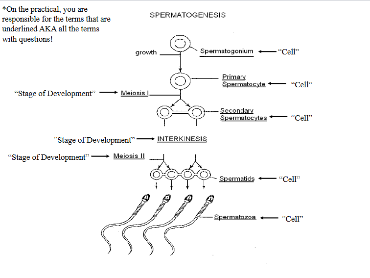
Identify the 1 - “Cell”
Spermatogonium

Identify the 2 - “Cell”
Primary Spermatocyte

Identify the 3 - “Cell”
Secondary Spermatocytes

Identify the 4 - “Cell”
Spermatids

Identify the 5 - “Cell”
Spermatozoa

Identify the 1 - “Stage of Development”
Meiosis I

Identify the 2 - “Stage of Development”
Meiosis I

Identify the 3 - “Stage of Development”
Interkinesis

Identify the 4 - “Stage of Development”
Meiosis II
Identify the 5 - “Stage of Development”
Meiosis II

Identify the 1 - “Cell”
Oogonium

Identify the 2 - “Cell”
Primary Oocyte

Identify the 3 - “Cell”
First Polar Body

Identify the 4 - “Cell”
Secondary Oocyte

Identify the 5 - “Cell”
Polar Bodies

Identify the 6 - “Cell”
Ovum

Identify the 7 - “Cell”
Second Polar Body

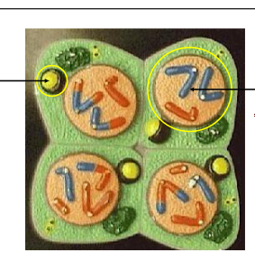
Identify the left circle indicated structure.
Golgi Body

Identify the right circle indicated structure.
Nucleus
Identify the entire cell.
Spermatids

Identify the entire cell.
Spermatozoa
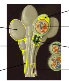
Identify the top left, large circular region.
Head

Identify the left middle, smaller rectangular shaped region.
Neck

Identify the bottom left, the skinny region.
Tail

Identify the top right yellow structure.
Acrosome

Identify the circular structure on the top right.
Nucleus

Identify the green structure on the right.
Mitochondria

Identify the bottom red structure.
Axial Filament

Identify the top indicator, the outermost layer.
Zona Pellucida

Identify the far left structure, the circle inside of the two circles.
Nucleus

Identify the two circular cells.
Polar Bodies
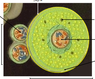
Identify the layer, the top arrow of the 3 on the right.
Yolk Rich Cytoplasm

Identify the center circle structure.
Nucleus

Identify the bottom indicator, the entire green outer layer.
Cortical Zone

Identify the entire larger cell.
Ovum
What do the cortical bodies do?
prevent polyspermy
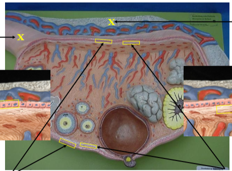
Identify the top X structure, the blue.
Mesovarium

Identify the left X, the pink structure.
Ovarian Ligament
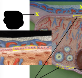
Identify the left layer, the pink cells with blue nucleus.
Germinal Epithelium
Identify the right layer, simulated by white paint surrounding ovary.
Tunica Albuginea

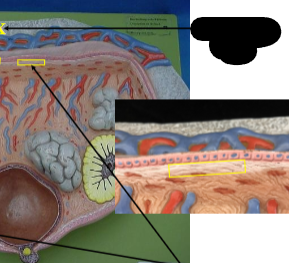
Identify the cell, the top rectangle indicating the blue and red branches.
Myoepithelial Cells
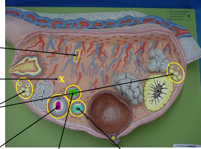
Identify the tissue, the X.
Stroma

Identify the two yellow circles, the multiple white circle structures inside.
Primordial Follicle

Identify the magenta highlighted structure.
Secondary Follicle

Identify the teal highlighted structure.
Primary Follicle

Identify the green highlighted structure.
Atretic Follicle

Identify the outer red layer.
Theca Folliculi Externa

Identify the outer blue layer.
Theca Folliculi Interna

Identify the inner blue layer.
Corona Radiata

Identify the white layer.
Zona Pellucida
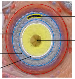
Identify the space on top.
Antrum

Identify the cell in the center, the yellow and brown structure.
Primary Oocyte

Identify the entire structure.
Secondary Follicle

Identify the brown structure, the top arrow.
Mature Follicle

Identify the space, X.
Antrum
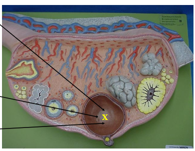
Identify the fluid that fills the space.
Liquor Folliculi

Identify the far left structure, #15.
Corpus Rubrum

Identify the 3 arrow structure.
Corpus Albicans

Identify the small yellow dot.
Secondary Oocyte
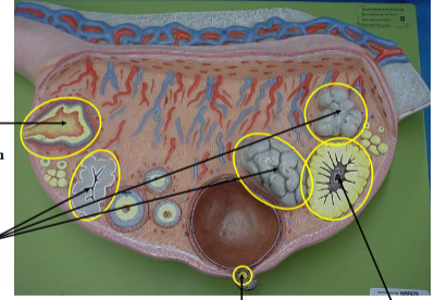
Identify the far right structure #22.
Corpus Luteum
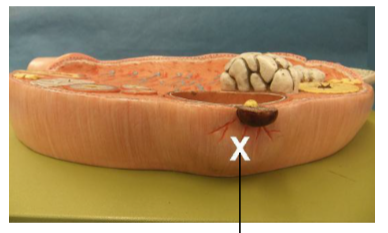
Identify the X structure.
Stigma