PSYCH1003 all cards
1/291
There's no tags or description
Looks like no tags are added yet.
Name | Mastery | Learn | Test | Matching | Spaced | Call with Kai |
|---|
No analytics yet
Send a link to your students to track their progress
292 Terms
Broca & Wernicke achievements
Identified language centres in the brain
What fields are within biological psychology
psychology: study of behaviour/mental processes
neuroscience: study of the nervous system
5 perspectives (DDDEMA)
Describing the behaviour
Evolution of the behaviour
Development of the behaviour
Development of the behaviour over the lifespan
Mechanisms of the behaviour
Application of biological psychology
the 3 approaches
Somatic intervention, behavioural intervention, correlation
What is somatic intervention?
approach involving manipulating body structure and function and looking for changes in behaviour
What is behavioural intervention?
approach that involves manipulating behaviour and looking for changes in body structure/function
What is correlation (the approach)
measures how much a body measure varies with a behavioural measure
Biological explanations for behaviour
physiological
ontogenetic (course of development in an individual organism)
evolutionary
Functional
What happened to Phinneas Gage?
He survived an injury where a metal rod pierced his scull, damaging his frontal lobe. This changed his personality and behaviour
What does the nervous system contain
CNS (central nervous system: brain, spinal cord)
AND
Peripheral nervous system (somatic nervous system, autonomic nervous system)
Somatic nervous system
VOLUNTARY.
concerns voluntary control of movement
sensory input and
motor output (muscle contraction)
Autonomic nervous system
INVOLUNTARY. Contains the:
parasympathetic nervous system: vegetative non emergency responses, energy conservation, calming body down, digestion
sympathetic nervous system: network of nerves that prepares organs for rigorous activity: fight or flight,
Goal: homeostasis (state of balance among all body systems)
Types of neurons in spinal chord
sensory neurons, interneurons, motor neurons
Sensory neurons
carry sensory info from sensory receptors to the CNS
transmit information from receptors to the brain
Interneurons
connect neurons to one-another
Receive the output by sensory neuron
Motor neurons
transmit commands from the brain to glands and muscles and the body
transmit the commands from interneurons to the glands and muscles of the body, often through the spinal cord
carry out voluntary actions like grabbing water, vital bodily functions like digestion and heartbeat
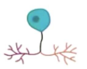
Draw & label a neuron
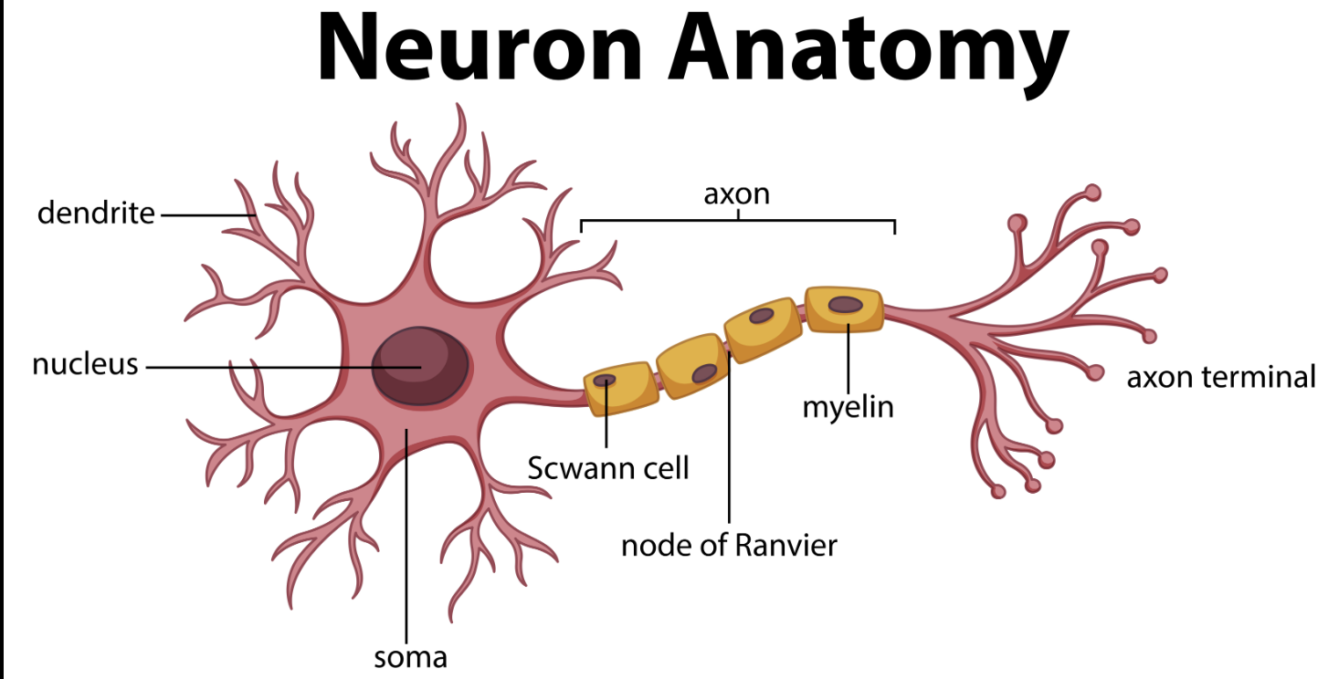
Dendrite: structure & function
Highly branched, tapering processes extending from the cell body of a neuron. | Receives the electrochemical signal from the previous neuron(s), sends to soma |
Cell body/soma structure and function
Contains nucleus, ribosomes and mitochondria | determines how the particular neuron will manipulate input from dendrites, maintains metabolism & health, receives and sends information |
Myelin sheath structure & function
Tight coat of cells primarily composing of lipids surrounding axon | Protects axon from other stimuli that may interfere with transmission of nerve impulses, and increases speeds of message transmission |
Node of ranvier structure and function
Between the cells that from the sheath are small spaces of bare wire | When a neuron fires, the electrical impulse is transmitted rapidly from node to node |
Axon structure and function
Long projection leading from the cell | Conducts the electrochemical signal away from the cell body |
Axon terminals structure and function
Finger-like projections leading from the axon to the next neuron(s) or target cells (such as muscle cells). | where the electrochemical signal leaves the neuron so it can be conducted to the next neuron(s) or target cell |
Afferent vs efferent axons
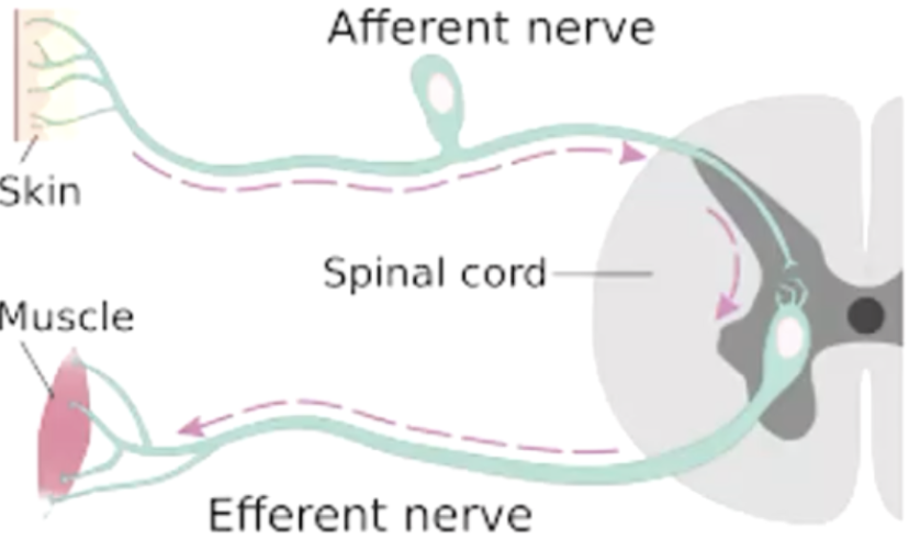
afferent axons bring information INTO the structure (a for admission)
efferent axon: carry information AWAY from structure (e f or exit)
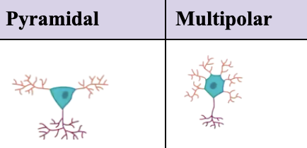
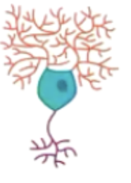
Pyramidal & multipolar neurons
Bipolar neuron appearance & purpose
Used in specialised sensory systems like vision, hearing

Purkinje neuron structure & role
In cerebellum,
motor coordination,
usually inhibitory neurons that fine tune voluntary movements and balance
Unipolar neuron appearance & role
Transmitting sensory information from the periphery to the cns
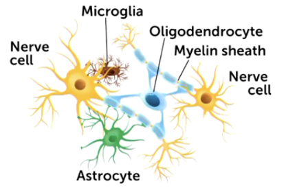
What are the main roles of glia cells
support and protect neurons
maintain homeostasis
forming myelin
modulating synaptic activity
cleaning up synaptic waste
Glia cell-neuron
was believed 10:1, now known its 1:1
Astrocyte role (5)
supportive / structural,
nutrient/waste exchange,
ion balance,
regulating synaptic activity,
repair scar formation
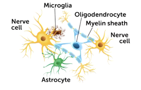
Oligendrocyte role
Found in cns, main role is myelination. One oli can myelinate several axons
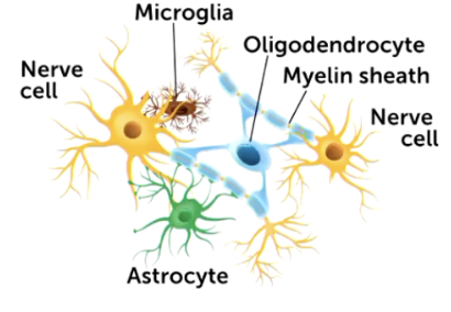
Schwann cells role
Found in periphery, also myelinate similar to olis
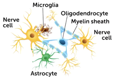
Microglia & radial glia role
Microglia
Located outside CNS, mylinate axons individually
radial glia
Developing brain, mainly embryo, neurogenesis and helping migration of neurons to correct locations. May differentiate into other cells
Firing of a neuron purpose & overview
Neurones mostly communicate at the synapse by converting an electrical charge in one neurone to a chemical message→ when this is released into the synapse it alters the charge of the next neurone and so forth
overall pattern of neural activation is distributed over thousands of neurones
neurones are covered by semi permeable lipid bilayer membrane
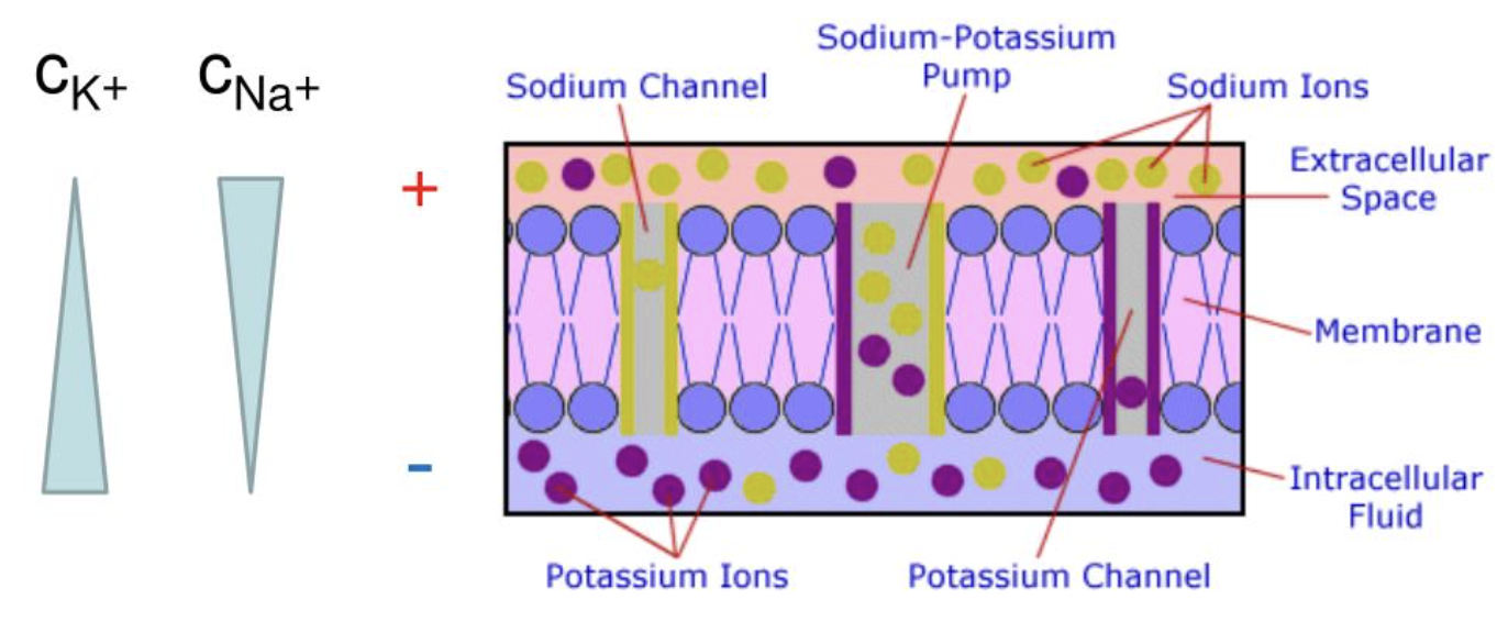
Resting polarisation of a neuron
when a neurone is at rest, its membrane is polarised between inside and outside
the inside of the membrane has a NEGATIVE charge whereas fluid outside the cell has a POSITIVE charge
Resting potential: Electrical gradient
Na+ and K+ is attracted to negative charge inside neuron
Resting potential: What is it?
Resting potential is the stable negative electrical charge across a cell membrane (~–70 mV)
When the neurone is not firing it is in a state of RESTING POTENTIAL (potential as the cell has stored up energy which has the potential to be used)
Resting potential: Chemical gradient
distribution of ions across the membrane
Na+ is more concentrated outside, more likely to be pulled into the neuron
K+ is more concentrated inside so are more likely to be pushed out of the neuron
Resting potential: The 3 factors maintaining
1. Sodium-potassium pump
2. Potassium leak channels
3. Trapped negative ions
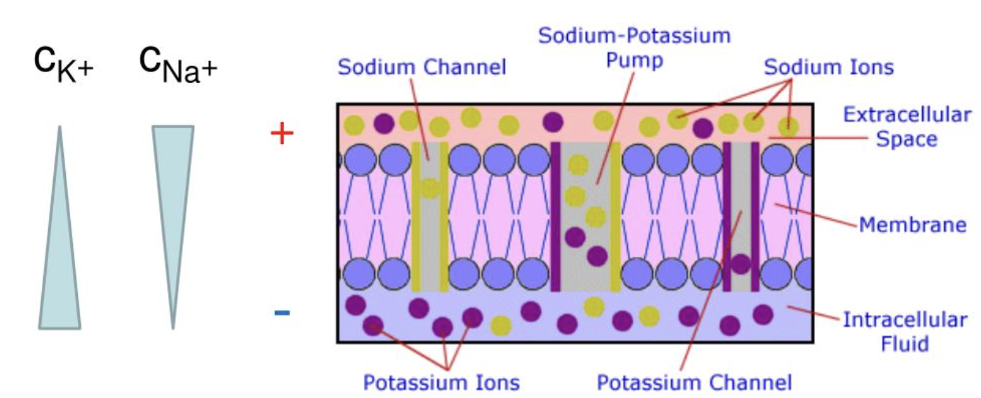
Resting potential: Sodium potassium pump
Actively transports ions using ATP. For every cycle it:
Pumps 3 Na+out of the cell
pumps 2K+ into the cell
hence creating a net loss of positive charge inside the cell and creating a negative resting potential
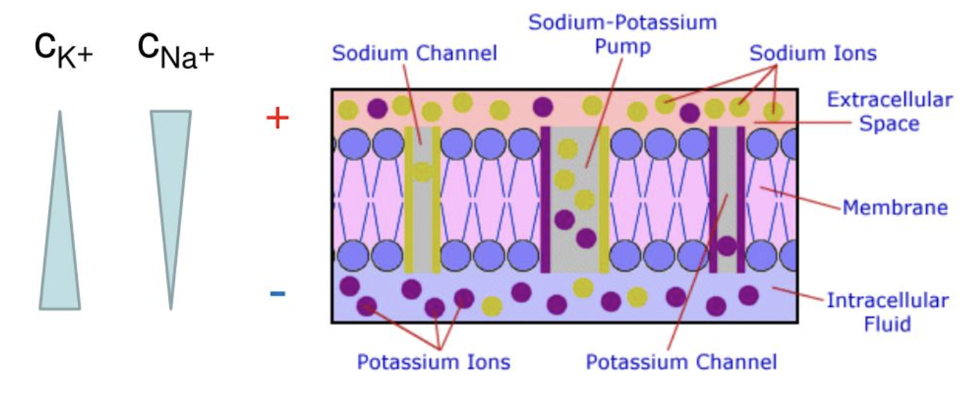
Resting potential: Potassium leak channels
Membrane is more permeable to K+ than NA+
K+ diffuses out of the cell through leak channels
as positive K+ leaves the inside becomes more negative
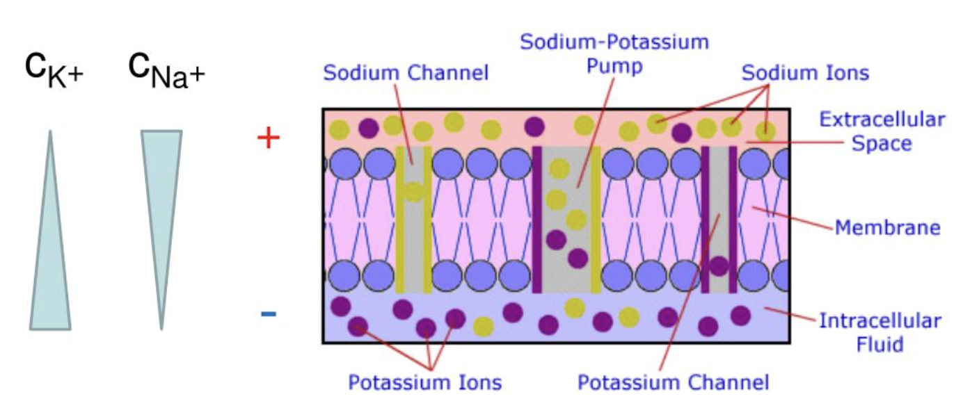
Resting potential: Trapped negative ions
large negatively charged proteins and molecules inside the cell cannot cross the membrane, contributing to negative charge
Graded potentials: What are they?
Small changes in the membrane potential of a neuron occurring when a stimulus affects the cell membrane
called graded because their size varies on the strength of the stimulus
if a threshold is reached, action potential will start
Graded potentials: 4 characteristics
Variable size
local signals
may be depolarising or hyperpolarising
can summate
Graded potentials: Variable size
the response of a graded potential is proportional to stimulus strength; weak stimulus→ small change in voltage and vice versa
Graded potentials: Local signals
graded potentials can occur in dendrites or the cell body (soma)
they spread a short distance along the membrane and gradually weaken
Graded potentials: Depolarising
stimulation can reduce polarisation (depolarise), decreasing voltage discrepancy between the outside and inside
excites neuron (renders it more likely to fire with further stimulation)
comes from an influx of positive sodium ions→ charge inside cell membrane becomes less negative, makes it more likely to fire if further stimulated
Graded potentials: Hyperpolarising
stimulation can increase polarisation (hyperpolarise)
inhibit neuron and make it less likely to fire
outflow of potassium ions, or influx of chloride ions→ potential across membrane becomes more negative, making less likely to fire
Graded potentials: Where do they occur?
most voltage changes occur at synapse along neurones dendrites and cell body, then spread down the cell membrane (like ripples in a pond)
Graded potentials: Summation
graded potentials are cumulative/additive
if a neurone is simultaneously depolarised by -2mV on a dendrite and hyperpolarised by -2mV at an adjacent point, the two graded potentials add up to zero and cancel each other out.
if the neuron is depolarised at multiple points, a greater influx of positive ions occurs, producing a ripple all the way down the cell body to the axon
Graded potentials: Types of summation
spatial: signals from multiple synapses add together
temporal: signals arrive rapidly from the same synapse and add up
Graded potentials: Threshold
If the combined graded potentials reach the -55mV threshold they trigger an action potential
Graded potentials: Event sequence
Stimulus activates ion channels in dendrites or soma
Small voltage change occurs (graded potential)
Multiple graded potentials may summate
If threshold is reached → action potential starts
Action potentials: What are they
A rapid temporary reversal of membrane potential that allows electrical signals to travel along the axon
signal used by neurons to communicate with other neurons, muscles and glands
Resting and activation potential requirements
at rest neurons have a resting membrane potential of -70mV
When stimulation reaches -55mV an action potential is triggered
Action potentials: Stages
Depolarisation
repolarisation
hyperpolarisation
return to resting potential
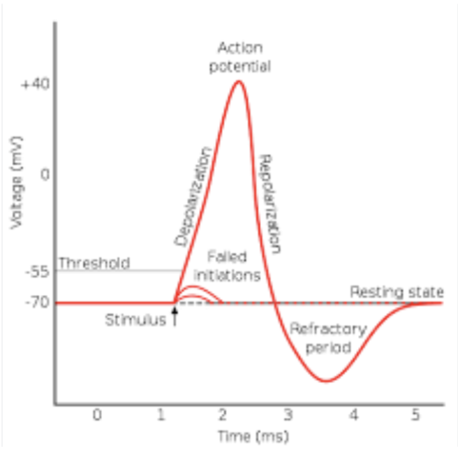
Action potentials: Depolarisation
once stimulus reaches -55mV threshold
voltage-gated Na+ channels open
→ Na+ rushes into the neuron
Hence:
membrane potential rapidly rises up to +30mV
→ inside of neuron becomes positive
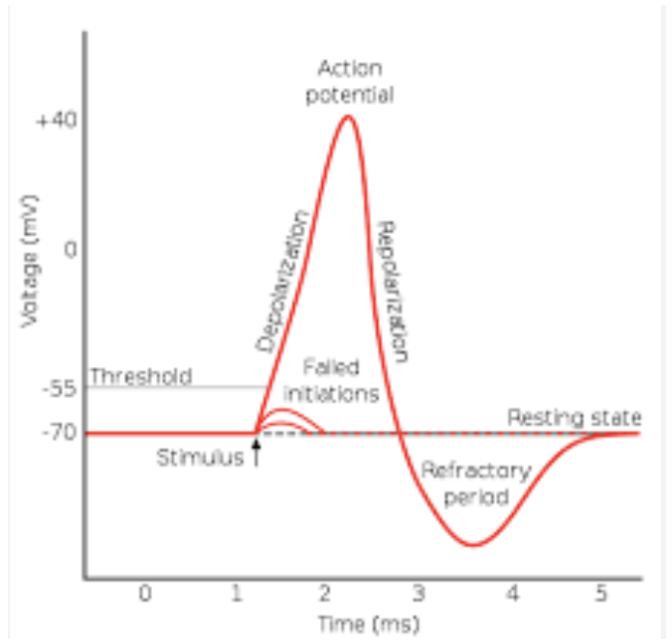
Action potentials: Repolarisation
Na+ channels close
voltage-gated K+ channels open
K+ ions leave the cell causing the membrane potential to become negative again
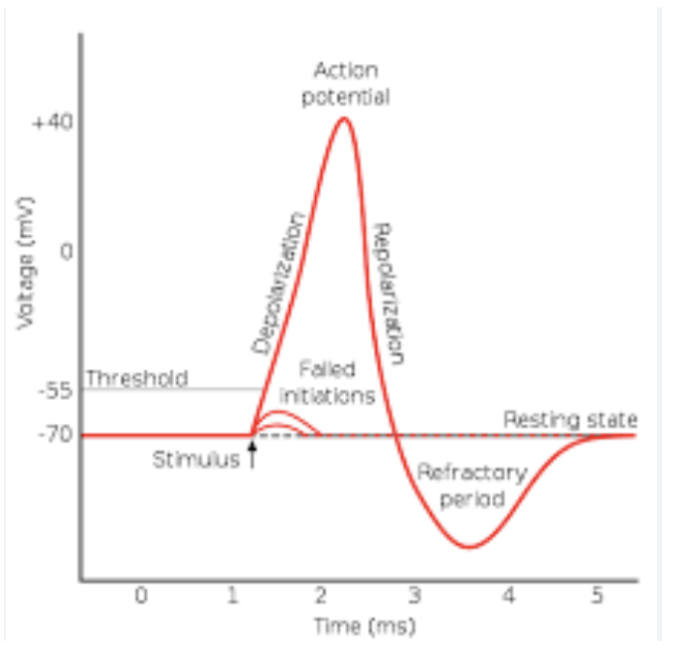
Action potentials: Hyperpolarisation
K+ channels close slowly→ extra K+ leaves the neuron
membrane potential becomes more negative than resting potential (about -80mV)
Action potentials: Return to resting potential
K+ channels close
sodium-potassium pump and leak channels restore normal ion distribution
neuron returns to -70mV
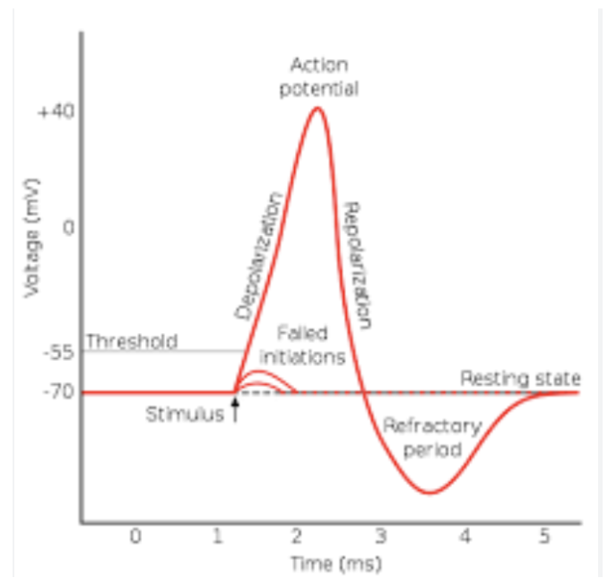
Action potentials: All or none
if threshold is reached, action potential always has same size
stronger stimulus doesn’t make it bigger, only more frequent
Action potentials: Refractory periods
neuron temporarily cant fire another signal)
Absolute refractory period: Na+ channels are inactivated, no new AP can occur
Relative refractory period: during hyperpolarisation a stronger stimulus is required to trigger another action potential
Action potential: Summary
An action potential is a rapid electrical signal in neurons caused by:
This process allows nerve impulses to travel along axons. Rest → Na⁺ In → K⁺ Out → Too Negative → Reset |
Neuron firing process
Neuron maintains resting potential across the entire neuron, including dendrites, cell body (soma) axon and axon terminals
Neuron receives a stimulus (usually via another neuron at a stimulus
opens ligand-gated ion channels to produce a graded potential (occurs in dendrites and cell body)
At the axon hillock all incoming graded potentials are added up.
if the combined signal reaches -55mV an action potential is triggered
action potential occurs along the axon membrane and is propagated along the axon to axon terminals
Once action potential reaches axon terminals, voltage gated Ca^2+ channels open, neurotransmitters are released into the synapse
next neuron begins its own graded potentials
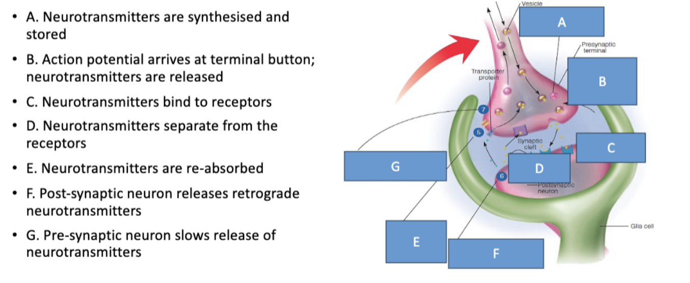
Neurotransmitter absorption and release
Neurotransmitters are synthesised and stored
synthesised at terminal buttons or soma, then stored in the terminal buttons
action potential arrives at terminal button neurotransmitters are realised
Action potential arrives at the terminal button, opens Ca+ channels and releases neurotransmitters (exocytosis
neurotransmitters bind to receptors
neurons release many types of neurotransmitters
neurotransmitters can bind to different receptor types, allowing for complex signalling
neurotransmitters separate from the receptors
neurons are reabsorbed by presynaptic neurons
postsynaptic neurons release retrograde neurotransmitters
negative feedback sites respond to retrograde neurotransmitters
Neurotransmitter receptors
Types of neurotransmitter receptors
lock and key model: neurotransmitters and receptors can have different shapes- not all can open to all receptors.
Two main types:
Ionotropic
metabotropic
Ionotropic receptors
receptor binding immediately opens ion channels
directly affects the membrane
rapid signalling (1-20ms)
short duration (100-500ms)
Metabotropic recceptors
receptor binding activates an intracellular messenger without immediately opening an ion channel
intracellular messenger opens ion channel
slower sequence of metabolic reactions >30ms
longer lasting (seconds to minutes+)
Glutamate neurotransmitter
One of most common
excitatory neurotransmitter
binds to NMDA/AMPA receptors
GABA neurotransmutter
Also very common
Inhibitory neurotransmitter
Binds to GABA receptors
Causes inhibitory postsynaptic potential
Excitatory post synaptic potential
post synaptic potential that makes the neuron more likely to produce action pot
action potential usually requires many excitatory postsynaptic potentials
spatial summation: summing potentials that arrive at different synapse
temporal summation: summing potentials that arrive at different times at the same synapse
Inhibitory postsynaptic potential
makes the postsynaptic potential less likely to produce an action potential
three major divisiions of the brain
forebrain (prosencephalon)
midbrain (mescencephalon)
hindbrain (rhombencephalon)
hindbrain and parts of midbrain constitute the brainstem
Hindbrain diagram
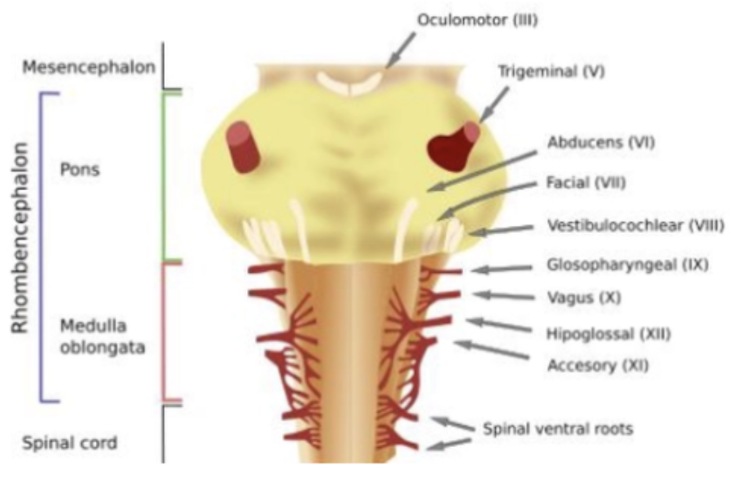
Medulla oblongata purpose
Controls vital autonomic functions such as:
Breathing
Heart rate
Blood pressure
Swallowing, coughing, and vomiting reflexes
Pons purpose
Acts as a relay station between different parts of the brain.
Helps regulate:
Sleep and arousal
Breathing
Facial movements and sensations
Cerebellum purpose
Coordinates voluntary movements.
Maintains:
Balance
Posture
Motor learning and fine motor control
Helps movements become smooth and accurate
Midbrain diagram
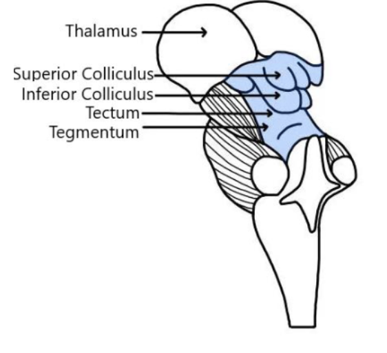
Superior colliculi purpose (tectum)
Visual reflexes and eye/head orientation
Inferior colliculi purpose (tectum)
Auditory processing and sound orientation
tegmentum purpose (4)
|
Forebrain structure
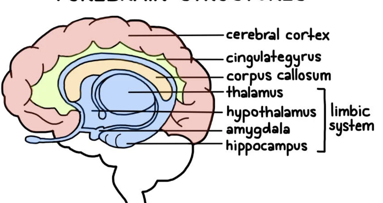
Forebrain: Cerebrum vs interbrain structure
Feature | Cerebrum (Telencephalon) | Interbrain (Diencephalon) |
|---|
Location | Largest, outermost part of the brain | Located beneath the cerebrum, surrounding the third ventricle |
Major Structures | Cerebral cortex, basal ganglia, limbic system | Thalamus, hypothalamus, epithalamus, subthalamus |
Forebrain: Main components
CCL: Corpus callosum, cerebral cortex, limbic system
Forebrain: Thalamus- where and what
Thalamus | above midbrain | Relays information to the cerebral cortex |
Forebrain: Hypothalamus- role
Maintains homeostasis by regulating hunger, thirst, body temperature, sleep, and hormone release.
Forebrain: pituitary gland- role
Releases hormones that control growth, metabolism, reproduction, and other endocrine glands.
Forebrain: basal ganglia role
Helps initiate and regulate voluntary movement, habit formation, and procedural learning.
Forebrain: hippocampus role
Essential for forming new long-term memories and spatial navigation.
Forebrain: Cingulate gyrus role
Involved in emotion, attention, motivation, decision-making, and pain processing.
Amygdala role
Processes emotions, especially fear, threat detection, and emotional learning.
Ventricles role
Fluid-filled spaces that contain and circulate cerebrospinal fluid (CSF), which cushions and nourishes the brain.
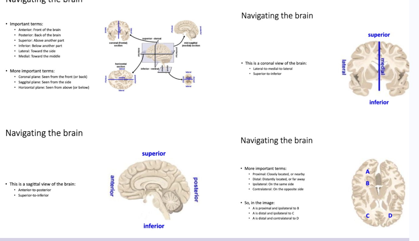
Naviagting the brain
Cerebral cortex lobes
Parts of the frontal cortex
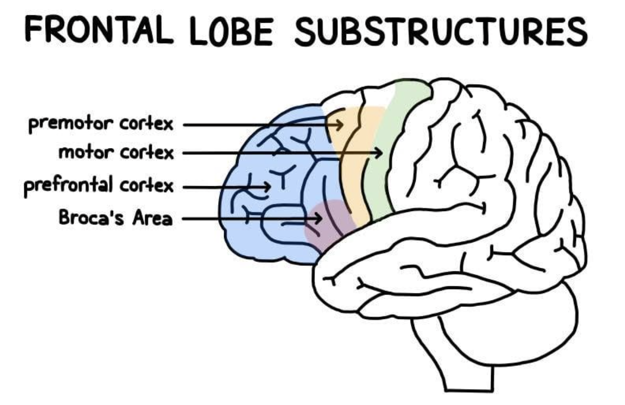
Primary motor cortex role
Voluntary movement
Premotor cortex role
Planning movements
Prefrontal cortex role
Decision-making, planning, personality
Broca’s area role
Speech production