anatomy of hearing-3
1/34
There's no tags or description
Looks like no tags are added yet.
Name | Mastery | Learn | Test | Matching | Spaced | Call with Kai |
|---|
No analytics yet
Send a link to your students to track their progress
35 Terms
_____ are the location of hair cells in semicircular canals
cupula
_______ are the location of hair cells in the vestibule or cochlea
otolithic membrane
cochlear duct
_____ is the sense of the room spinning
vertigo
_____ is the rapid, rocking movement of the eyes
nystagmus
you will always have __________ when you have vertigo
nystagmus
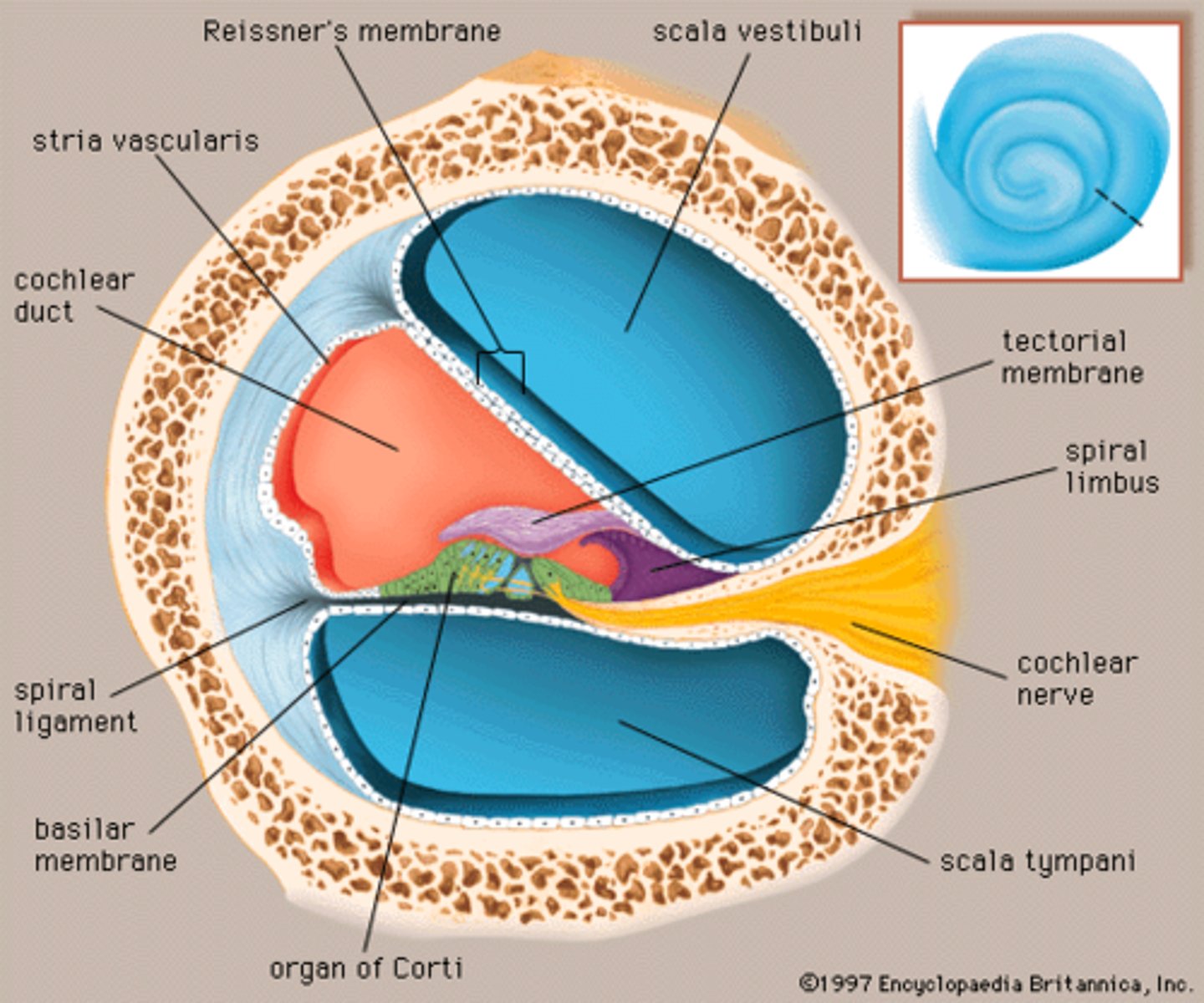
the cochlear duct is made of _____ membranes (#)
2
membranes that make up the cochlear duct
vestibular (Reissner) membrane
basilar membrane
the ___________ lines up with the spiral lamina
basilar membrane
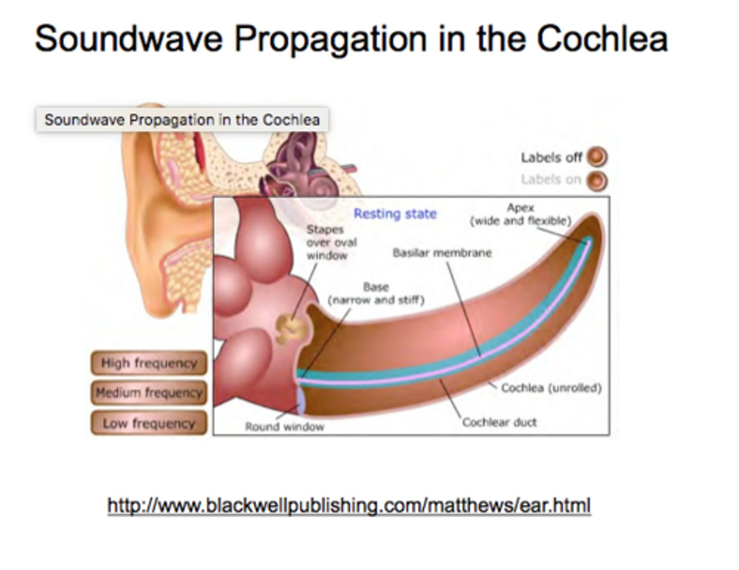
descriptions of apex of cochlea
wide and flaccid
frequencies at the apex of the cochlea are _____
low
descriptions of base of cochlea
narrow and tense
frequencies at the base of the cochlea are ______
high
________ is in the bony cochlea
perilymph
__________ is in the cochlear duct
endolymph
the ____________ is tonotopic
basilar membrane
different frequencies in different parts of cochlea
scala media v cochlear duct
space v structure
unrolled cochlea
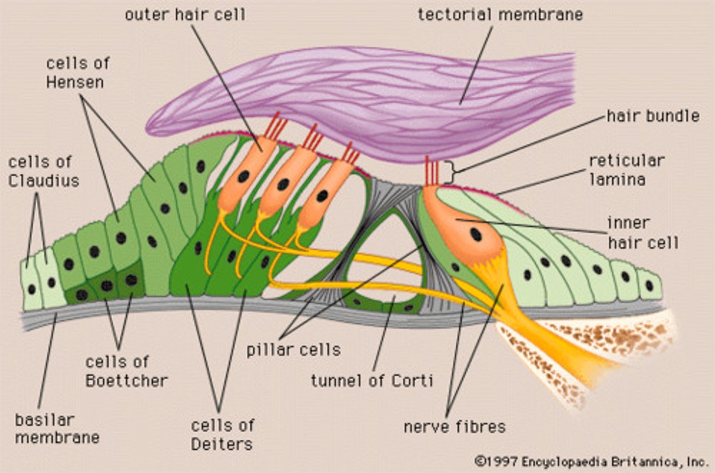
organ of corti
center part of the cochlea, containing hair cells, canals, and membranes
aka spiral organ
the _______ is the sensory organ for hearing
organ of corti
supporting cells in spiral organ of corti
phalangeal cells (inner phalangeal cells)
rods of corti (pillar cells)
cells of Deiters(outer phalangeal cells)
supporting cells of Hensen
the receptor (sensory) cells in spiral organ of corti
inner hair cells
outer hair cells
the __________ hold inner hair cells
phalangeal cells (inner phalangeal cells)
the _________ form the inner tunnel of corti and separate the inner and outer phalangeal cells
rods of Corti (pillar cells)
the __________ hold the outer hair cells
cells of Deiters (outer phalangeal cells)
the _________ form the outer tunnel of corti
supporting cells of Hensen
the ___________ are where the action happens; where mechanical energy changes to electrochemical
receptor (sensory) cells
organ of corti is located in ______
cochlear duct
organ of corti diagram
4 differences between outer and inner hair cells
1. shape
2. arrangement of stereocilia (hairs)
3. nerve supplies
4. connection to the tectorial membrane
the ________ is a gelatinous mass hair cells stick into
tectorial membrane
shape of inner and outer hair cells
inner: teardrop/gourd shape
outer: test tube shape
arrangement of stereocilia inner v outer hair cells
inner: in a u-shaped pattern
outer: in a v- or w-shaped pattern
nerve supplies inner v outer hair cells
inner: as many as 10 fibers to 1 hair cell
outer: as many as 10 outer hair cells for 1 nerve fiber
connection to tectorial membrane inner v outer hair cell
inner: tenuous or brushing against it, slight connection
outer: firmly embedded in tectorial membrane