Inflammation (MOD)
1/25
There's no tags or description
Looks like no tags are added yet.
Name | Mastery | Learn | Test | Matching | Spaced | Call with Kai |
|---|
No analytics yet
Send a link to your students to track their progress
26 Terms
What are the clinical (cardinals) signs of acute inflammation?
Sign | Medical Term | Cause |
|---|---|---|
Redness | Rubor | Vasodilation |
Heat | Calor | Increased blood flow |
Swelling | Tumor | Fluid leakage into tissues |
Pain | Dolor | Bradykinin & prostaglandins |
Loss of function | Functio laesa | Tissue damage/swelling |
What is Inflammation?
A protective response of the body to eliminate the cause of cell injury, remove damaged tissue, and begin healing.
What are the two types of inflammation?
Acute and Chronic
What is acute inflammation?
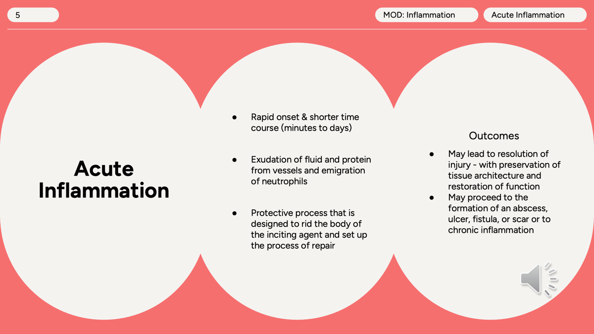
Acute Inflammation
Rapid onset (minutes to days)
Short duration
Main cell: Neutrophils
Examples:
Cut on your finger
Bacterial infection
Sprained ankle
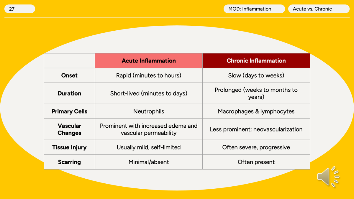
Acute inflammation is a rapid, short-duration response characterized by fluid exudation and neutrophil migration that aims to eliminate the cause of injury and initiate tissue repair, ending in resolution, scar formation, abscess, ulcer, fistula, or chronic inflammation.
What is chronic inflammation?
Chronic Inflammation
Lasts weeks, months, or years
Main cells:
Macrophages
Lymphocytes
Plasma cells
Examples:
Rheumatoid arthritis
Tuberculosis
Chronic hepatitis
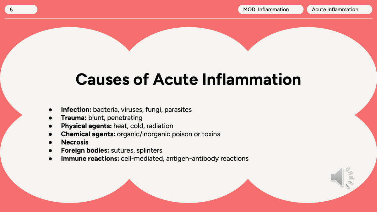
What are the common causes of acute inflammation?
Acute inflammation can be triggered by infection, trauma, physical or chemical injury, necrosis, foreign bodies, or immune-mediated reactions, all of which cause tissue damage and activate the inflammatory response.
What happens in the vascular phase of Acute inflammation?
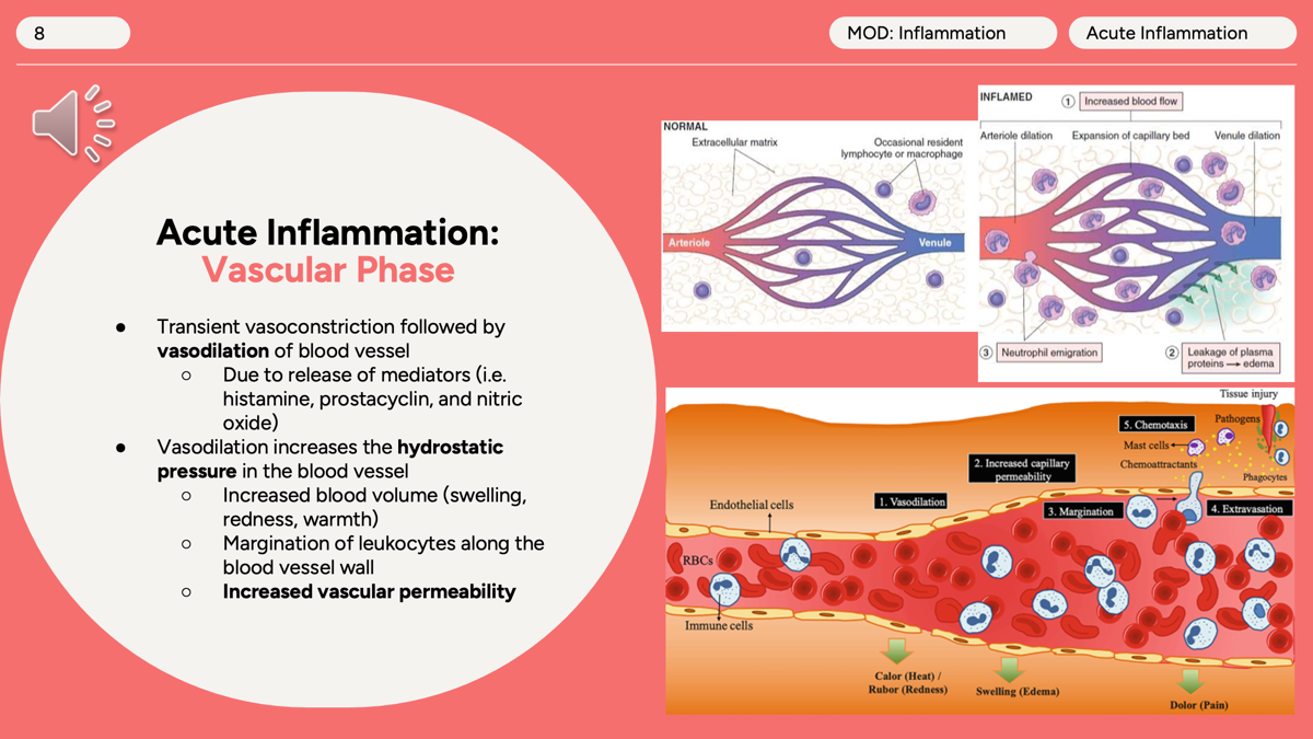
The vascular phase is the first stage of acute inflammation and involves changes in blood vessels.
Brief vasoconstriction occurs immediately after injury.
Vasodilation follows, caused by histamine, prostacyclin, and nitric oxide, increasing blood flow to the area.
Increased blood flow raises hydrostatic pressure, causing redness (rubor) and heat (calor).
Blood vessels become more permeable (leaky), allowing fluid and proteins to enter tissues.
This leakage causes edema (swelling/tumor).
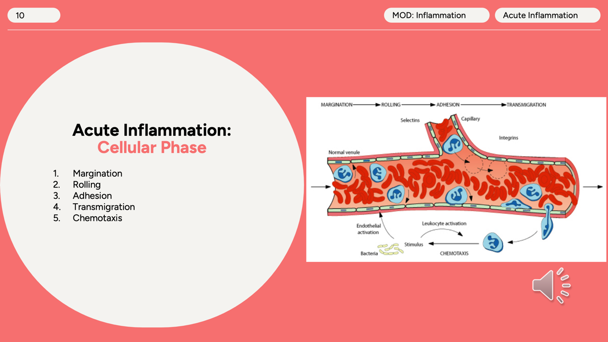
As fluid leaves the vessels, neutrophils move toward the vessel wall (margination), preparing for migration into the injured tissue.
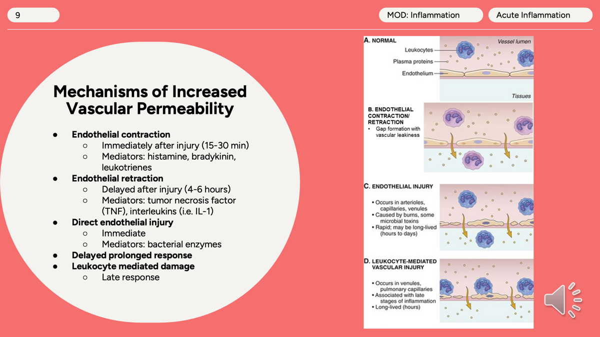
What are the the 5 different types of mechanism of increased vascular permeability:
endothelial contraction
endothelial retraction
direct endothelial injury
delayed prolonged response
leukocyte mediated damage
What is vascular permeability?
Vascular permeability means blood vessels become "leaky," allowing fluid and proteins to move from the bloodstream into tissues.
Normal vessel
[Cell][Cell][Cell]
After histamine
[Cell] [Cell] [Cell]
↑ gaps
What are the steps of cellular Phase in Acute Inflammation?
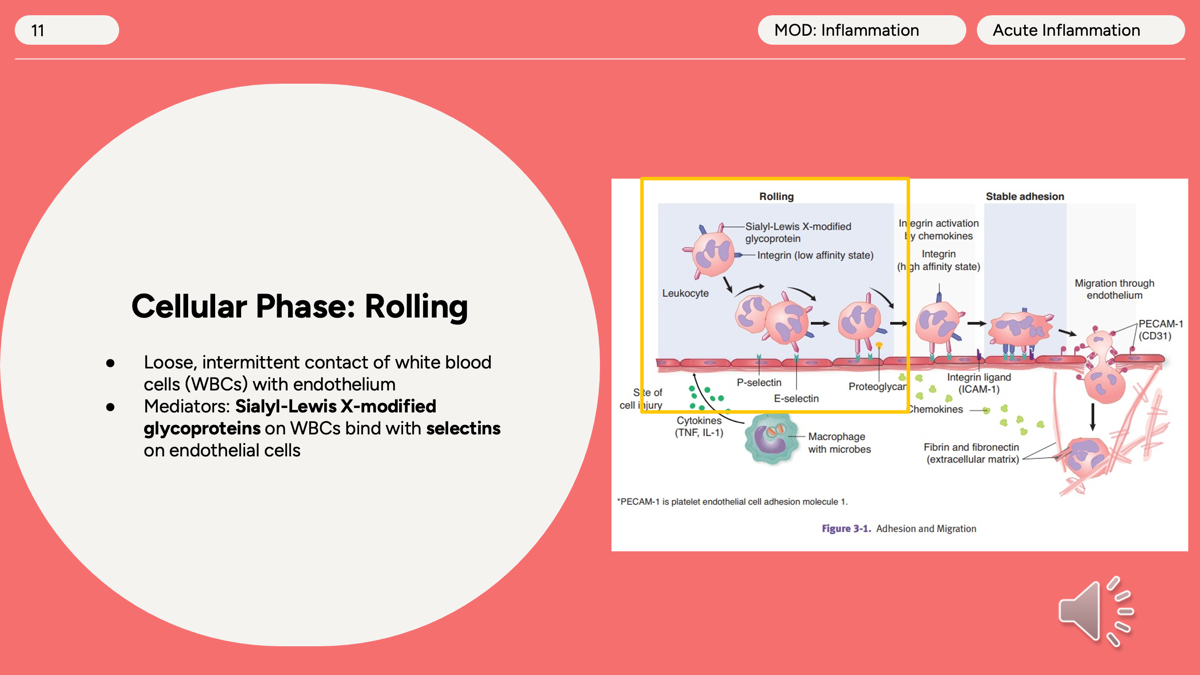
What is rolling in cellular Phase
Rolling is the first step of leukocyte (WBC) movement from the blood vessel to the site of injury.
Rolling is the first step of leukocyte recruitment in acute inflammation, where selectins on endothelial cells form weak bonds with Sialyl-Lewis X on leukocytes, causing the WBC to slow down and roll along the vessel wall.
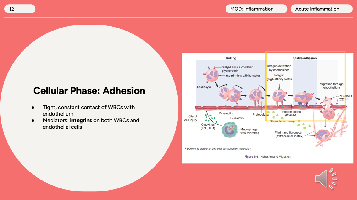
Cellular Phase: Adhesion
After rolling, the WBC needs to stop completely so it can leave the blood vessel and enter the tissue.
Adhesion is the second step of leukocyte recruitment, where activated integrins on WBCs bind tightly to ICAM-1 and VCAM-1 on endothelial cells, causing the leukocyte to stop and prepare to exit the bloodstream.
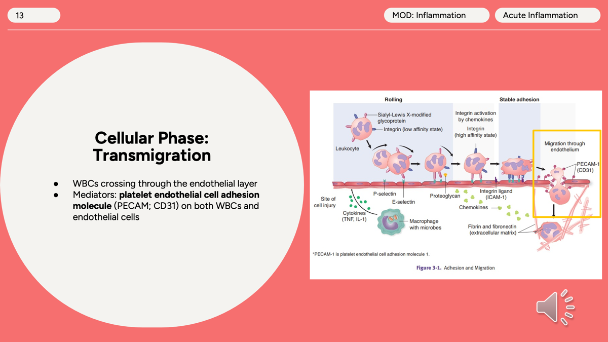
Cellular Phase: Transmigration (Diapedesis)
This is the third step of leukocyte recruitment.
After the WBC has:
Rolled (selectins)
Adhered (integrins)
Transmigration (diapedesis) is the movement of leukocytes through the endothelial layer into tissues, mediated primarily by PECAM-1 (CD31) on both leukocytes and endothelial cells.
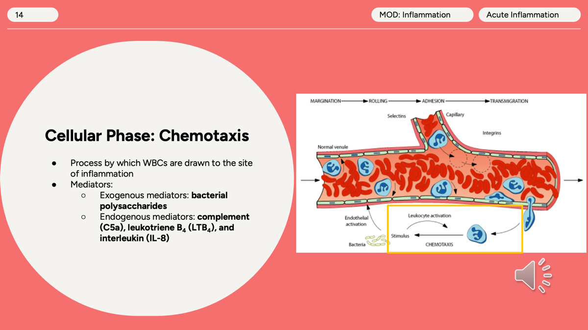
Cellular Phase: Chemotaxis
Chemotaxis is the directed movement of leukocytes toward a site of infection or injury, guided by chemical attractants such as C5a, IL-8, LTB₄, and bacterial products.
Step | Main Molecule |
|---|---|
Rolling | Selectins |
Adhesion | Integrins |
Transmigration | PECAM-1 (CD31) |
Chemotaxis | C5a, IL-8, LTB₄ |
What are the mediators of Chemotaxis?
Endogenous (Made by the Body)
C5a (Complement)
Powerful chemoattractant
LTB₄ (Leukotriene B4)
Attracts neutrophils
interleukin IL-8
Cytokine that recruits neutrophils
Exogenous (From Bacteria)
Bacterial products
Especially bacterial peptides and polysaccharides
example of Phagocytotic cells
○Neutrophils (early in acute inflammatory response)
○Monocytes and macrophages
○Dendritic cells
Outcomes of Acute Inflammation
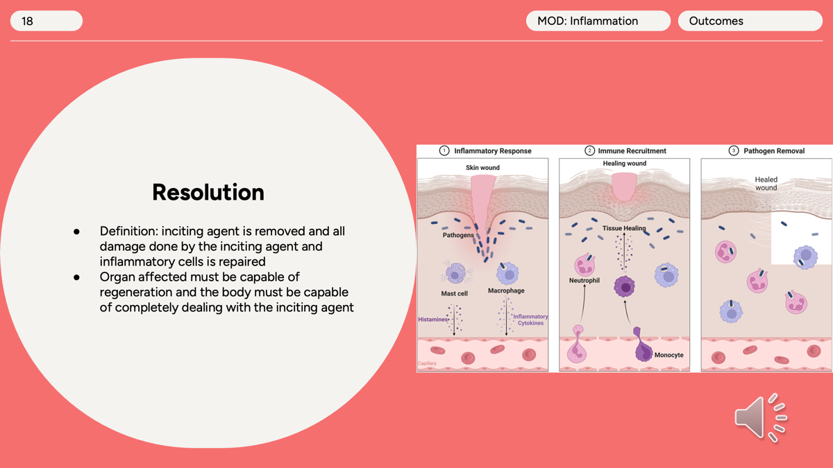
Resolution
Resolution = Complete healing and return to normal tissue.
For resolution to occur:
✅ The inciting agent (bacteria, virus, toxin, injury) must be removed.
✅ The tissue must be able to regenerate (replace damaged cells).
✅ Inflammatory cells (especially neutrophils) and debris must be cleared away.
✅ Normal tissue structure and function are restored.
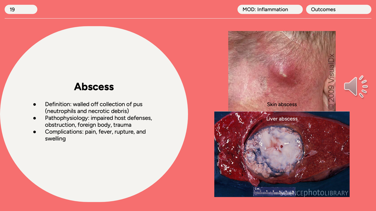
A localized collection of pus surrounded by a wall of inflammatory tissue.
Abscess
Impaired host defenses, obstruction, foreign body, trauma.
pain, fever, rupture, and swelling
What's in the pus?
🟢 Neutrophils (dead and alive)
🟢 Bacteria (if infection is present)
🟢 Necrotic tissue debris
🟢 Fluid
●Requirements for abscess formation: The body cannot rid itself of the inciting agent, or the process of repair and scarring is occurring more rapidly in the tissue around the site of the abscess.
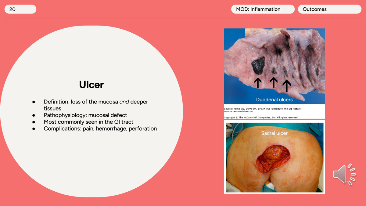
loss of the surface lining (epithelium/mucosa) and deeper underlying tissue.
Ulcer
Mucosal defect
seen in GI tract
pain, hemorrhage, perforation
●If only the mucosa is lost, the correct term is an erosion.
●Microscopic morphology of an ulcer: The ulcer has four layers, which recapitulate steps from acute inflammation to repair. The layers, from superficial to deep, are fibrin, neutrophils, granulation tissue, and fibrosis.
●Complications of an ulcer: Pain; hemorrhage, if the ulcer involves a vessel; and perforation, resulting in hemorrhage within a cavity or the lumen of the gastrointestinal tract, or seeding of the peritoneal cavity with the contents of the gastrointestinal tract, causing peritonitis.
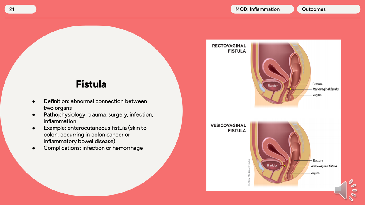
An abnormal passage (tunnel) connecting two organs or an organ to the skin.
Fistula
trauma, surgery, infection, inflammation.
enterocutaneous fistula (skin to colon, occurring in colon cancer or inflammatory bowel disease)
Infection or hemorrhage
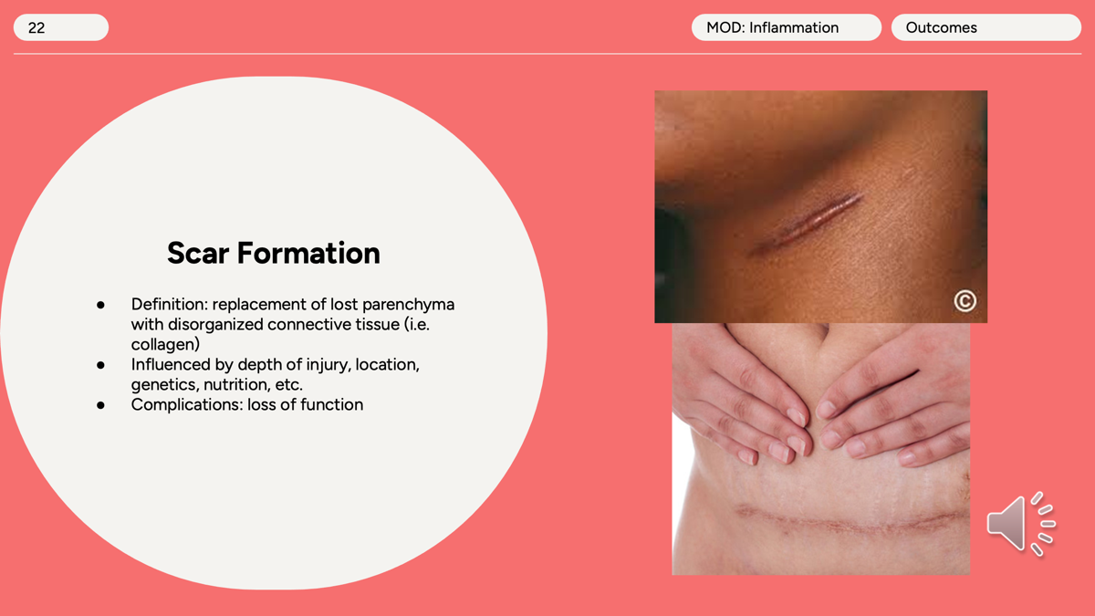
Scar formation
Replacement of lost parenchyma with disorganized connective tissue (i.e. collagen).
Loss of function.
●Influenced by depth of injury, location, genetics, nutrition, etc.
●Requirements for scar formation: Loss of tissue in an organ not capable of regeneration or loss of basement membrane or other framework required for successful regeneration.
What are the causes of chronic inflammation?
Viral infection, persistent microbial infection, prolonged exposure to a toxin, and autoimmune dysfunction/disease
What are the cells involved in chronic inflammation?
Macrophages and Lymphocytes
What is the process of granulomatous inflammation (specific type of chronic inflammation?
collections of activated macrophages i.e. epithelioid histiocytes), can have multinucleated giant cells.
●Immune response designed to wall off irritant, infection, foreign body, or autoimmune conditions
●Associated conditions:
○Tuberculosis, sarcoidosis, silicosis, Crohn’s disease, foreign body granuloma (i.e. splinter, glass, suture), fungal infections (i.e. Histoplasmosis, Cryptococcosis)
Acute vs Chronic inflammation