ASU BIO 201: Special Senses Eyes and Ears (Lab)
1/72
There's no tags or description
Looks like no tags are added yet.
Name | Mastery | Learn | Test | Matching | Spaced | Call with Kai |
|---|
No analytics yet
Send a link to your students to track their progress
73 Terms
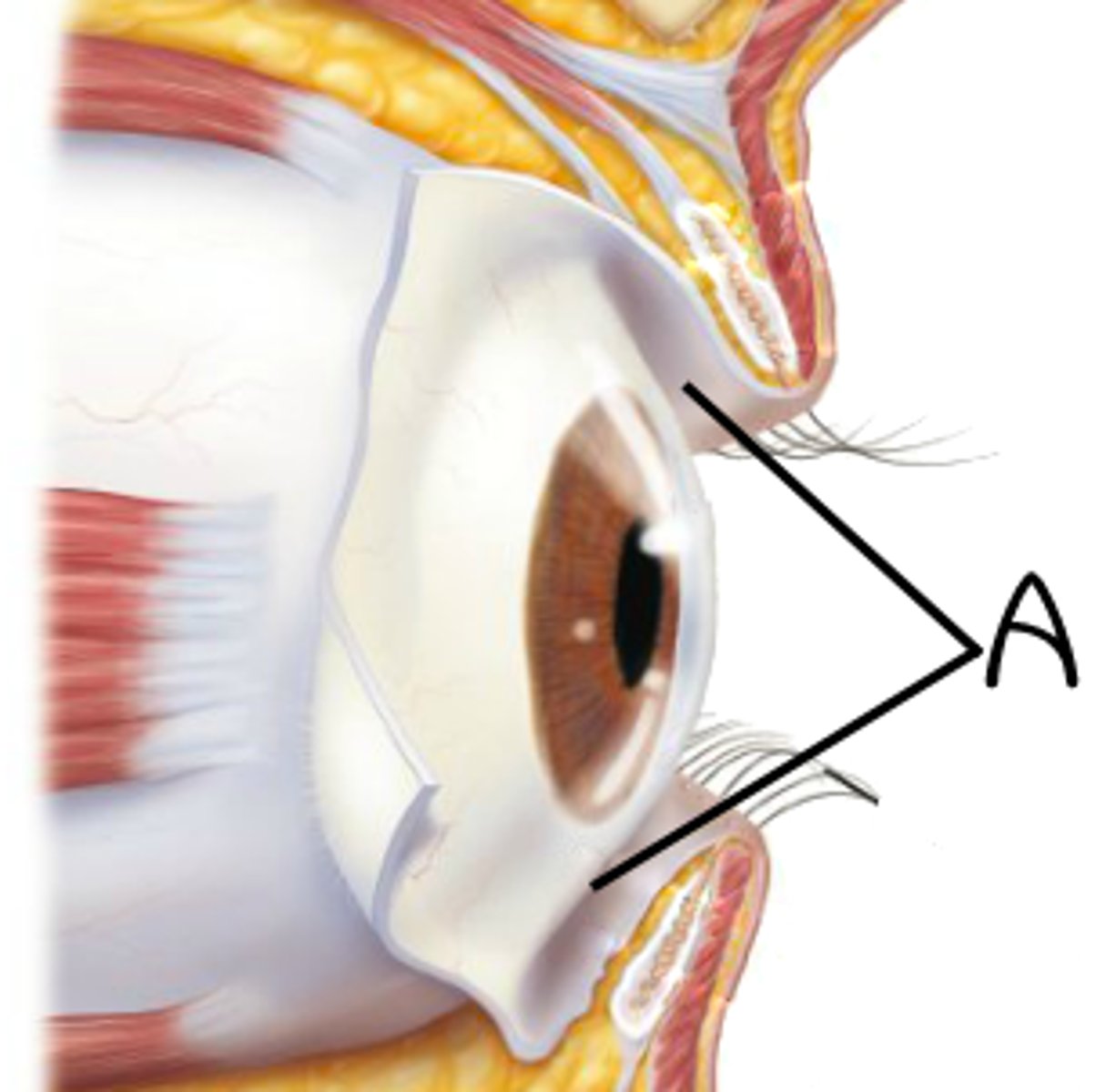
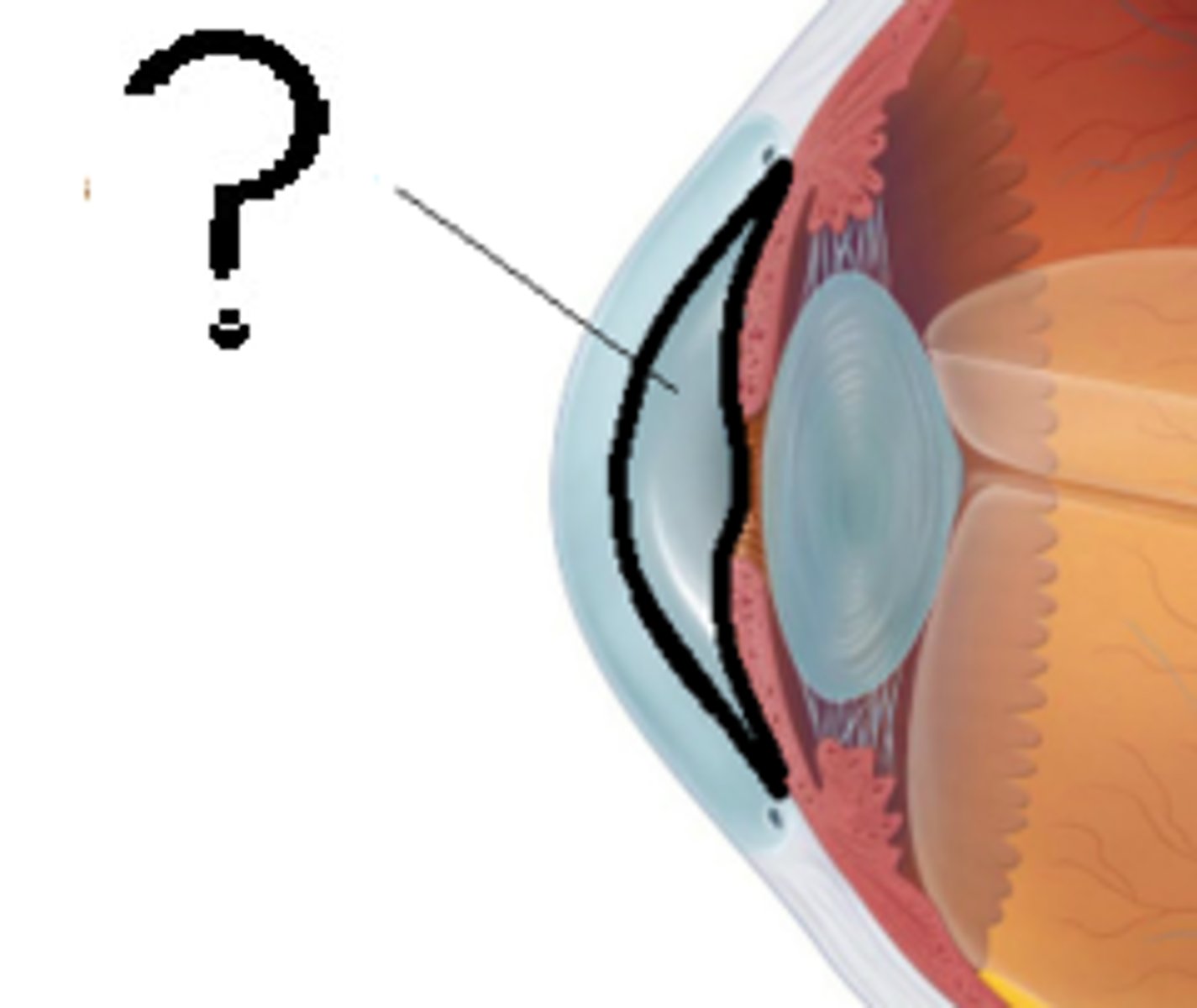
Conjunctiva
transparent mucus membrane that covers the inner surface of the eyelid and anterior surface of the eye, with the exception of the Cornea
Lacrimal apparatus
lacrimal gland produces tears; tears flow through lacrimal punctum, lacrimal canals, lacrimal sac, nasolacrimal duct; tears cleanse and lubricate the eye, deliver oxygen and nutrients to the conjunctiva, and prevent bacterial infection via lysozyme
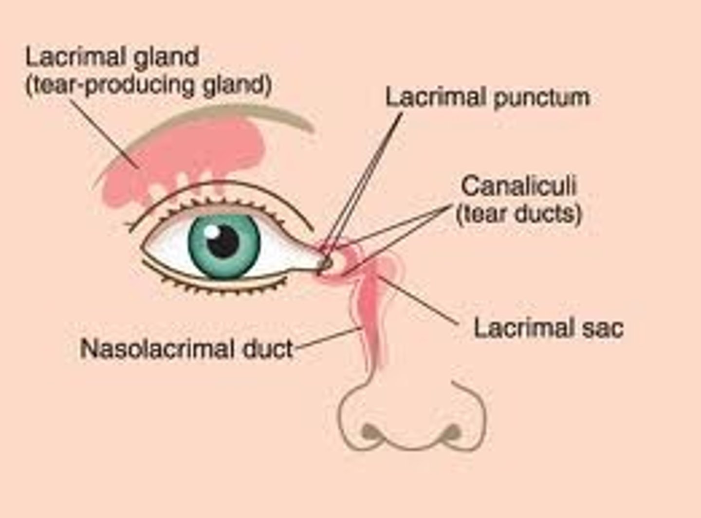
Flow of tears
Lacrimal gland -> surface of eye -> lacrimal punctum -> lacrimal canals -> lacrimal sac -> nasolacrimal duct
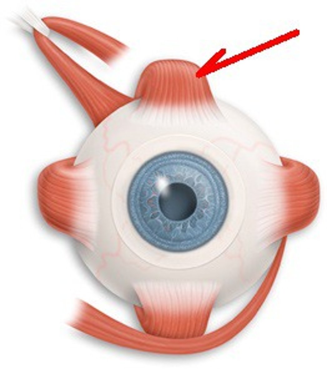
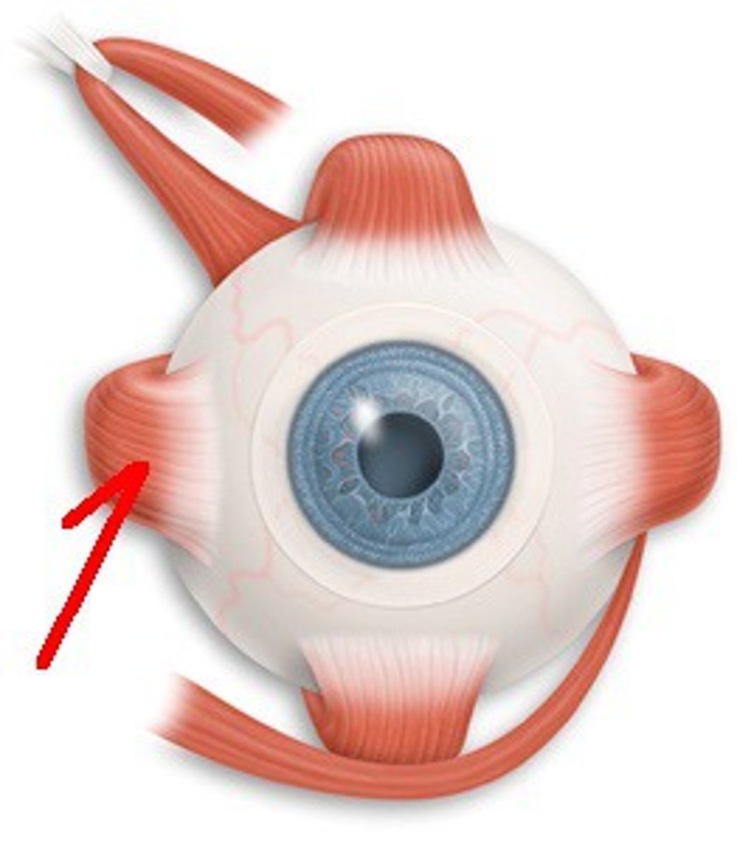
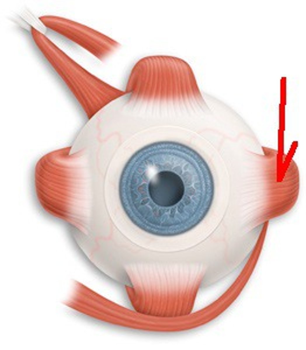
Extrinsic muscles of the eye
Superior, Inferior, Medial, and Lateral rectus
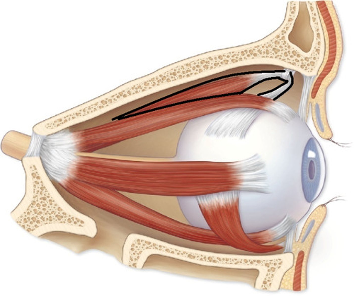
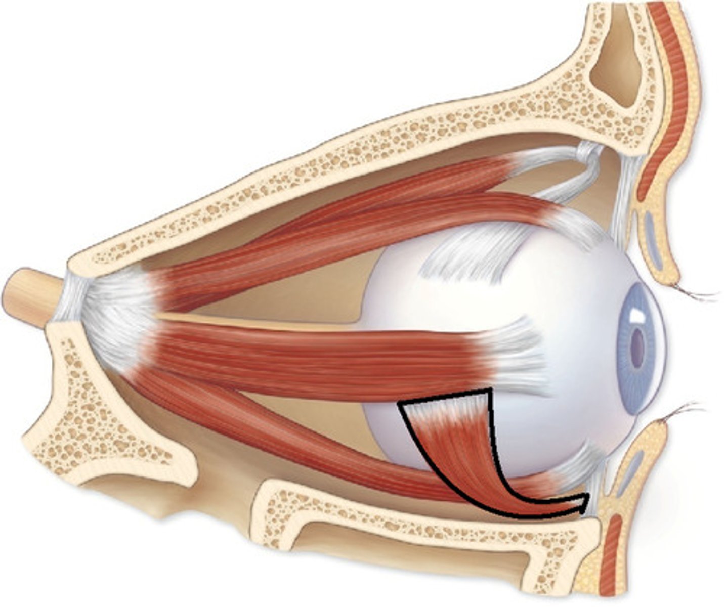
Superior and Inferior Oblique
Superior rectus
elevates eye, innervated by oculomotor III
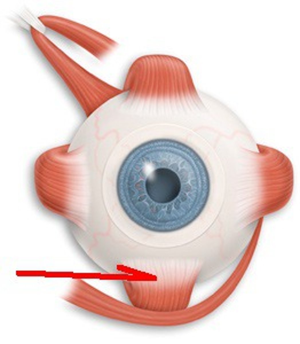
Inferior rectus of eye
depresses eye, innervated by oculomotor III
Medial rectus
moves eye medially, innervated by oculomotor III
Lateral rectus
moves eye laterally, innervated abducens VI
Superior oblique
depresses and turns eye laterally, innervated by trochlear VI
Inferior oblique
elevates and turns eye laterally, innervated by oculomotor III
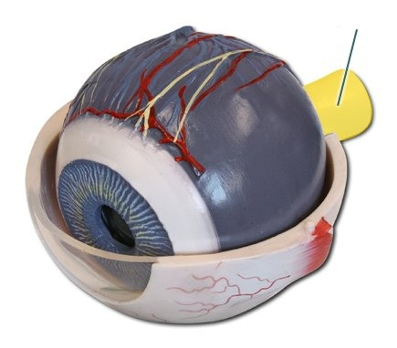
Layers of the eye
fibrous, vascular, inner
fibrous layer
tunica fibrosa:
sclera, cornea
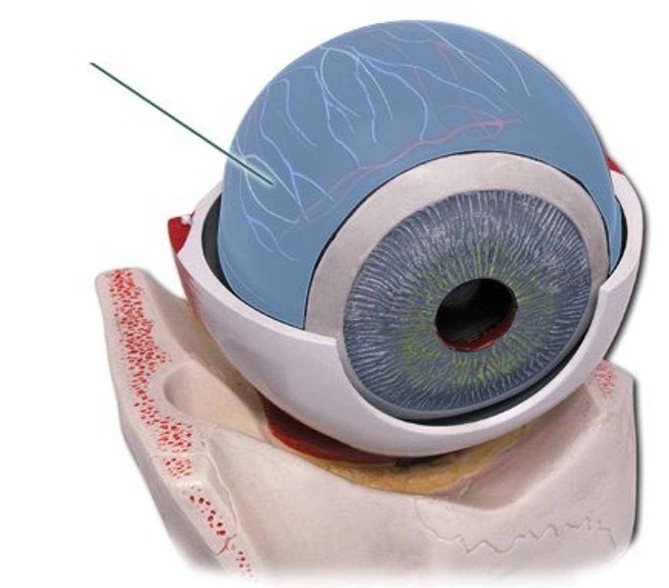
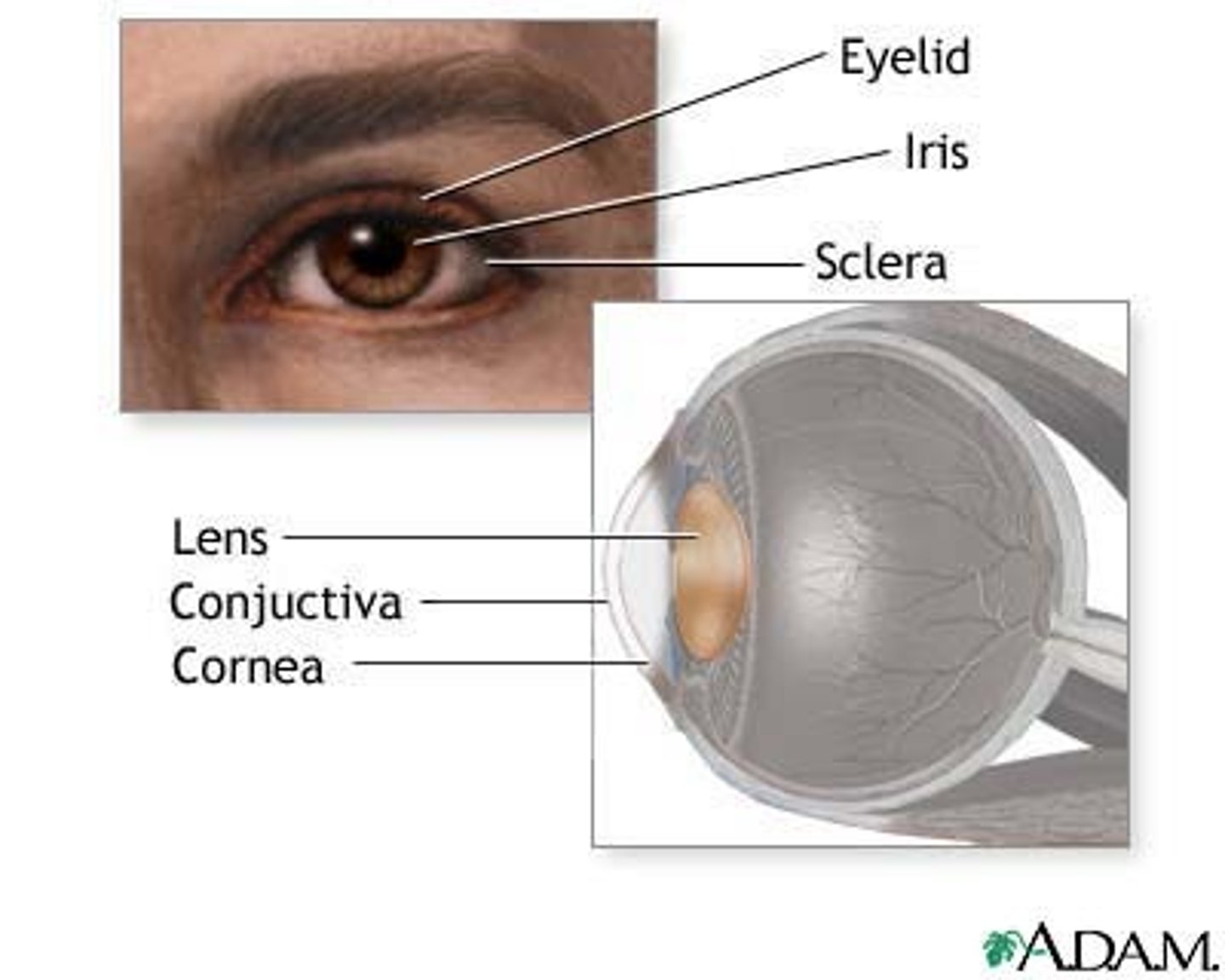
Sclera
white of the eye, formed from dense collagenous CT; shapes eye and anchors extrinsic muscles
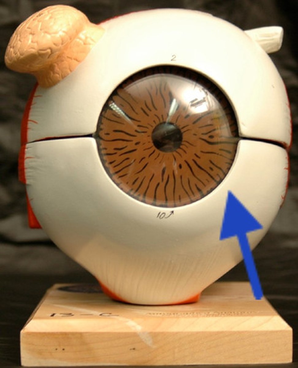
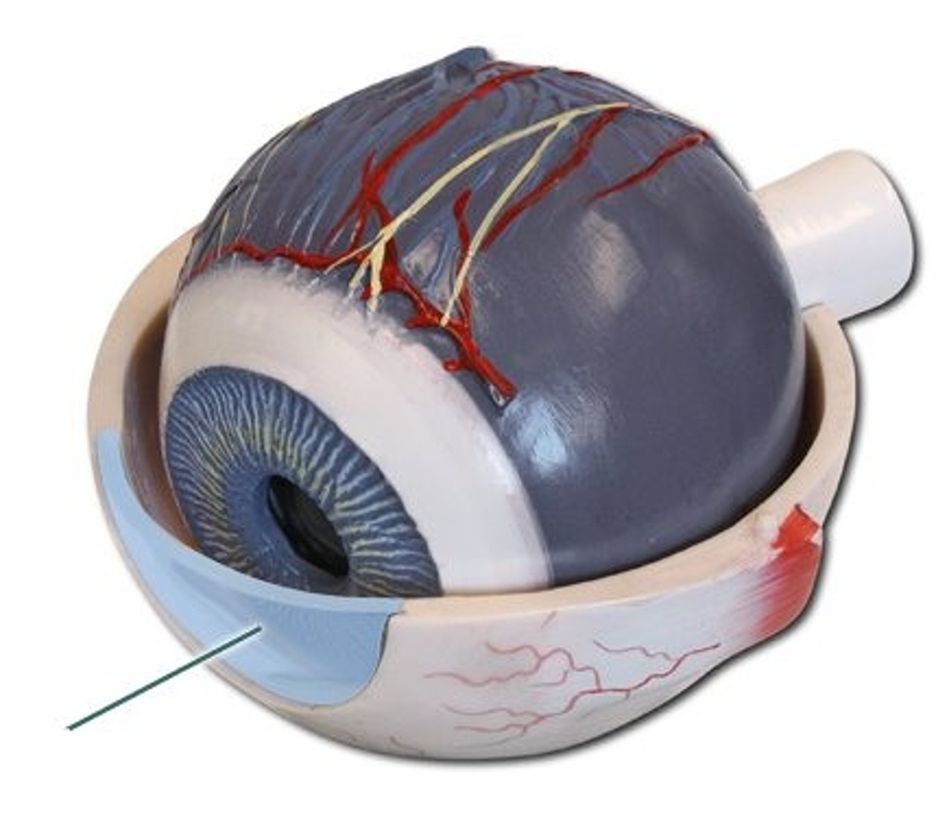
Cornea
anterior transparent region that admits light that covers the pupil/iris
Vascular layer
tunica vasculosa:
choroid, ciliary body, iris, pupil
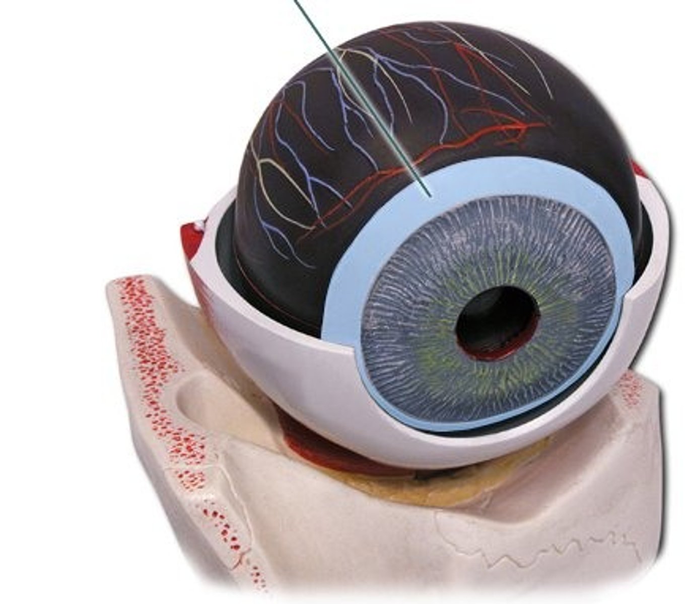
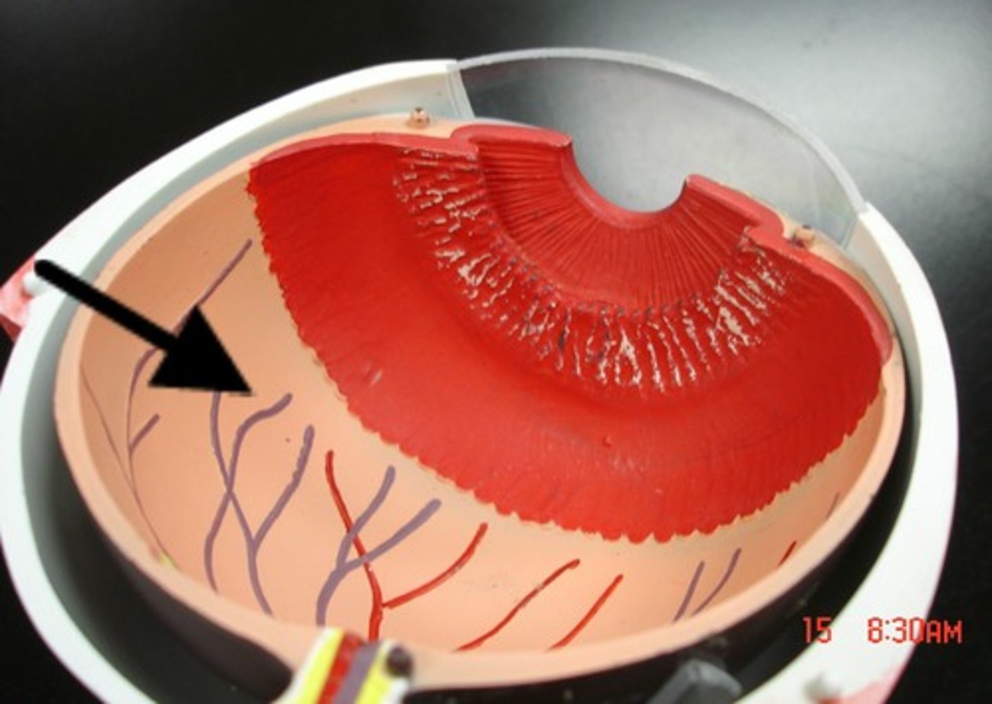
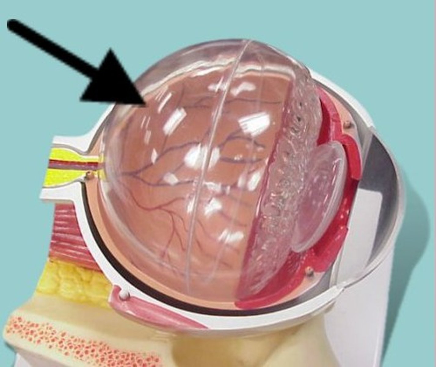
Choroid
highly vascular, deeply pigmented layer behind retina, helps absorb light and provide nutrients to retina
Ciliary body
thickened extension of choroid forming a muscular ring around the lens, and shapes the lens via suspensory ligaments. Supports iris and secretes aqueous humor
Iris
adjustable diaphragm that controls the diameter of the pupil
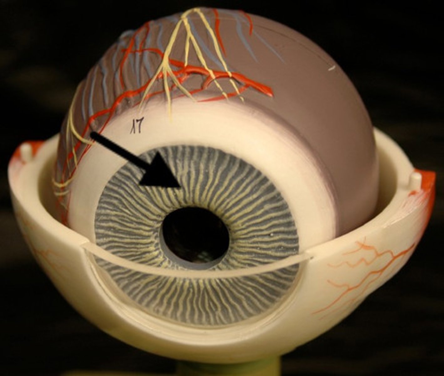
Pupil
opening created by the iris which allows light to pass into the eye

Inner layer
tunica interna:
retina, optic nerve
Retina
cup-shaped extension of the diencephalon; contains a pigmented and a neural (sensory) layer
Optic nerve
Cranial Nerve II which innervates the eye; joins the retina at the optic disc, crosses at the optic chiasm
Composition: sensory
Function: vision
Damage: blindness
Optic components
transparent components of the eye that admit light rays, bend lights, and focus them on the retina
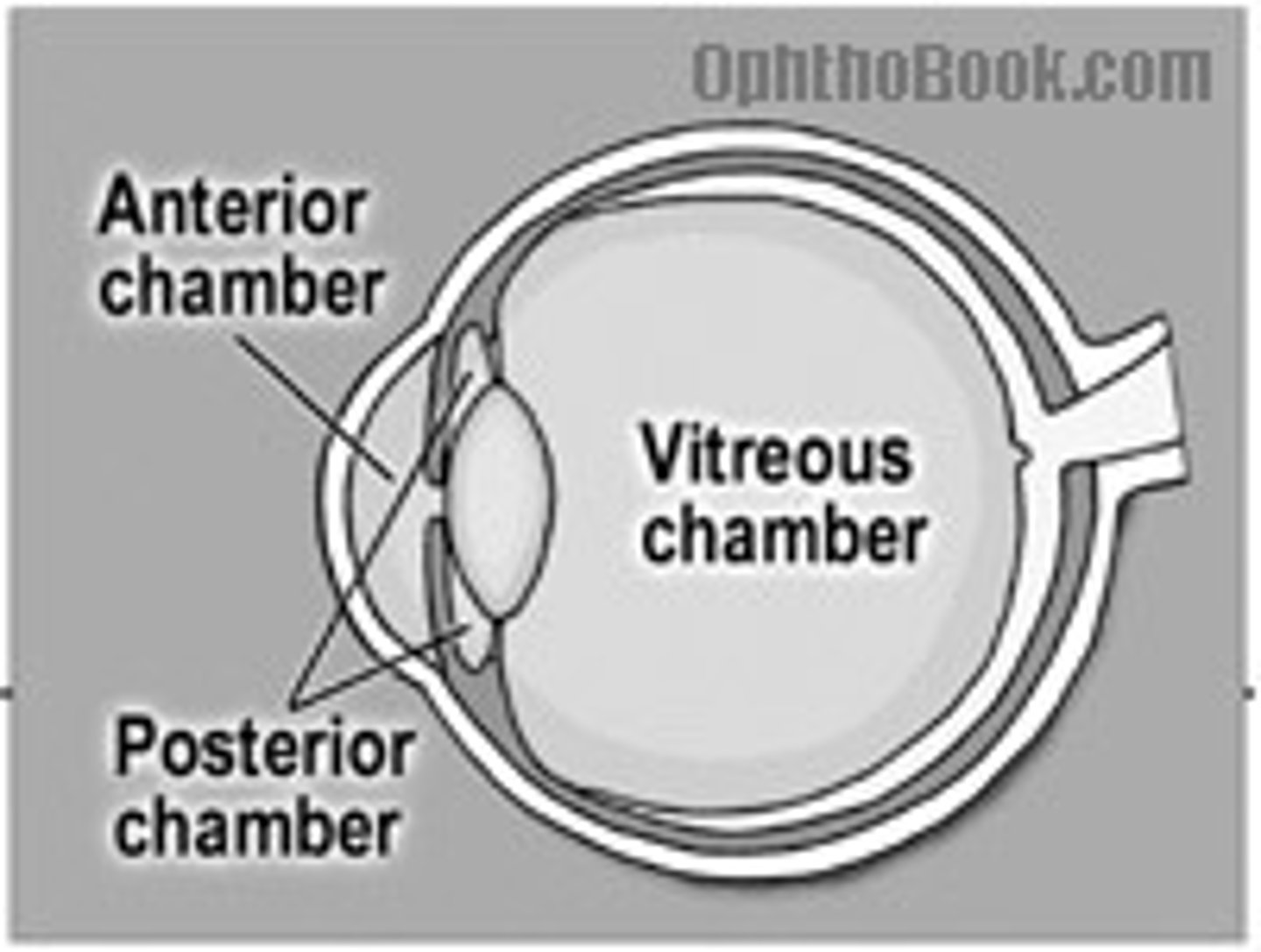
posterior and anterior chamber, aqueous humor, lens, vitreous humor
Posterior chamber
lies between the iris and lens

Anterior chamber
lies between the cornea and the iris
Aqueous humor
clear fluid secreted by ciliary body; flows from posterior chamber through pupil to anterior chamber and replenishes throughout life
Lens
can be bent by the ciliary body to focus passing light
Vitreous humor
transparent jelly that lies in vitreous chamber behind the lens the holds the retina in place; is never replaced
Neural components
retina - pigmented layer and neural layer (optic nerve, fovea centralis, rods/cones)
Pigmented layer
absorbs light and prevents it from scattering; stores Vitamin A which is needed by photoreceptors
Fovea Centralis
produces finely detailed images on retina
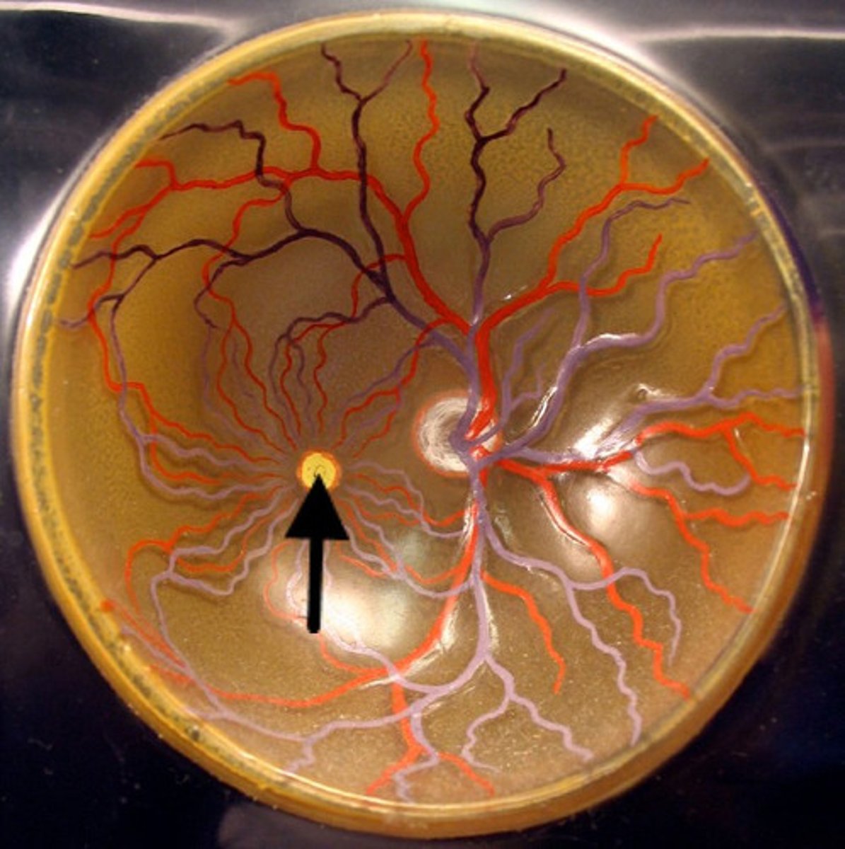
Optic disc
Blind spot, visual filling
Photoreceptors
cells that absorb light and produce a chemical or electrical signal, rods and cones
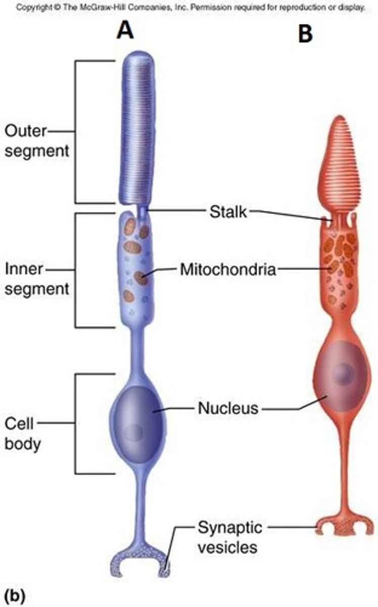
Rods
responsible for night vision and produces images in shades of gray, most numerous photoreceptor, animals have higher density allowing for night vision
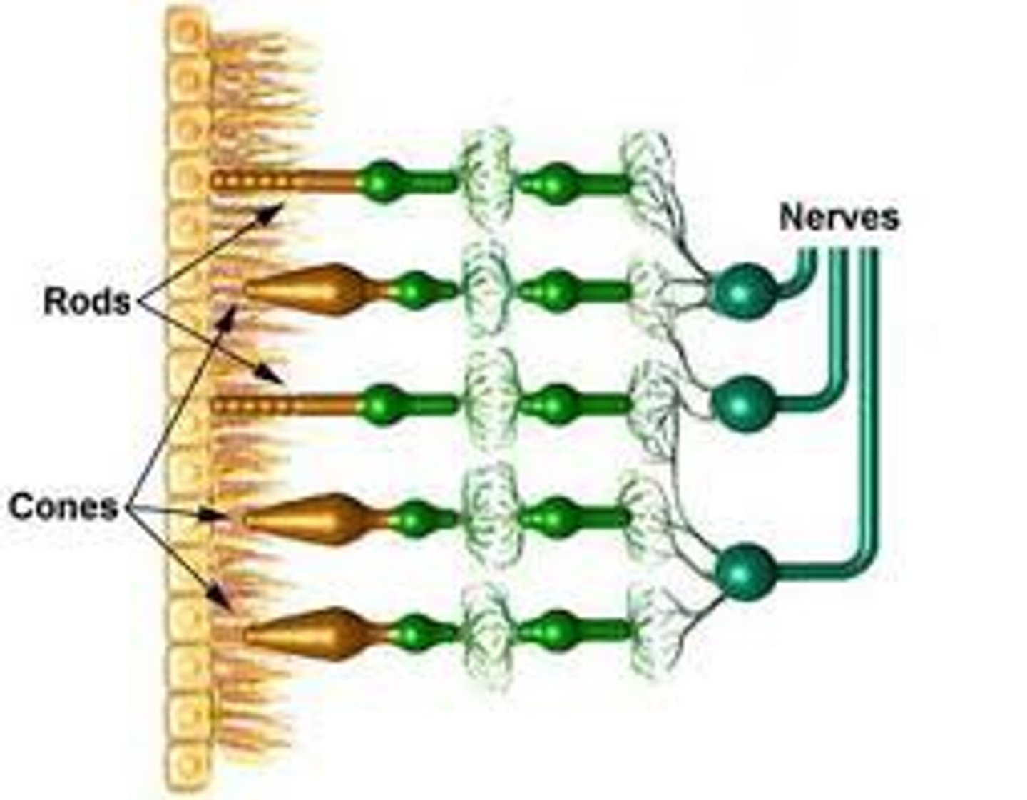
Cones
responsible for color vision and respond to bright light, provide higher detail, humans have high density

Color blindness
sex-linked recessive trait which results in a lack of various cone cells. Occurs in 8% of men and 0.5% of women
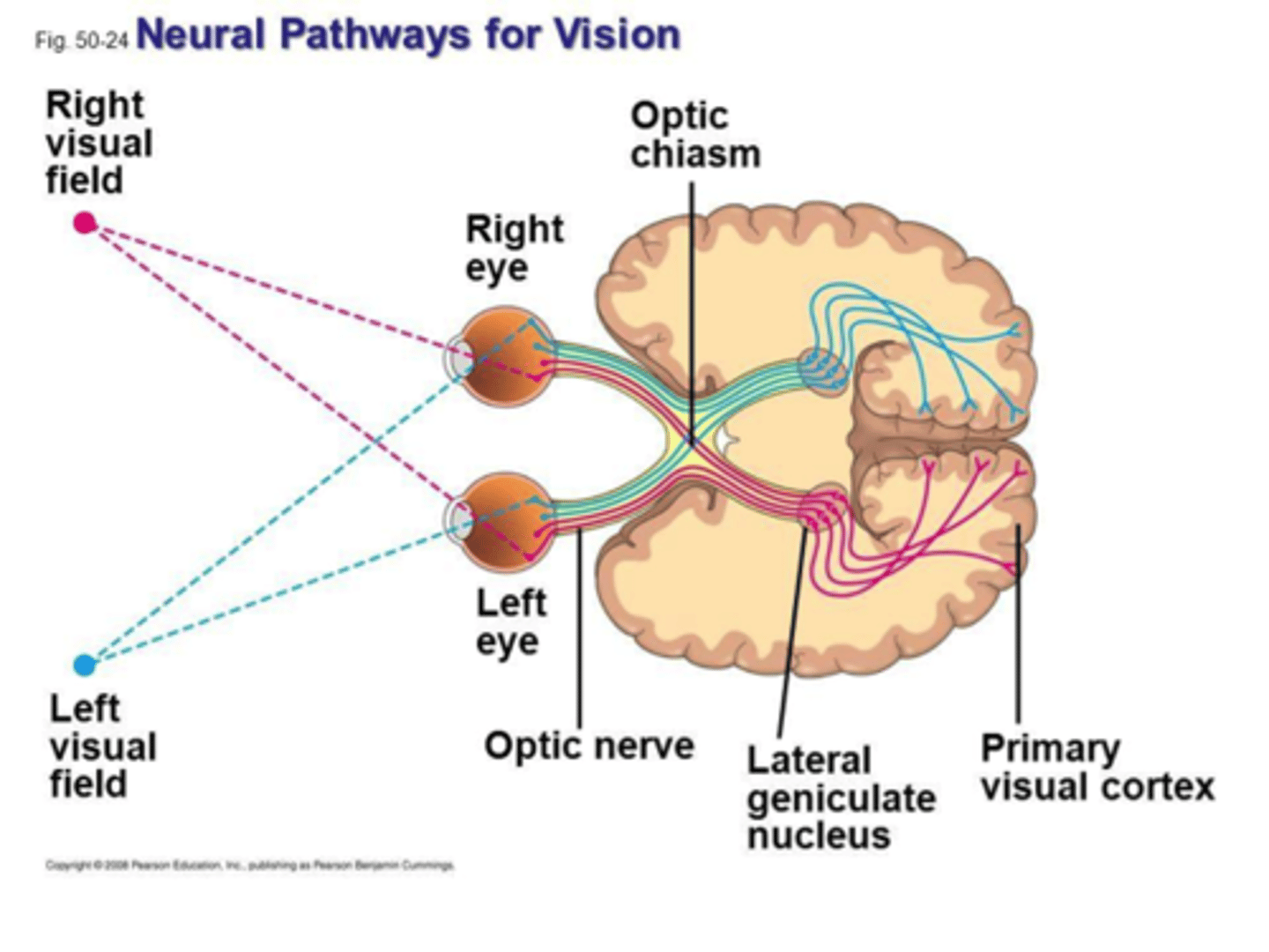
Visual pathway
cornea -> anterior chamber -> iris (pupil) -> posterior chamber -> lens -> vitreous humor -> retina -> optic nerve -> optic chiasm -> occipital lobe
optic nerves cross at the optic chiasm therefore the right occipital lobe monitors the left side of the visual field, while the left occipital lobe monitors the right side of the visual field
Anatomy of the ear
outer, middle, inner ear
Outer ear
funnels vibrations to the tympanic membrane (eardrum)
Auricle (pinna), external auditory canal
Auricle
Pinna, shell-shaped structure formed by elastic cartilage that funnels vibrations to the external auditory canal
External Auditory Canal
passage leading from the auricle, through the temporal bone, to the tympanic membrane
Middle Ear
found in tympanic cavity
Tympanic membrane, pharyngotympanic (auditory) tube, auditory ossicles, stapedius, tensor tympani
Tympanic membrane
"ear drum" that separates the external auditory canal form the tympanic cavity. Transfers vibrations to the auditory ossicles
Pharyngotympanic (auditory) tube
passageway to the nasopharynx and functions to equalize pressure between the middle and outer ear
Auditory ossicles
smallest bones in the human body; function to transfer vibrations from the tympanic membrane to the inner ear
malleus, incus, stapes
Malleus
"hammer" that is found between tympanic membrane and incus, connected to tensor tympani muscle
Incus
"anvil" found between malleus and stapes
Stapes
"stirrup" that is found between incus and oval window; connected to stapedius muscles
Stapedius
reduces motion of stapes to lessen effect on inner ear
Tensor Tympani
pulls malleus away from eardrum
Inner ear
housed in the temporal bone passageways called the bony labyrinth. The bony labyrinth houses the membraneous labyrinth
Perilymph, Endolymph, cochlea
Perilymph
cushioning fluid found between the bony and membranous labyrinth
Endolymph
fluid found within the membranous labyrinth
Cochlea
coiled, snail-shell-like organ for hearing that arises from the vestibule;
oval window, round window, scala vestibuli, scala media, scala tympani, vestibular membrane, tectorial membrane, basilar membrane, spiral organ of corti
Oval window
connected to stapes; transfers vibrations to cochlea
Round Window
found at the end of the cochlea acting as a pressure relief
Scala Vestibuli
filled with perilymph, begins near the oval window
Scala Media
Cochlear duct; filled with endolymph, contains spiral organ of corti
Scala Tympani
filled with perilymph, ends at the round window
Vestibular membrane
separates the scala vestibuli from the cochlear duct
Tectorial membrane
feather-shaped gelatinous substance that acts to open channels for depolarization; part of the spiral organ of corti
Basilar membrane
separates the cochlear duct from the Scala Tympani
Spiral Organ of Corti
structure that converts vibrations to neural impulses; stereocilia vibrate pressing up against the tectorial membrane in order to open channels allowing for depolarization
Vestibule
chamber that contains the organs of equilibrium
oval window, saccule, utricles, maculae
Saccule
portion of the vestibule that leads to the cochlea; contains maculae and is responsible for linear acceleration
Utricle
portion of the vestibule that leads to the semicircular canals; contains maculae and is responsible for linear acceleration
Maculae
equilibrium receptors found in the vestibule that respond to gravitational pull and changes in head position
-small crystals called otoliths add to the weight and inertia to enhance a sense of gravity and motion
Static Equilibrium
the body is stationary but the head is tilted; perceived by macula found in the saccule and utricle
Semicircular Canals
responsible for sense of equilibrium
made up of anterior, posterior, and lateral ducts which respond to angular movement of the head
Dynamic equilibrium
perception of motion or angular acceleration, perceived by the semicircular canals
ex) turning in a car
Vestibulocochlear Nerve
CN VIII
Formed from vestibular nerve and cochlear nerve
Composition: Sensory
Function: Hearing and balance
Damage: deafness and impaired balance
Step 1 of hearing
vibrations are captured by the auricle -> funneled by the external auditory canal -> hit tympanic membrane -> transfers vibrations through the auditory ossicles -> oval window
Step 2 of hearing
vibrations travel -> perilymph -> up scala vestibule -> vibrates the vestibular membrane -> vibration of scala vestibuli vibrates the endolymph -> causing movement of stereocilia against the tectorial membrane (depolarizing neural cells) -> vibrations travel through basilar membrane -> scala tympani -> out the round window