Paranasal Sinuses
 Call Kai
 Learn
Learn Practice Test
 Spaced Repetition
 Match
Match Flashcards
 Knowt Play
1/65

There's no tags or description
Looks like no tags are added yet.
Name | Mastery | Learn | Test | Matching | Spaced | Call with Kai |
|---|
No analytics yet
Send a link to your students to track their progress
66 Terms
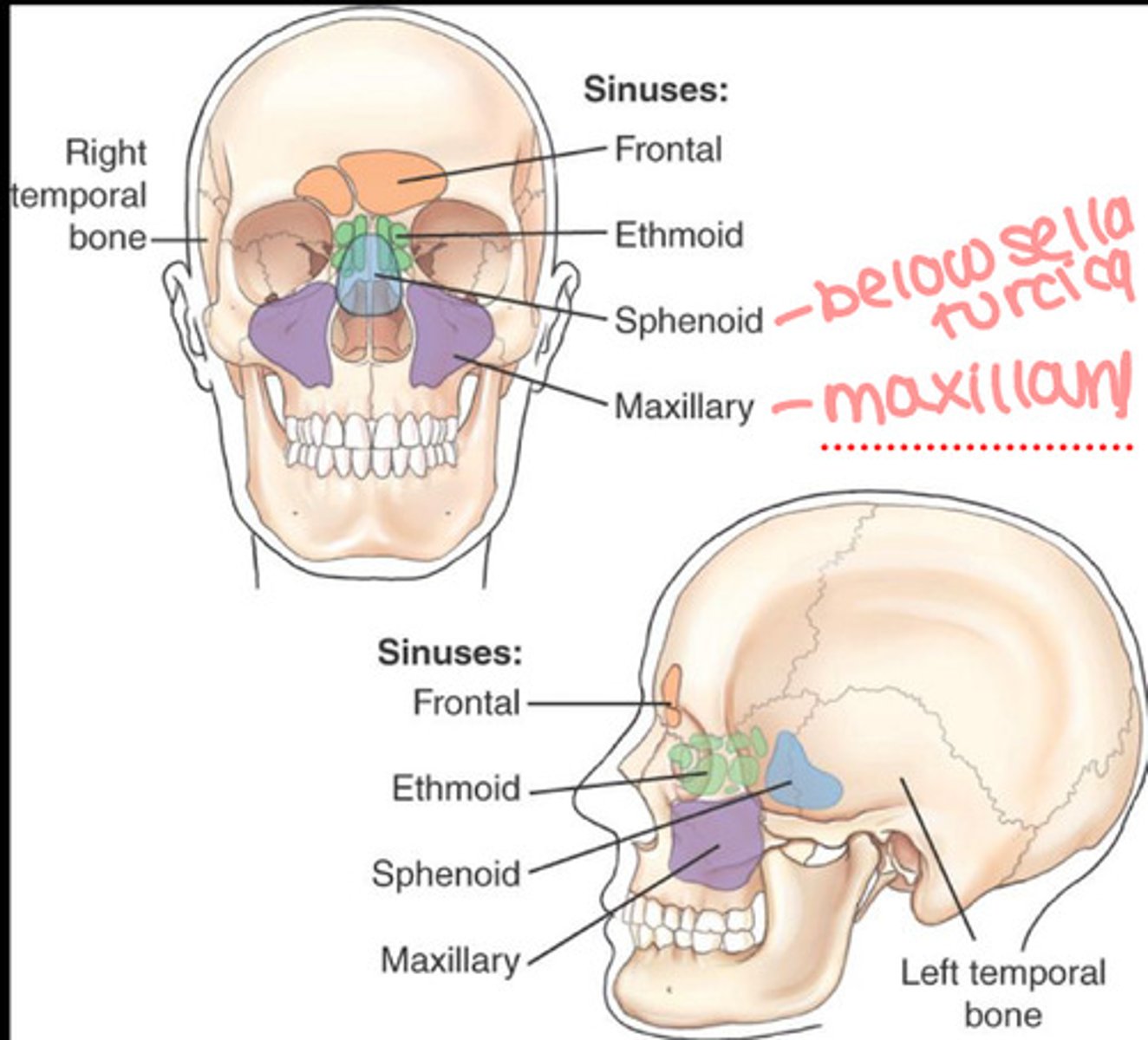
Paranasal sinuses are
large air filled cavities lined with mucous membranes
What is the function of paranasal sinuses
Communicate with nasal cavity
Produce mucus to moisten nasal passageways
The four groups of paranasal sinuses
Maxillary
Front
Ethmoid
Sphenoid
Which paranasal sinus is the last to develop?
Ethmoid around 17-18
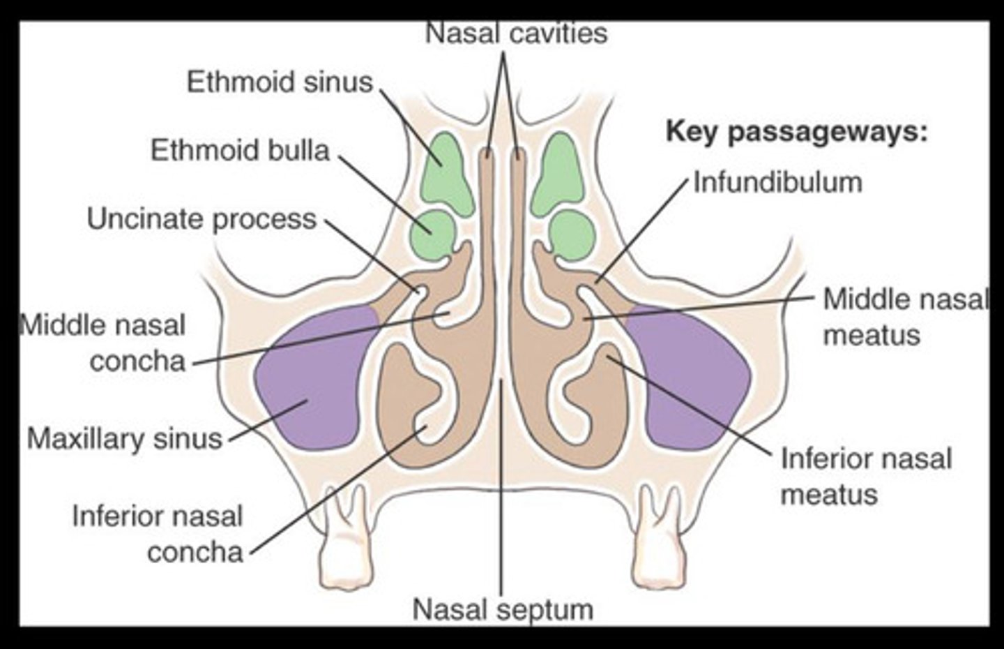
Coronal view of sinuses
Osteomeatal complex
Pathways of communication between sinus and nasal cavity once all sinuses are fully developed
Maxillary sinuses are where
within facial bone, maxilla
Shape of maxillary sinuses
2 pyramid shape
Maxillary sinus orientation
In maxilla
Above upper teeth
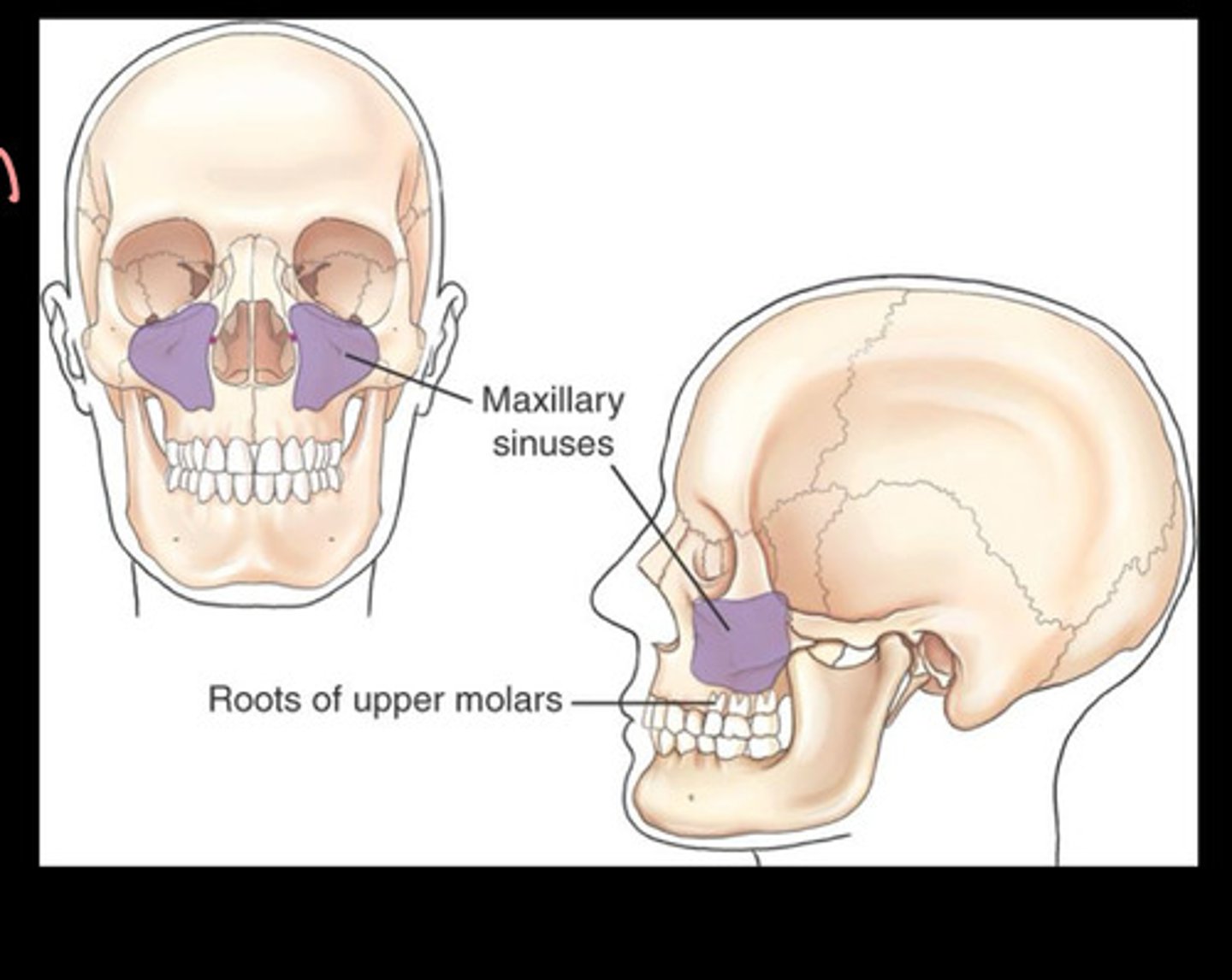
For maxillary sinuses, since it is above upper teeth
infection can travel to sinuses
Frontal sinus are located where
Inner and outer tables of frontal bone
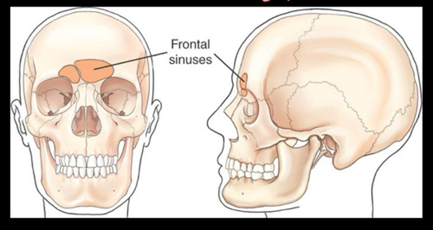
The frontal sinuses are rarely
symmetric
Number of frontal sinuses
1-2
sometimes may even be absent
The frontal sinus is larger in
males
Ethmoid sinuses are located where
within the lateral masses of the ethmoid bone
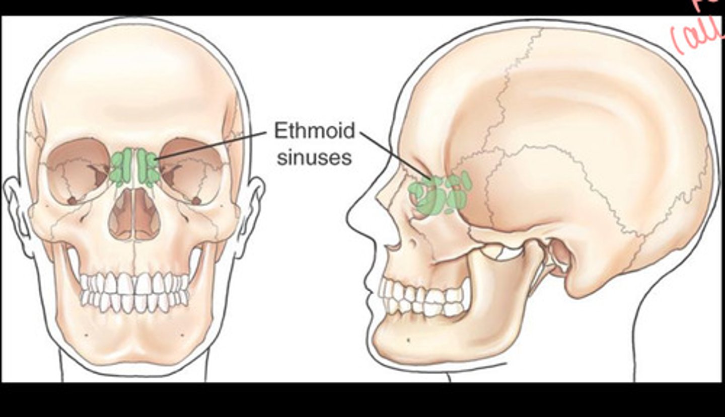
How many collections of ethmoid sinuses
3
anterior, middle, and posterior
The sphenoid sinuses are located where
Below the sella turcica
at the base of the skull
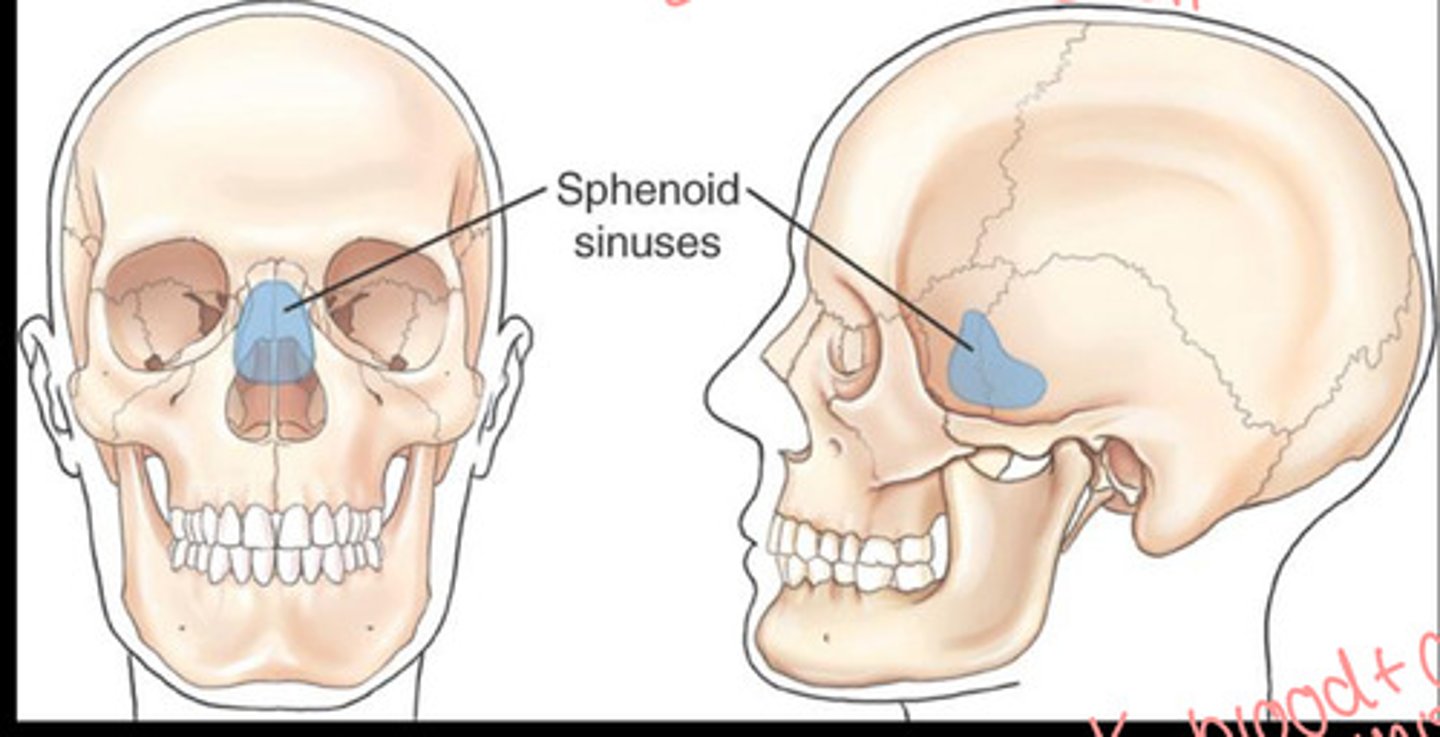
How many sphenoid sinuses
1-2
Sphenoid sinus may indicate what type of fracture
Basal skull fracture/sphenoid effusion
A basal skull sinus is when
blood and CSF leaks into the sphenoid sinus
sphenoid effusion
Paransal Sinus Basic Routine
Lateral
PA Caldwell
Parietoacanthial (waters)
Submentovertex (SMV)
Special views for paranasal sinuses
Parietoacanthial transoral (open mouth Waters)
Paranasal sinus exams should be
erect to demonstrate air fluid levels
Sinus radiograph demonstrations
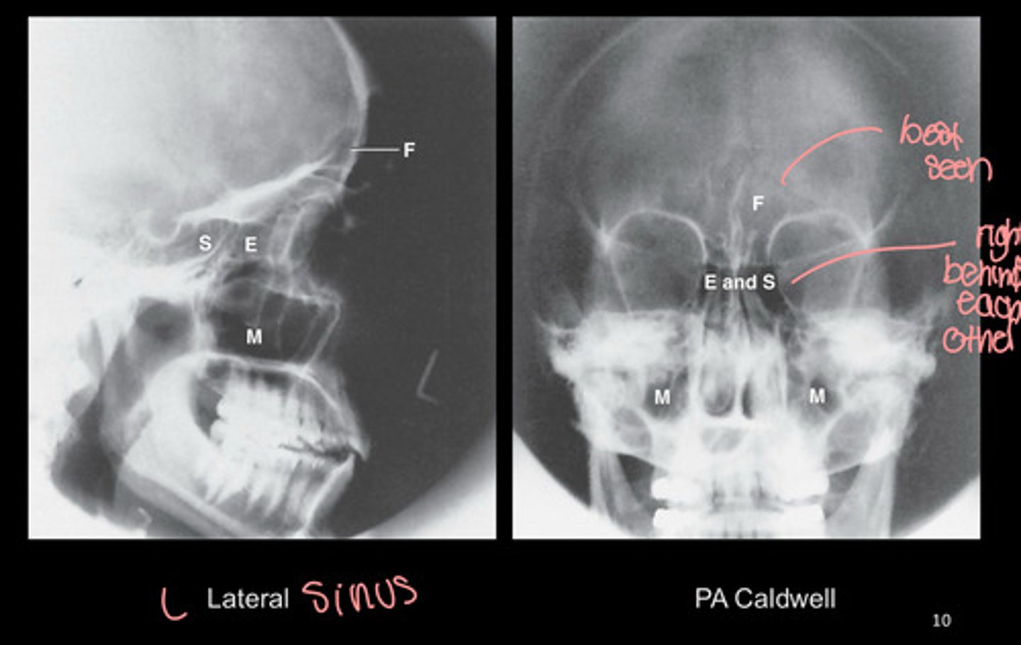
Lateral Sinus patient positioning
upright in oblique position
What lines are involved in lateral sinus
IOML
IPL
MSP
In lateral sinus, IOML is
parallel to IR or perpendicular to the front edge of the IR
In lateral sinus, the IPL is
perpendicular to the IR
MSP in lateraal sinus is
parallel to IR
Collimation for lateral sinus
1/2-1'' beyond nose
3'' above nasion
Inferior to include occlusal plane
Central ray for lateral sinus
1/2-1'' posterior to outer canthus
What sinuses do you see on lateral sinus projection
All 4 sinuses
Superimposed facial bones
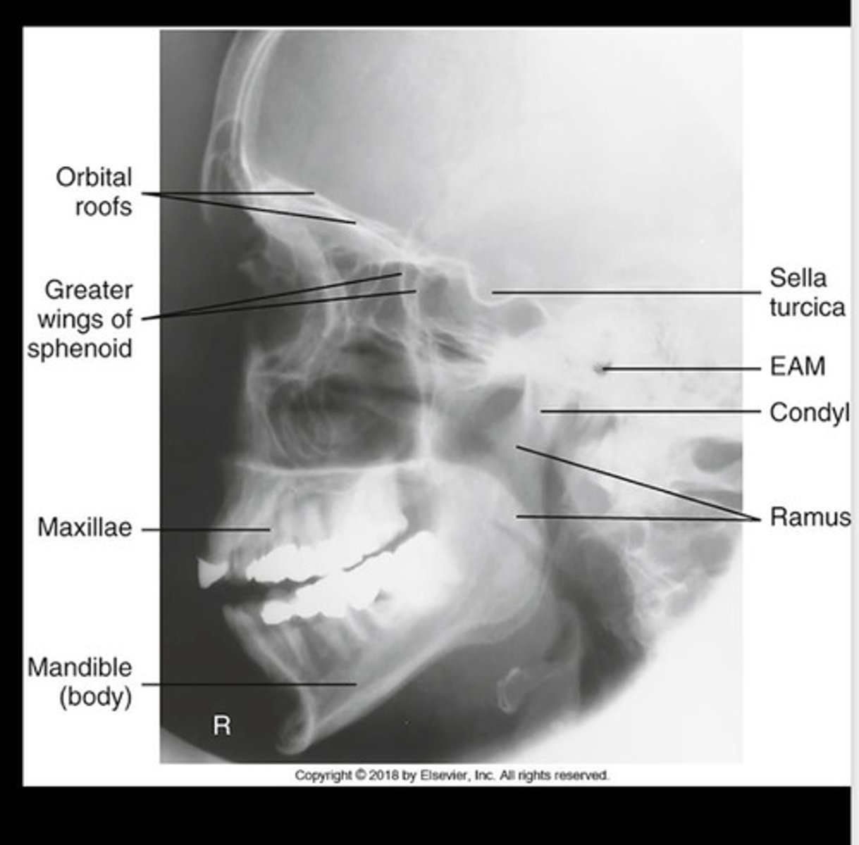
How can you tell rotation on lateral sinus
Greater wing not superimposed
Mandibular rami not superimposed
How can you tell tilt on lateral sinus
Orbital roofs not superimposed
Optimal exposure on lateral sinus if
able to see maxillae and sella turcica in profile
For a PA Axial Caldwell for sinuses, there is no
tube angulation
The OML for PA Axial Caldwell for sinuses needs to be
15 degree superiorly from horizontal
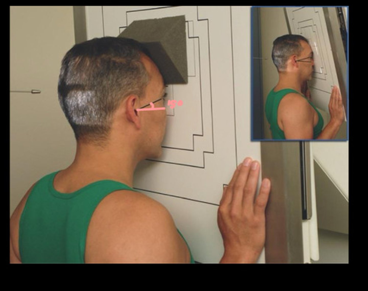
Collimation for PA Axial Caldwell for sinuses
1'' beyond lateral border of skull
Shadow of top of head and the occlusal plane
CR for PA Axial Caldwell Sinuses
Horizontal exiting through the nasion
The PA Axial Caldwell Sinus shows which sinuses
frontal sinus
anterior ethmoid sinuses
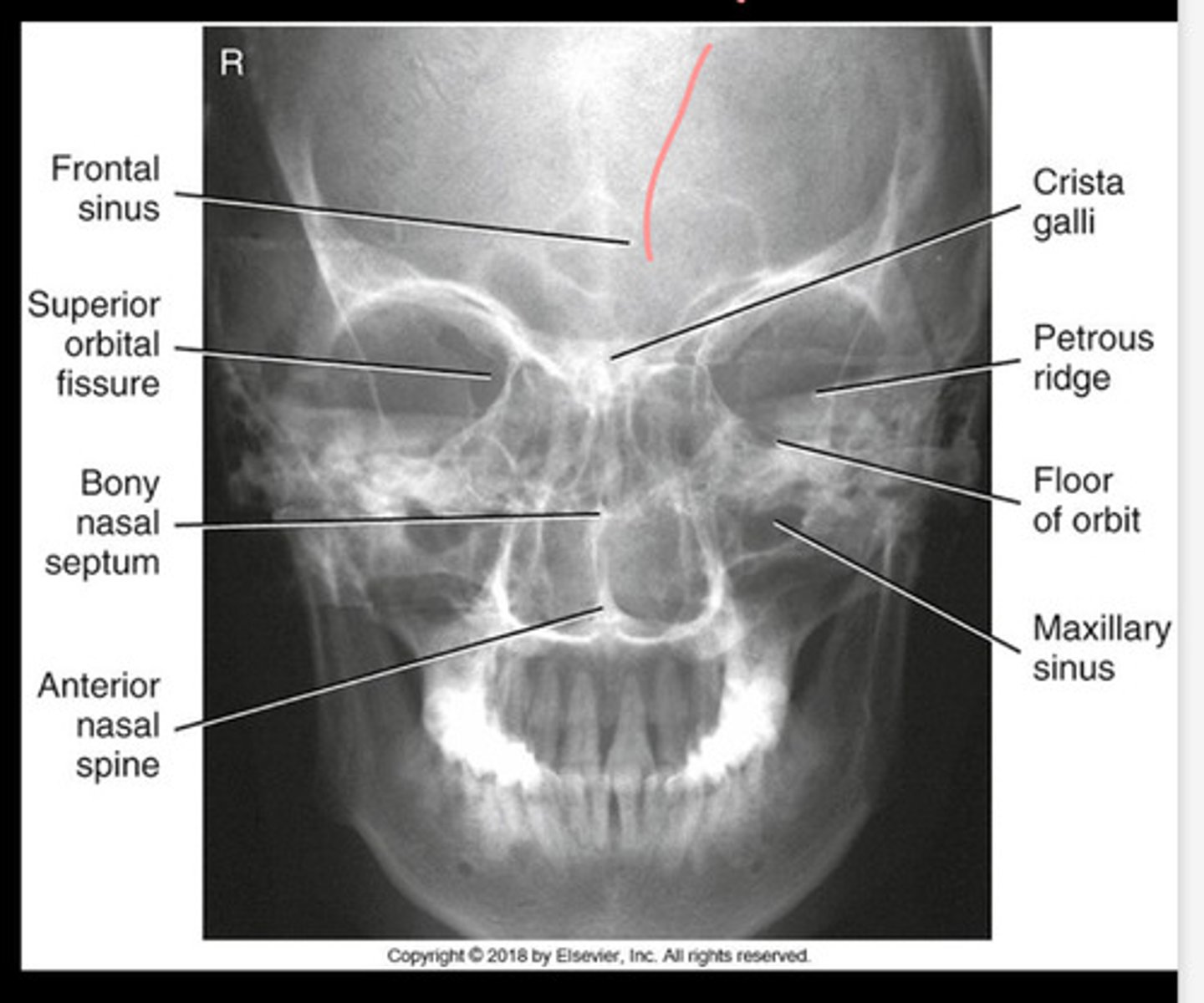
The petrous ridges in PA axial caldwell sinus should be placed
in lower 1/3 of orbit
How can you tell rotation on PA Axial Caldwell Sinus
Equal distance from orbit to lateral cortex of cranium
How can you tell tilt on PA Axial Caldwell Sinus
orbits on the same plane
You dont want to tilt down because
you do not want to distort the air fluid levels
PA Parietoacanthial Waters patient positioning
upright in the middle of the IR
Collimation for Parietoacanthial Waters view
1'' lateral to the skull border, shadow of the top of head, and inferiorly to the occlusal plane
What lines are involved in Parietoacanthial (waters)
MML-perpendicular to the IR
or OML-37 from horizontal
MSP
The MML for parietoacanthial (waters) should be
perpendicular to the IR
In a parietoacanthial (waters)
hyperextend the neck to make the MML perpendicular to the IR
or
OML 37 degrees from horizontal!!
central ray for parietoacanthial (waters)
perpendicular to the IR to exit at acanthion
What sinuses do you see on parietoacanthial waters?
Maxillary sinuses
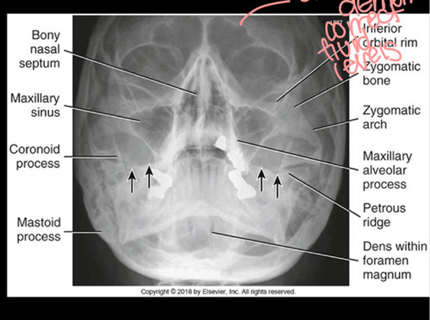
In the parietoacanthial waters view, the petrous ridges
should be below the maxillary sinuses
How can you tell rotation on parietoacanthial waters view
equal distance from lateral border of orbit to cranium
symmetrical maxillary sinuses
Parietoacanthial Transoral Waters
Special view to see sphenoid sinuses
Patient positioning for parietoacanthial transoral waters
upright with MSP in the middle of the IR
What lines are involved for parietoacanthial transoral waters
MML perpendicular to IR
OML 37 degrees from horizontal
Collimation for parietoacanthial transoral waters
1'' lateral to skull border, shadow of the top of head, and inferiorly the occlusal plane
PA Transoral waters shows what sinuses
SPHENOID
front and maxillary sinuses also included
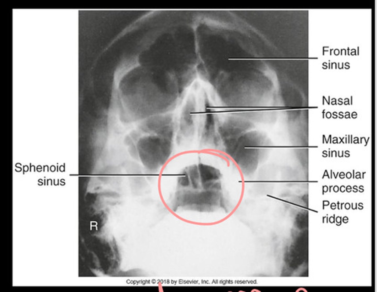
In transoral waters, the petrous ridges should be
below the maxillary sinuses
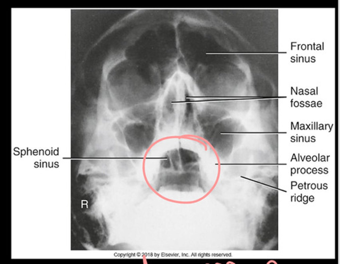
How can you tell a PA transoral waters is not rotated
Equal distance between lateral border of skull and lateral border of orbit
Symmetrical orbits
Symmetrical maxillary sinuses
What is the best view to see the sphenoid sinus?
PA transoral waters
Patient positioning for SMV sinuses
erect
What lines are involved with SMV
IOML-parallel to the IR
MSP-perpendicular to the IR
Central ray for SMV sinus
1.5'' below mandibular symphysis between angles of the mandible (3-4'' anterior to EAM)
What sinuses should you see in SMV
Ethmoid and sphenoid sinuses
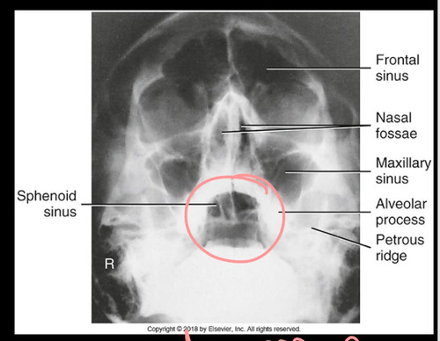
In an SMV sinus, the mandibular condyles should be
anterior to petrous ridges
How can you tell tilt on SMV
Equal distance between mandibular rami and lateral cranium