Unit 9 - Digestive tract
1/79
There's no tags or description
Looks like no tags are added yet.
Name | Mastery | Learn | Test | Matching | Spaced | Call with Kai |
|---|
No analytics yet
Send a link to your students to track their progress
80 Terms
Alimentary canal
GI tract, a muscular tube that extends from mouth to anus
mouth, pharynx, esophagus, stomach, small intestine, large intestine and anus
GI tract organs
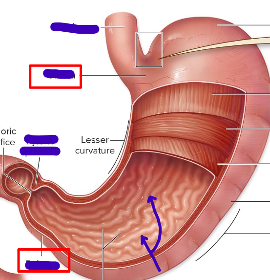
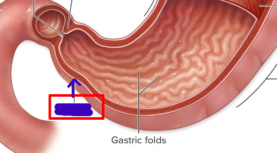
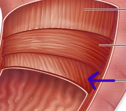
Rugae
gastric folds that increase the SA of the stomach
Accessory digestive organs
lie external to the GI tract and are connected by ducts
tongue, salivary glands, liver, gallbladder, and pancreas
Accessory digestive organs
Secondary retroperitoneal
partially inside and outside of the peritoneal
duodenum, pancreas, ascending and descending colon, and rectum
Secondary retroperitoneal organs
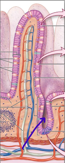
Mucosa
epithelial lining, connective tissue, lamina propria, muscularis mucosae, lightly folded
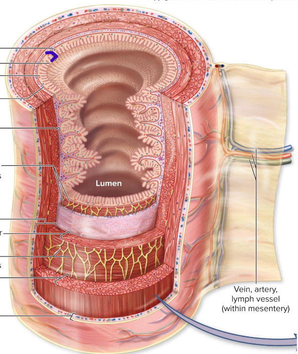
Submucosa
connective tissue, major blood vessels, mucosa associated lymphoid tissue
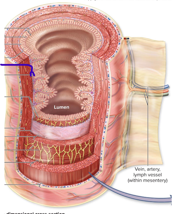
Muscularis
smooth muscle layer, there for motility (adaptive)
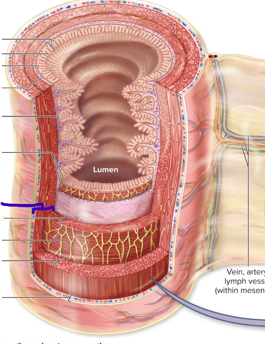
Serosa
lubrication of pieces and tunics
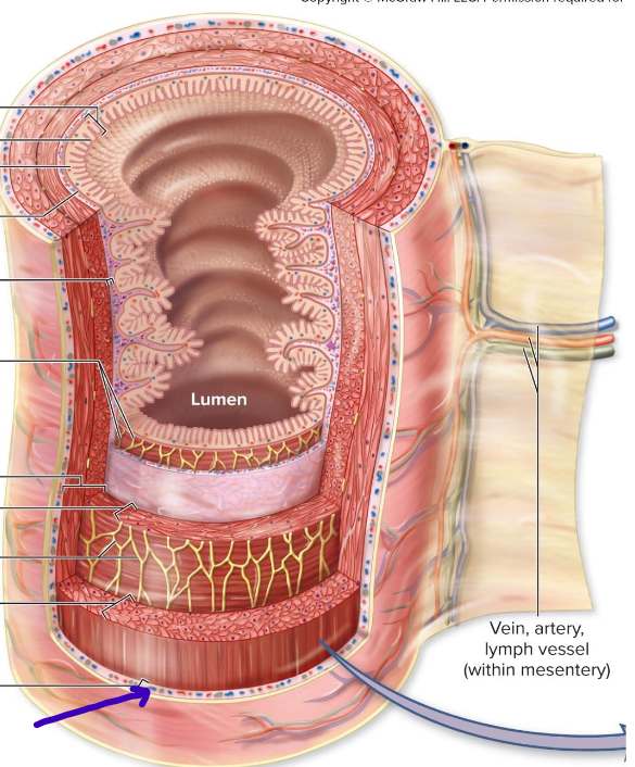
MSMS: Mucosa, Submucosa, Muscularis, Serosa
4 Tunics order (deep to superficial)
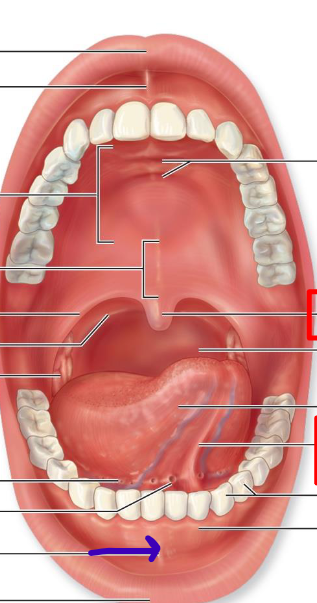
Superior labial frenulum
connects top lip to gums
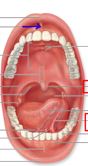
Hard palate
forms anterior roof of mouth
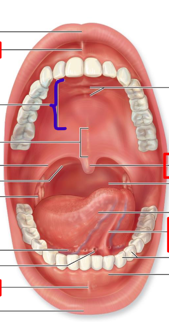
Soft palate
forms posterior roof of mouth
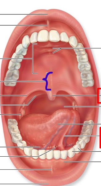
Uvula
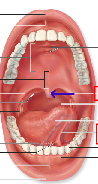
Lingual frenulum
connects tongue to floor of mouth
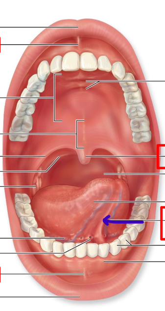
Inferior labial frenulum
connect lower lip to gums
Intrinsic salivary glands
exocrine; secretes small continuous amounts of saliva from tongue, palate, lips and cheeks
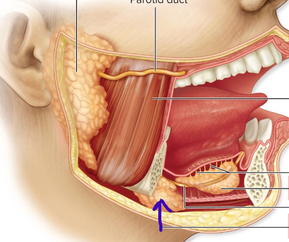
Extrinsic salivary glands
produces saliva while eating
Parotid salivary glands
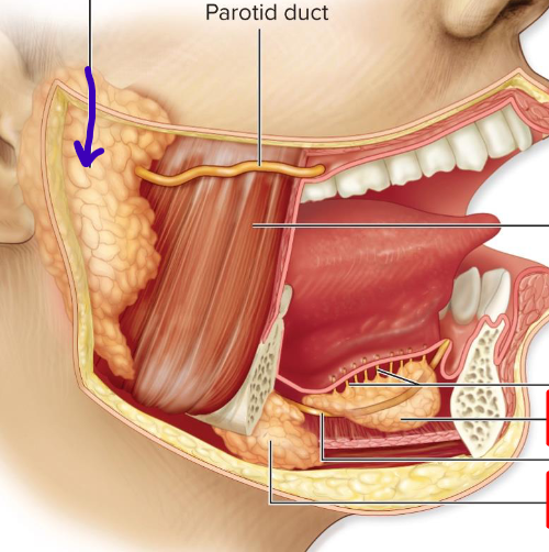
Sublingual salivary gland
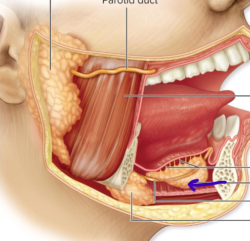
Submandibular salivary glands
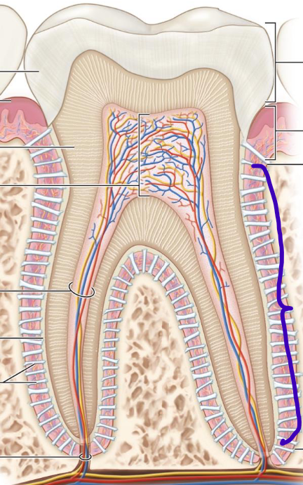
Enamel
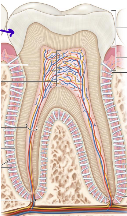
Gingiva
epithelial
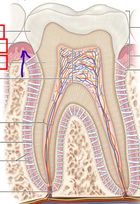
Dentin
primary mass; much harder than bone
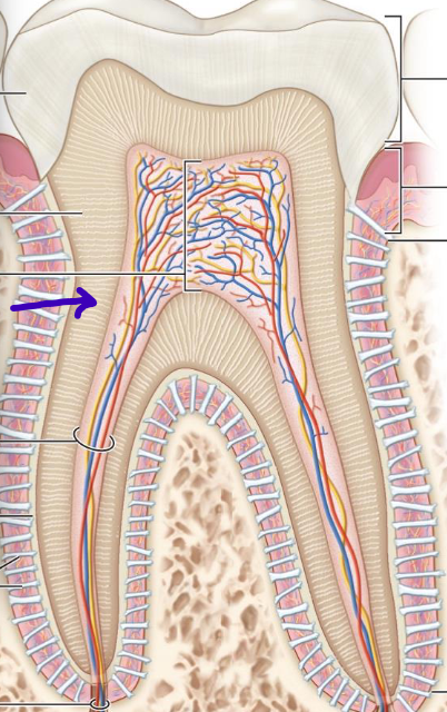
Crown
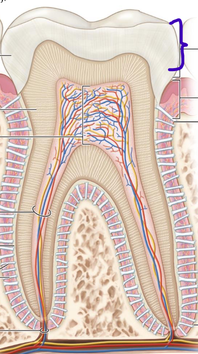
Neck
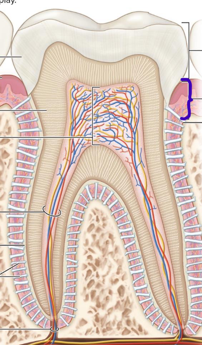
Root
Pharynx
inferior two thirds involved in respiration and digestion
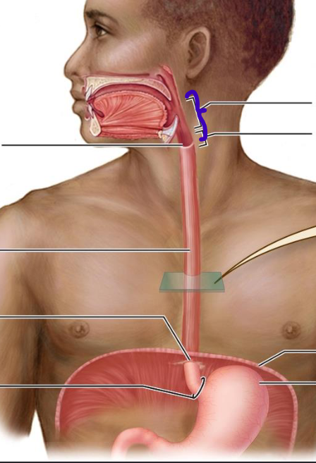
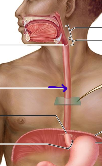
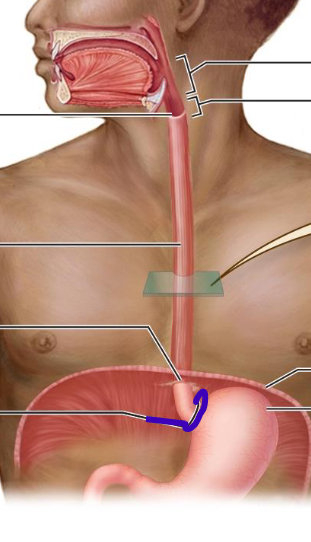
Esophagus
muscular tube that joins the pharynx to the stomach; completely collapsible
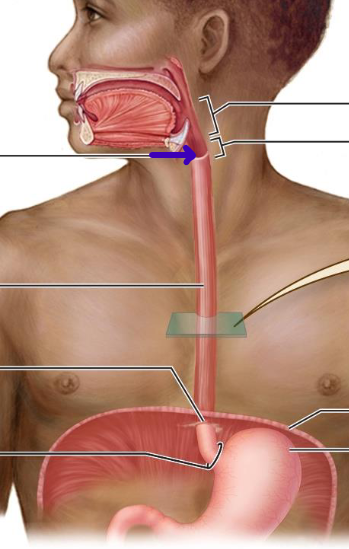
Superior esophageal sphincter
Esophageal hiatus
passes through diaphragm
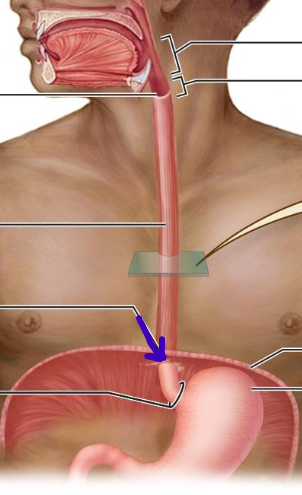
Inferior esophageal sphincter
closes lumen to prevent stomach acid from entering esophagus
Cardia
entryway to stomach
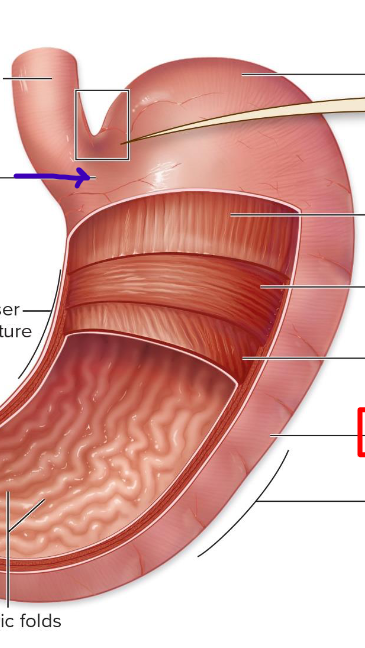
Fundus
round top to collect gas
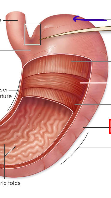
Body
largest portion with alternating muscle layers
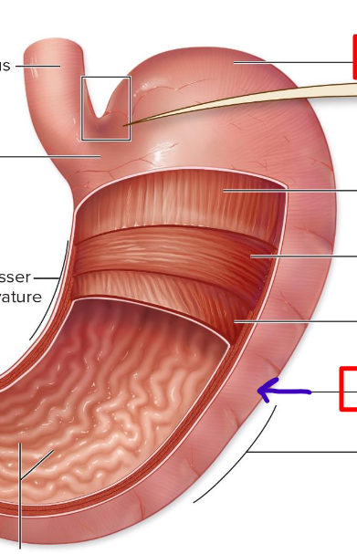
Pyloric
funnel-shaped, empties contents to duodenum
Circular muscle
middle muscularis layer
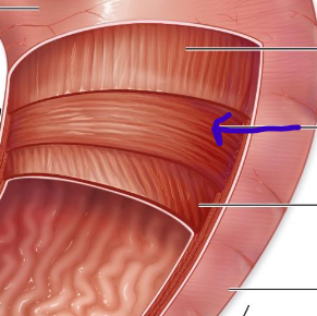
Longitudinal muscle
most superficial muscularis layer
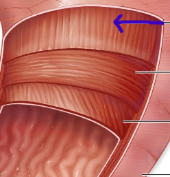
Oblique muscle
deepest muscularis layer
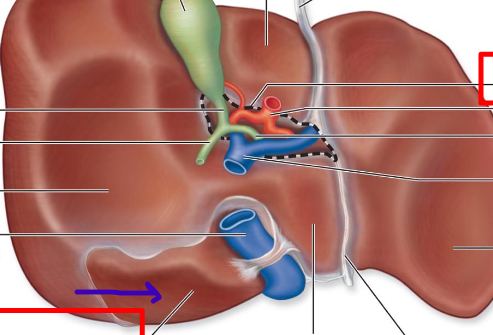
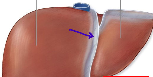
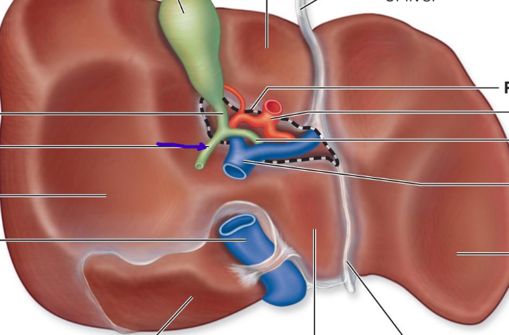
Liver
largest gland in the body with two surfaces
Diaphragmatic
anterior and superior surface of liver
Visceral
posterior and inferior surface of the liver
Right lobe
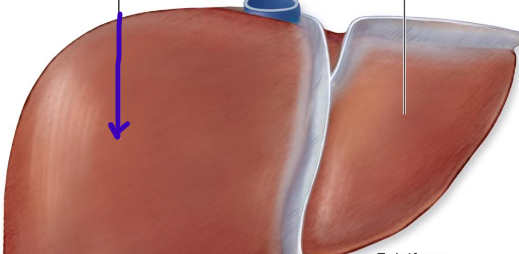
Left lobe
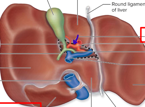
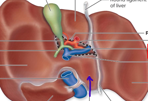
Porta hepatis
hilum of liver
Bare area
where the liver physically touches the diaphragm
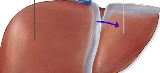
Falciform ligament
divides right and left liver lobes
Caudate lobe
Quadrate lobe
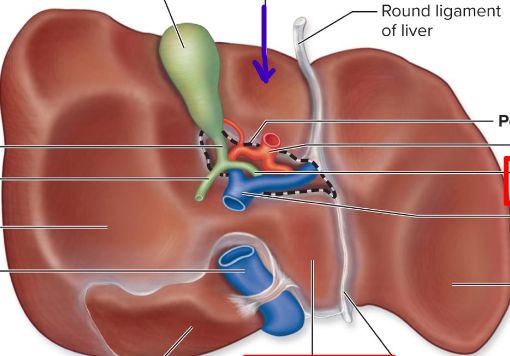
Gallbladder
a muscular sac that lies of the visceral surface of the right lobe of the liver
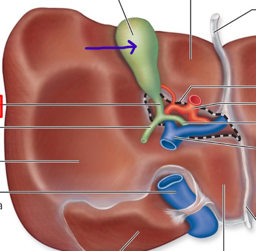
Common hepatic duct
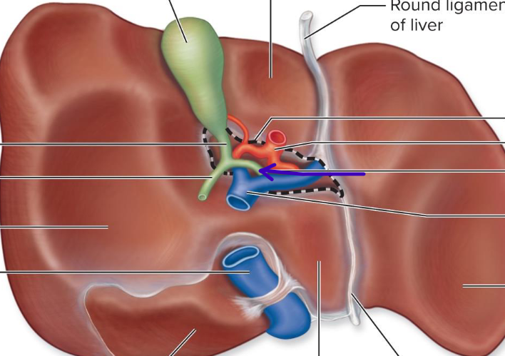
Cystic duct
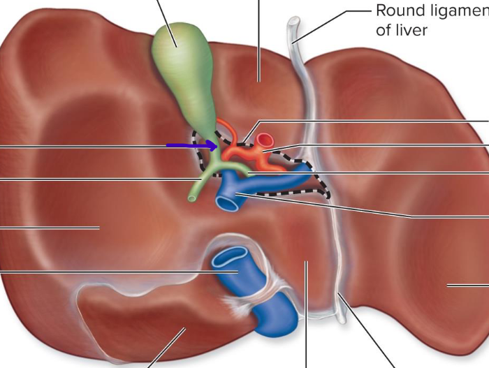
Common bile duct
empties into duodenum
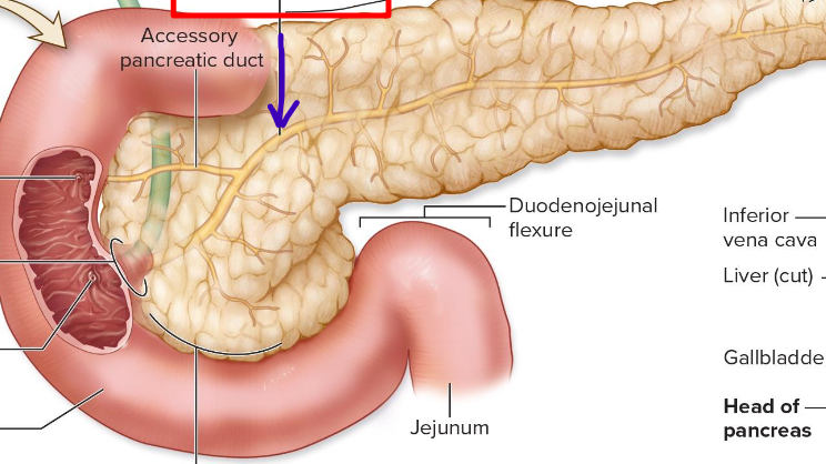
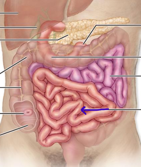
Pancreas
both an endocrine and exocrine gland
Main pancreatic duct
joins the bile duct and accessory pancreatic duct to empty into duodenum
Hepatopancreatic ampulla
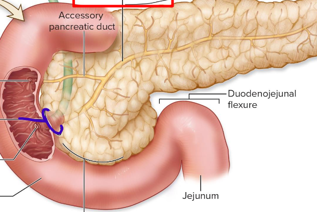
Duodenum
receives digestive enzymes and bile
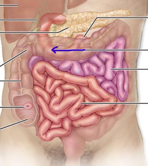
Jejunum
superior left part of coiled intestinal mass
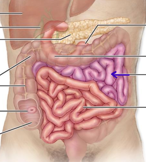
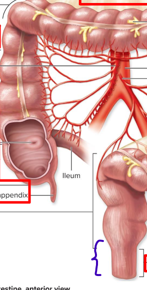
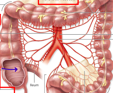
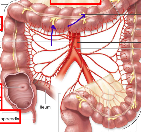
Ileum
inferior right part of coiled intestinal mass
Intestinal crypts
filled with stem cells
Large intestine lacks vili, have deeper crypts and have two mucous layers
Differences between small and large intestine
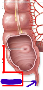
Cecum
a blind pouch that is the beginning of large intestine
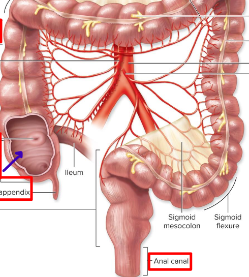
Vermiform appendix
contains lymphoid tissue to neutralize pathogens
Colon
divided into distinct segments
Ascending colon
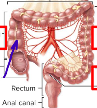
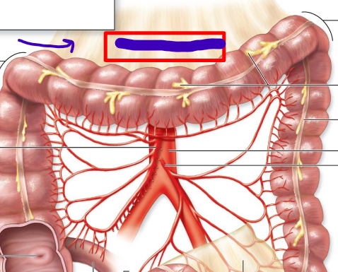
Transverse colon

Descending colon
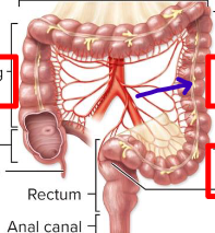
Sigmoid colon
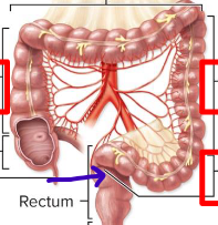
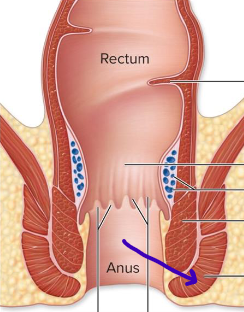
Rectum
descends along the inferior half of the sacrum
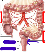
Anal canal
the last subdivision of the large intestine
Ileocecal valve
Haustrum
sacs that help push fecal matter
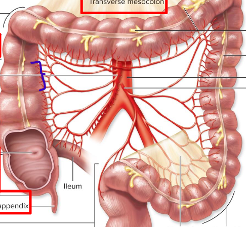
Transverse mesocolon
Omental appendicies
fat storage
Teniae coli
muscles that ribbons the large intestine

Rectal valve
allows gas to leave separate from fecal matter
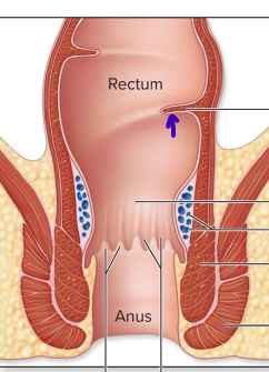
Internal anal sphincter
smooth involuntary muscle
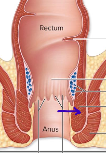
External anal sphincter
skeletal voluntary muscle