HL Biology Unit 6.3 - Gas Exchange
1/16
Name | Mastery | Learn | Test | Matching | Spaced | Call with Kai |
|---|
No analytics yet
Send a link to your students to track their progress
17 Terms
What does gas exchange occur between?
The capillaries and lung tissue
What are characteristics of tissues/surfaces used in gas exchange?
moist, thin, permeable, large surface area
What are alveoli?
Thin sacs in the lungs (the ends of bronchioles) that facilitate gas exchange and hold type I and type II pneumocytes. They have branching chambers that increase surface area.
Describe the structure of an alveolus and how oxygen/carbon dioxide is exchanged from it
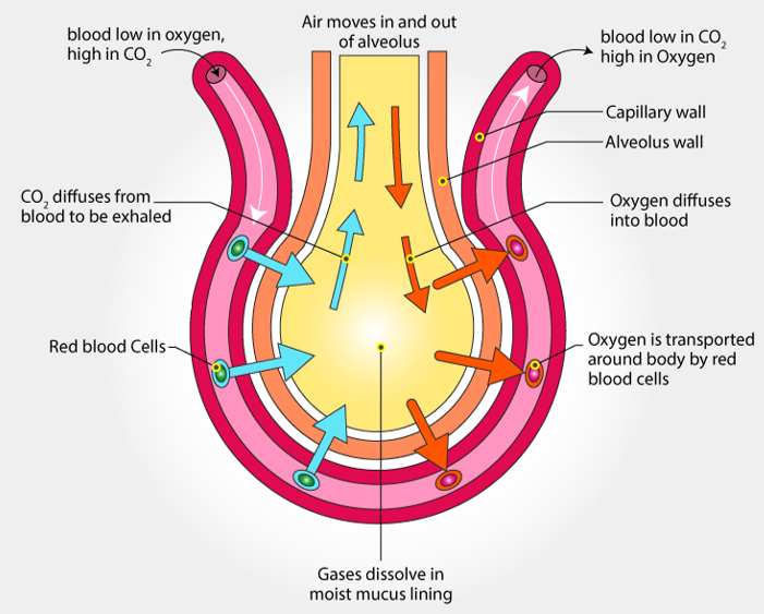
What do type I pneumocytes do?
Thin, flat cells used to facilitate quicker gas exchange in alveoli.
What do type II pneumocytes do?
Secrete fluid to make a moister environment for gas exchange and secretes a surfactant to prevent alveolar collapse by decreasing surface tension to alveoli don’t cohere to each other.
What is ventilation?
Expiration + Inspiration, moving air in/out of the lungs to maintain a concentration gradient of gases
What movements occur during Inspiration?
Diaphragm contracts
Abdominal muscles relax
External intercostal muscles contract, pulling ribcage up and out
Internal intercostal muscles relax, pulling back into their elongated state
What movements occur during Expiration?
Diaphragm relaxes
Abdominal muscles contract
External intercostal muscles relax, pulling back into their elongated state
Internal intercostal muscles contract, pulling ribcage inward and downward
What term describes the intercostal muscles?
Antagonistic - they work in pairs to perform opposite movements
What is hemoglobin?
An oxygen binding protein in red blood cells that helps transport oxygen
What is the structure of hemolglobin?
Made of 4 polypeptide chains, each with an iron-containing heme group that reversibly binds to oxygen - hemoglobin can bind to up to 4 oxygen molecules
What is cooperative binding?
When it gets easier for oxygen to bind to hemoglobin as more oxygen is binded because each molecule added alters that conformation of it, leading hemoglobin to also have a higher oxygen affinity in oxygen-rich areas (promotes oxygen loading) and not in oxygen-starved areas (promotes oxygen unloading)
What does a standard hemoglobin oxygen dissociation curve look like, and why?
It’ll have a Sigmoid “S” curve because cooperative binding alters oxygen affinity (which impacts %saturation, the y-axis) and causes it to be non-linear, and eventually heme groups become saturated and the graph plateaus
Compare the oxygen dissociation graphs of hemoglobin, fetal hemoglobin, and myoglobin
Fetal arteries have lower pressure, so their hemoglobin has a higher affinity at lower pressures, meaning the graph will increase faster and rest above a normal hemoglobin graph overall.
Myoglobin is an oxygen bonding molecule found in skeletal muscles that holds onto it for longer at lower concentrations to delay anaerobic respiration AND is not capable of cooperative binding, so it very quickly builds a high saturation and retains it for longer at maximum capacity. It rests above both graphs and is more like an exponential curve.

What is the Bohr Shift?
Tissues with a higher metabolic rate will release more CO2. Since hemoglobin has an allosteric binding site for CO2, when it binds it lowers O2 affinity. Blood cells will also release O2 in the presence of high CO2.
This means when theres a lower pH (more CO2), the graph will lower, and a higher pH, the graph will rise.

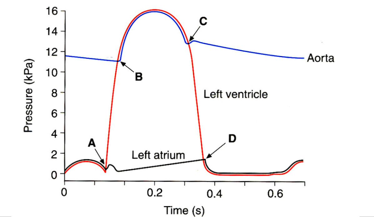
What occurs at each letter?
(Before A) AV Valves open
A) Ventricles contract, the SA valve is closed
B) Semilunar valve opens
C) Semilunar valve closes
D) AV Valve opens