PATH1 Neuropathology
1/232
There's no tags or description
Looks like no tags are added yet.
Name | Mastery | Learn | Test | Matching | Spaced | Call with Kai |
|---|
No analytics yet
Send a link to your students to track their progress
233 Terms
- trauma
- vascular events
what causes CNS hemorrhage and edema
- ataxia
- behavioral changes
- seizures
- head presssing
- circling (toward lesion)
- blindness
common clinical signs in cerebrum/forebrain injury (central signs)
- ataxia
- hypermetria
- hypertonicity
- head tilt
- circling
- nystagmus
- tremors
- cranial nerve deficits
common clinical signs in midbrain/cerebellum/brainstem injury
- paralysis
- weakness
- spasticity
common clinical signs with spinal cord injury
coup
injury on the same side as impact
contracoup
injury on the opposite side of impact
concussion
head injury leading to loss of consciousness without gross evidence of injury
contusion
head injury that results in hemorrhage, +/- tearing of brain parenchyma, +/- skull fracture
fracture
- happens to skull or vertebrae
- when this happens, bone can act as foreign body or mechanism for laceration of neurological tissue it surrounds
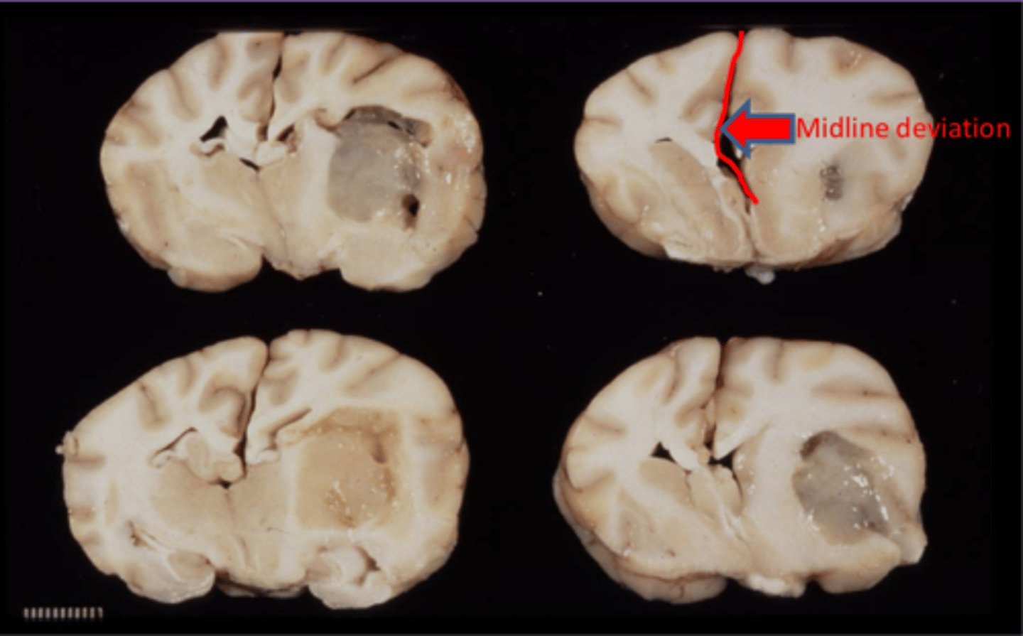
- can have effects as space occupying lesion
- increased intracranial pressure
- compression
- +/- herniation (cerebellum most common)
- can be delayed
hematoma
- dura mater
- pia mater
- arachnoid mater
3 layers of meninges
epidural hematoma
hematoma outside of the meninges
subdural hematoma
hematoma inside the meninges
intracerebral hematoma
hematoma within the brain
cerebral herniation
cerebral coning
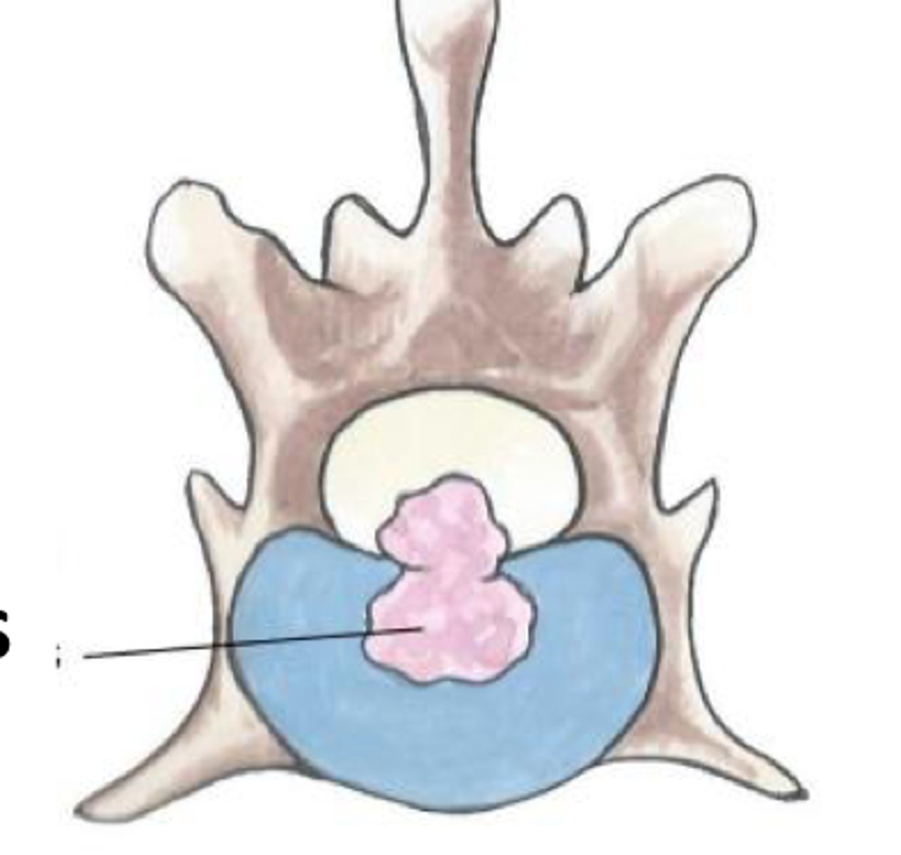
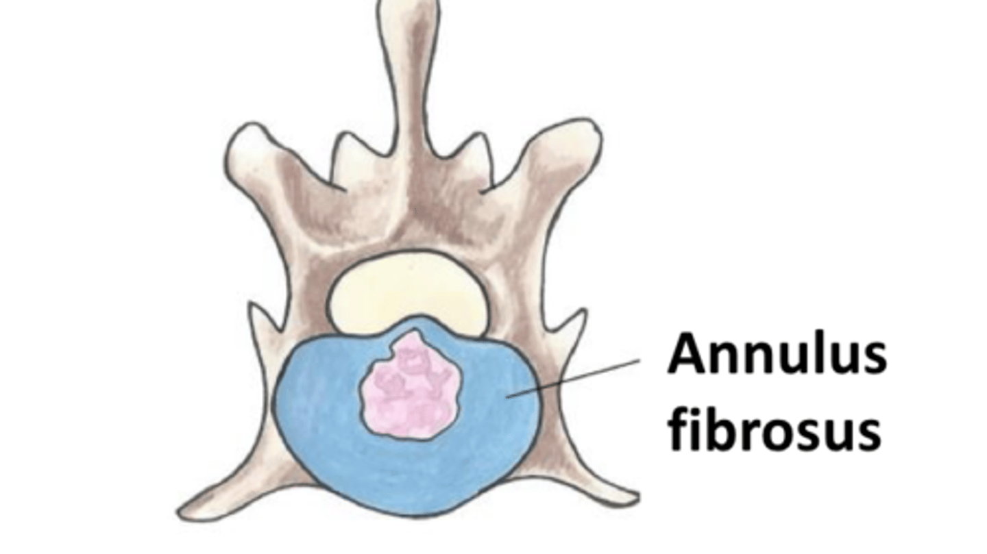
- nucleus pulposus
- annulus fibrosis
layers of intervetebral disc
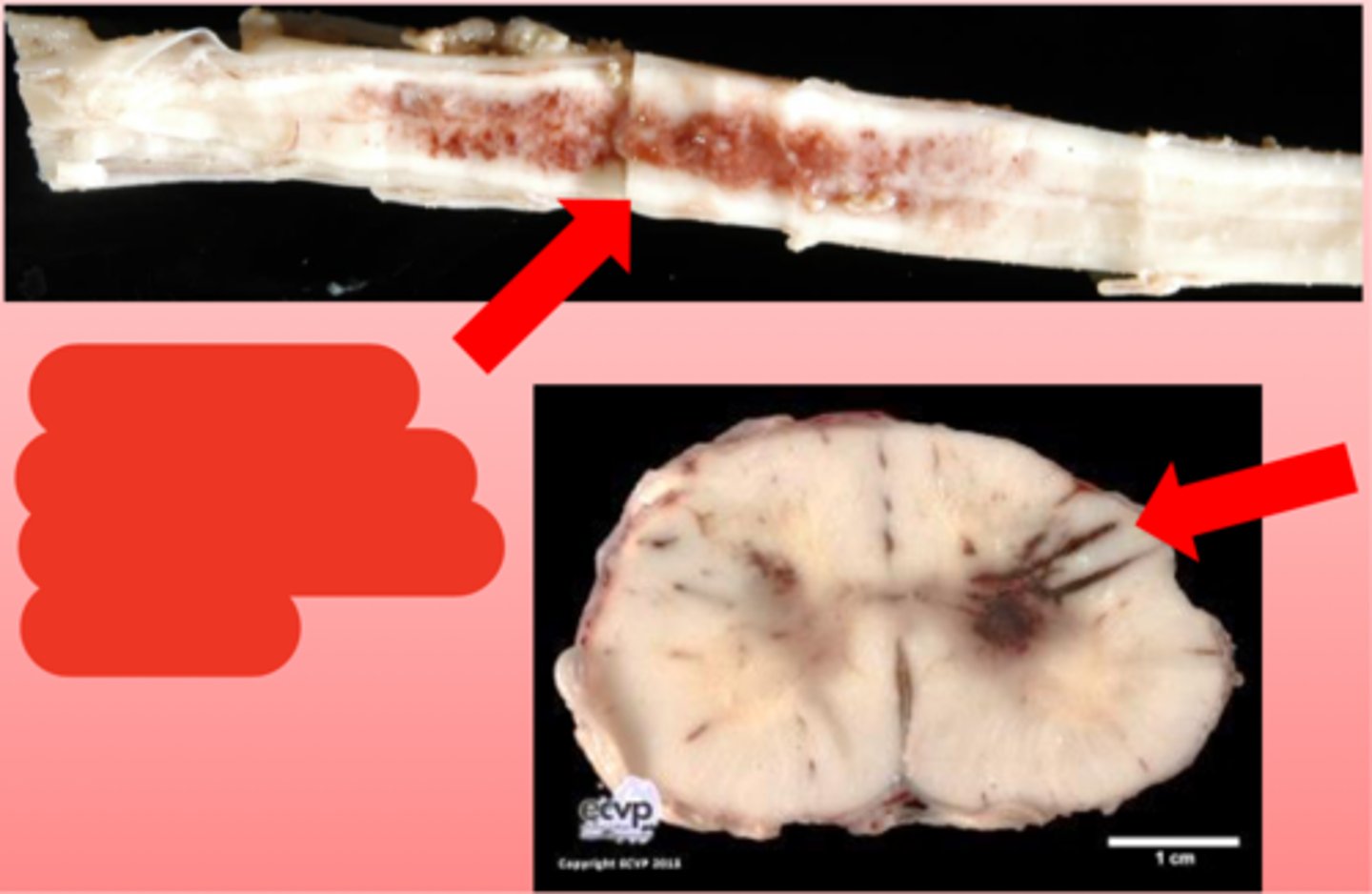
Intervertebral discs can herniate into the spinal cord, traumatizing the spinal cord and cause hemorrhage, edema, and necrosis
intervetebral disc degeneration (IVDD) and herniation
dogs
IVDD is common in which species
Type I extrusion disc herniation
Type II protrusion disc herniation
acute
onset of Type I disc hernation (extrusion)
Chondrodystrophic (dwarf) breed dogs like Dachshunds, frenchies, basset hounds, corgis
which breeds is IVDD type I more commonly seen
gradual onset
onset of Type II disc herniation (protrusion)
large breed dogs
which breeds is IVDD Type II more commonly seen in
Type II
disc hernation is uncommon in cats but if they do get it, which type do they usually suffer from?
hemorrhagic myelomalacia
ascending and/or descending hemorrhage and necrosis within the spinal cord
disc hernation or other trauma to the spinal cord
cause of hemorrhagic myelomalacia
ascending or descending paralysis and sensory deficits 24 hours after spinal cord injury
presentaiton of hemorrhagic myelomalacia
hemorrhagic myelomalacia
strokes
sudden onset of focal neurological deficits from intracranial vascular event
local ischemia from infarction or hemorrhage
causes of strokes
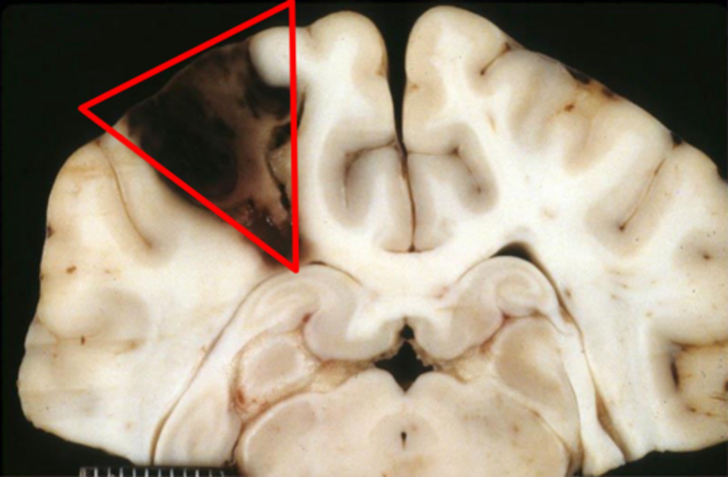
acute cerebral infarct
red to black, sharply demarcated, wedge-shaped
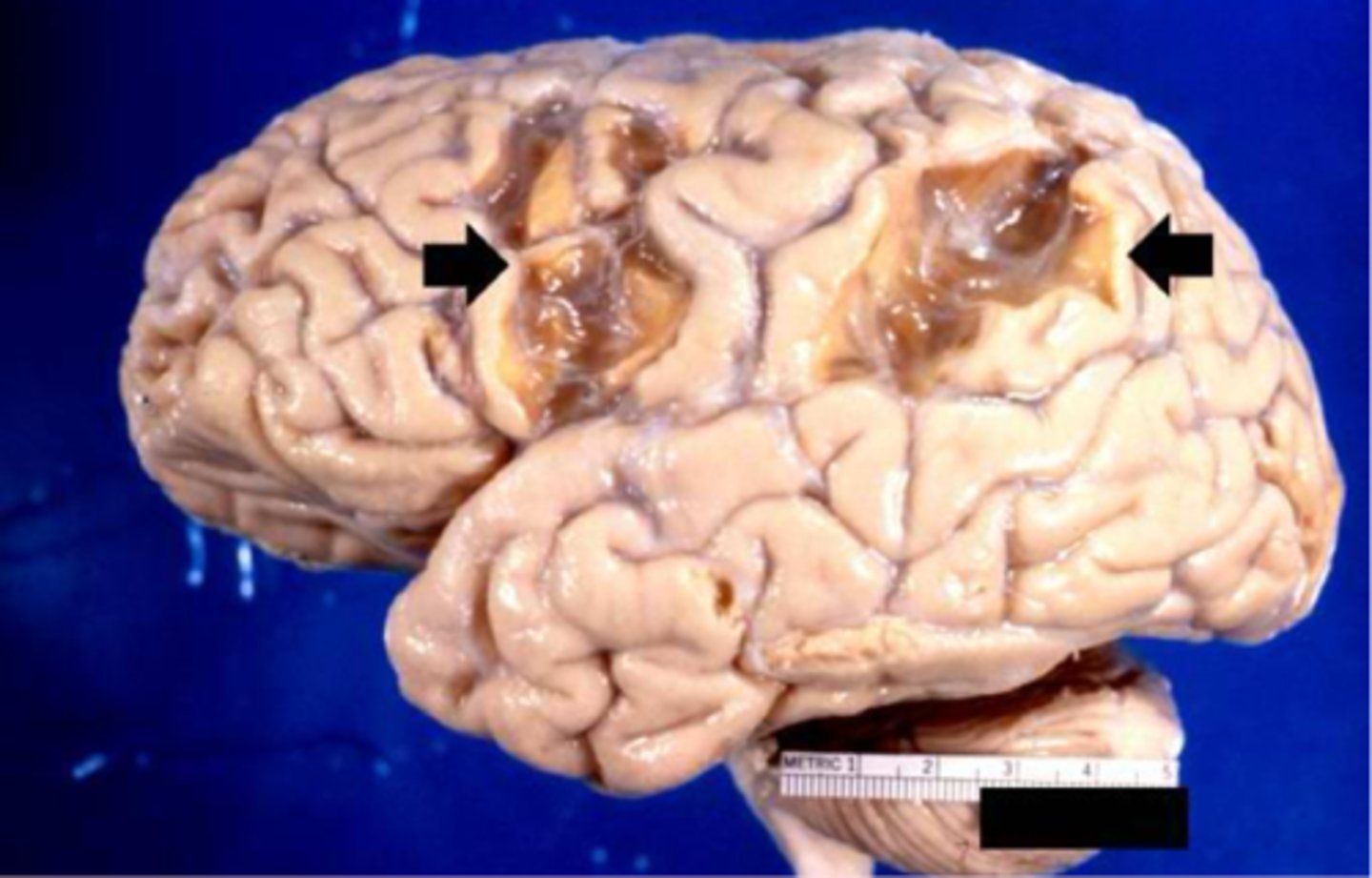
chronic cerebral infarct
golden brown, depressed, may resolve as shrunken pale scars
- uncommon, affects cats of all ages
- ataxia/circling, seizures, blindness, and postural deficits
- unilateral ischemic necrosis and atrophy of cerebral cortex
Feline Ischemic Encephalopathy (FIE)
middle cerebral artery
damage to which vessel in the brain can cause feline ischemic encephalopathy
fibrocartilaginous emboli (FCE)
degenerative intervetebral disc material extruded into spinal blood vessels and travels to parenchyma of spinal cord, causing infarction
fibrocartilaginous emboli (FCE)
- hemorrhage
- edema
- softening of parenchyma (malacia)
what gross lesions would make you suspect there was a vascular/traumatic injury
dogs
which species is CNS neoplasia most commonly seen in
brachycephalic breeds (boxers or boston terriers)
which dog breed has a increased incidence of CNS tumors
gliomas
which CNS tumors are brachycephalic breeds more common to suffer from?
meningioma
most common CNS neoplasia in dogs and cats
- astrocytoma
- oligodendroglioma
which CNS neoplasia are 2nd most common in dogs
choroid plexus (epithelial tumors) like papilloma, adenoma, carcinoma)
3rd most common CNS neoplasia
leptomeninges
arachnoid and pia mater are collectively referred to as
in choroid plexus within ventricles
pia mater is adhered to the surface of the brain and also
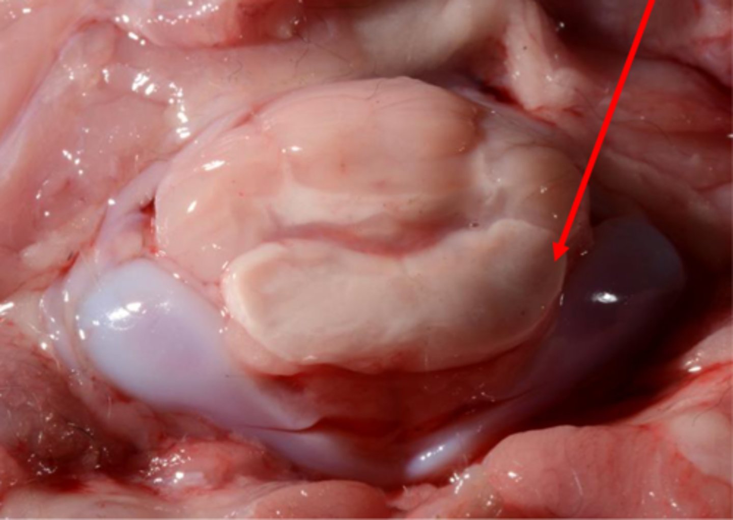
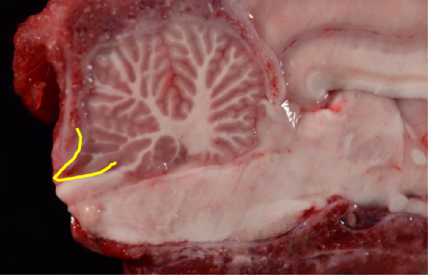
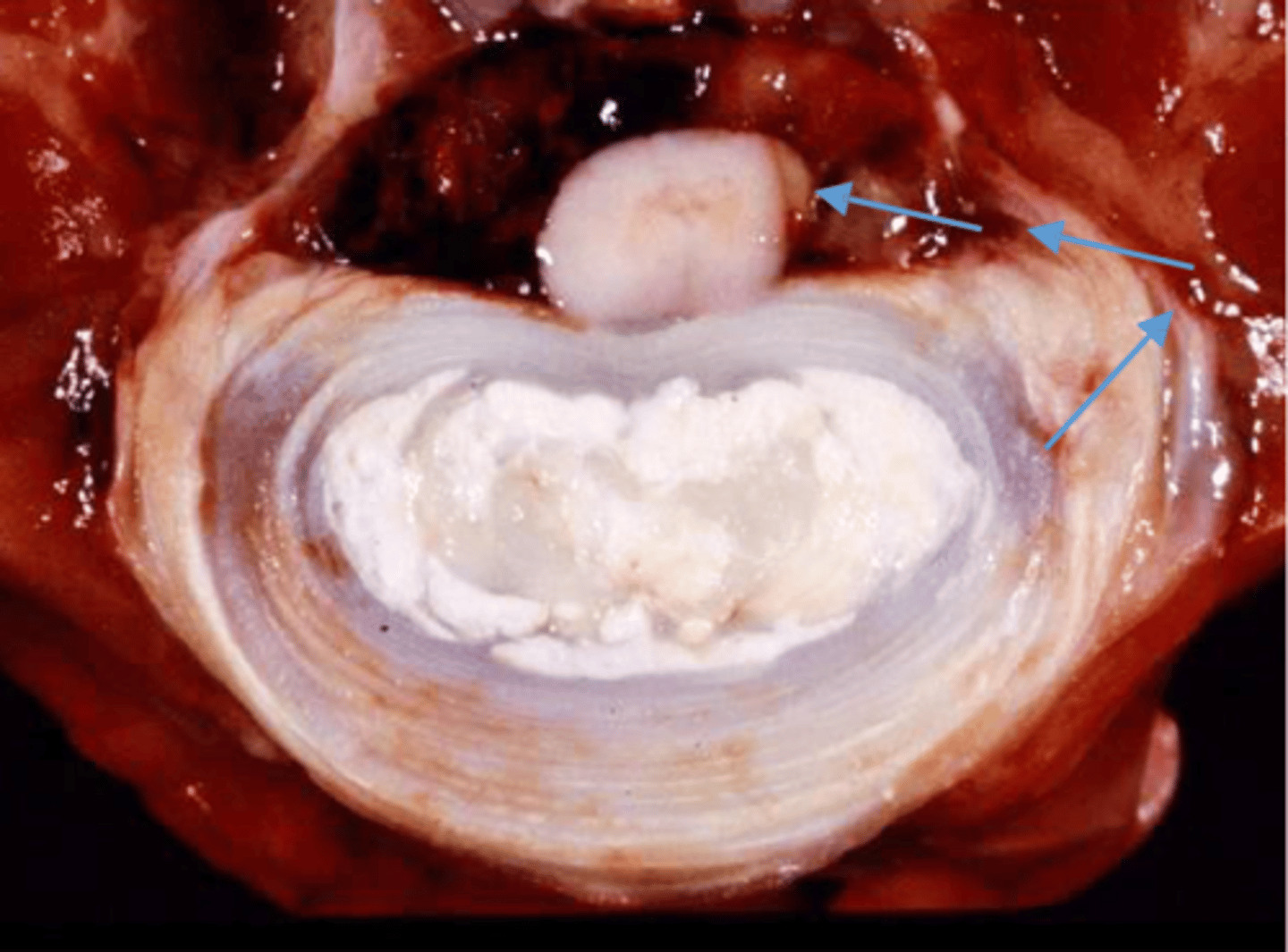
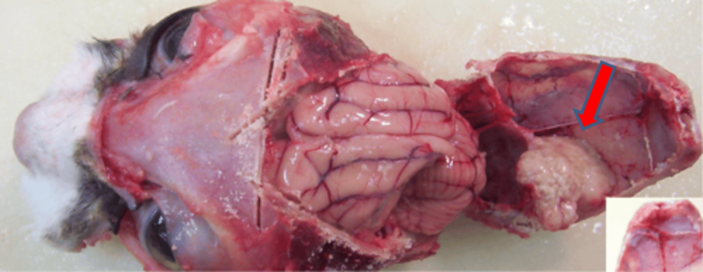
- most common primary intracranial brain tumor of dogs and cats
- can arise from any of 3 meningeal layers
- usually superficial or dorsal on brain or spinal cord, intraventricular also possible (pia mater)
- benign but space occupying
- low recurrence rate following surgical excision
meningioma
discrete, firm to rubbery with meningeal attachment. can find compression of neural tissue beneath
meningioma gross appearance
meningioma
5 times
how much more abundant are astrocytes than neurons in the CNS
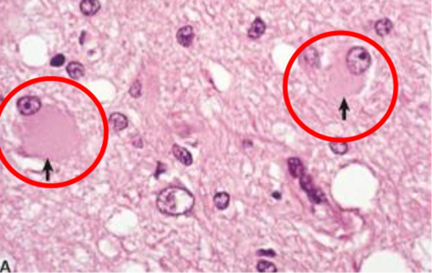
gemistocytes
astrocytes that respond to injury by increasing their cytoplasm
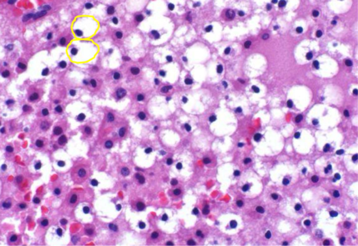
alzheimer type 2 astrocytes
astrocytes that appear as clusters of astrocytic nuclei that are swollen and clear. associated with hepatic and renal encephalopathy
form myelin sheath around axon
function of oligodendrocytes
malignant
are glial tumors (astrocytomas and oligodendrogliomas) malignant or benign
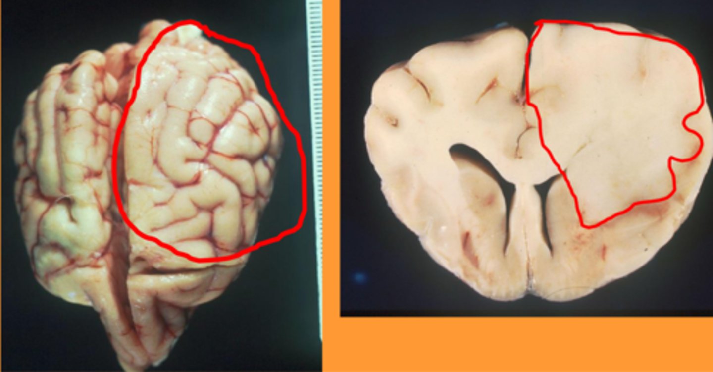
- very infiltrative with indistinct margins
- most often found in cerebrum (temporal lobes)
astrocytoma
astrocytoma
- mature dogs
- males
- cerebrum (frontal lobe)
oligodendroglioma
- pink to gray
- soft and gelatinous
- indistinct or sharply demarcated margins
gross appearance of oligodendrogliomas
oligodendroglioma
fried egg appearance
microscopic appearance of oligodendrogliomas
oligodendroglioma
phagocytosis
main function of microglia
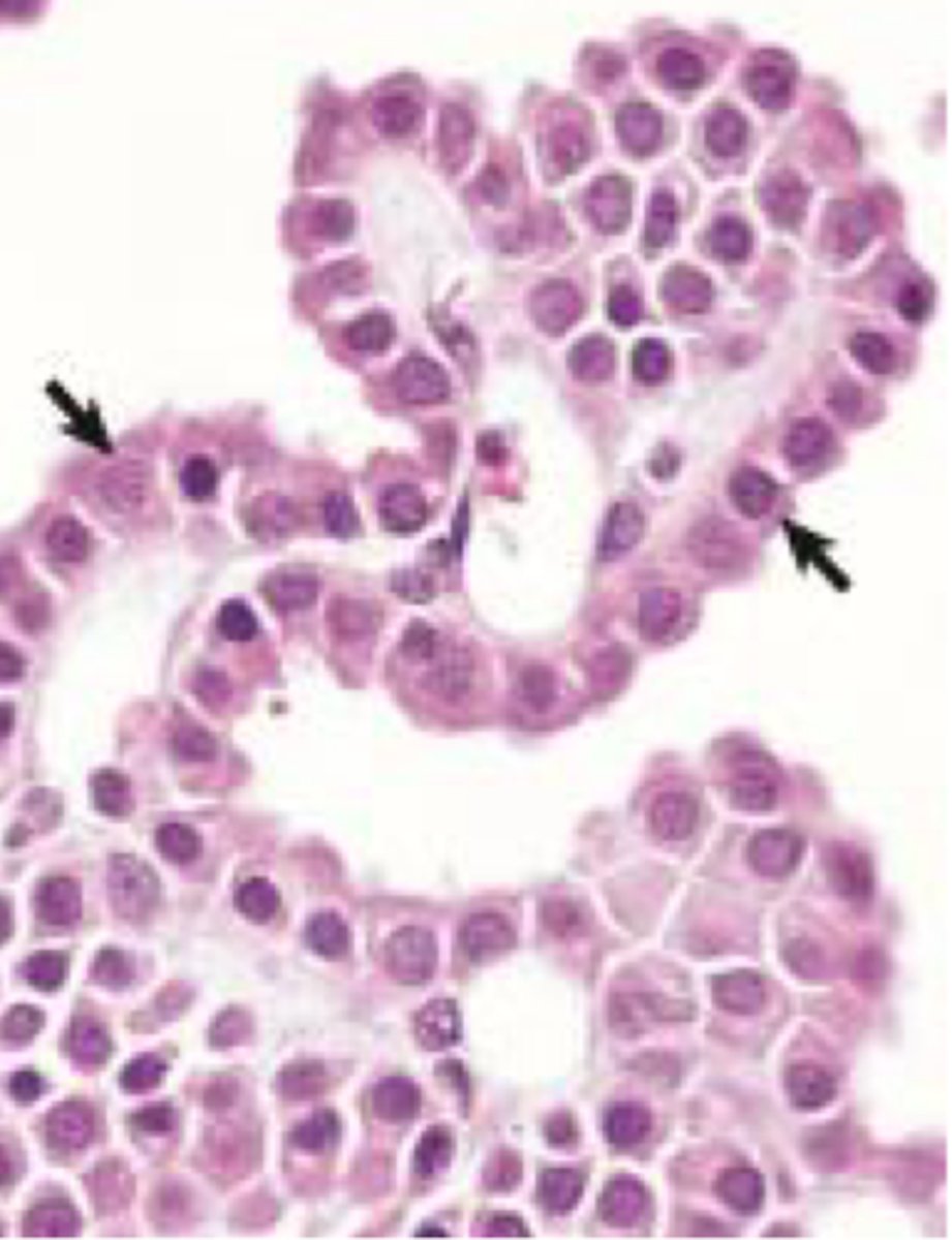
choroid plexus
ciliated (modified) cuboidal ependymal epithelium that produces CSF
- within ventricles
- supported by pia mater
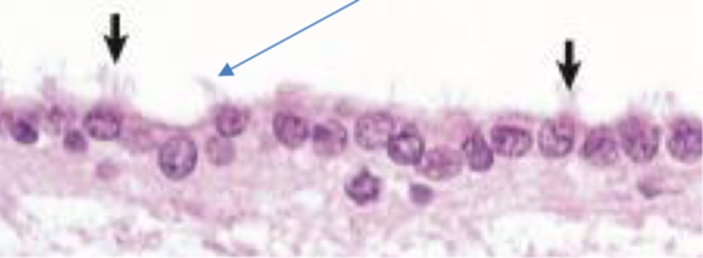
ependymal cells
- ciliated cuboidal epithelium
- line ventricles and central canal of spinal cord
- important part of brain/CSF barrier
4th ventricle
most common location for choroid plexus tumors
- vomiting
- positional nystagmus
- head tilt
- tetraparesis
presentation of choroid plexus tumors
rare in all species
ependymoma
- tan to red/grey and nodular
- within ventricles
gross appearance of choroid plexus tumors
hydrocephalus
what can papillomas, adenomas, and carcinomas cause in the brain due to ventricular obstruction
external granular cell layer
which cell layer in the cerebellar cortex is only present in utero or neonates (2-4 week period after birth)
medulloblastomas
which CNS neoplasia is highly malignant and is composed of undifferentiated cells of the neural tube
young animals
medulloblastomas arise from the external granular cell layer of the cerebellum and occur almost exclusively in what kind of animal
calves
medulloblastomas occur most commonly in this species
- grey masses, often in cerebellum
- +/- secondary hydrocephalus
gross appearance of medulloblastomas
- perineural cells
- fibroblasts
- schwann cells
cells in the peipheral nervous system that commonly give rise to neoplasms
schwannoma
benign tumor of schwann cells
neurofibroma
benign schwann and perineural cell tumor (collective term)
peripheral nerve sheath tumor (PNST)
malignant version of schwann or perineural cell tumor
- benign or malignant
- locally invasive
- rarely metastasize
peripheral nerve sheath tumors
neurofibromatosis
syndrome in cattle where single or multiple peripheral nerve sheath tumors develop in multiple anatomic sites
meningitis
inflammation of the meninges
encephalitis
inflammation of the brain
myelitis
inflammation of the spinal cord
meningoencephalomyelitis
inflammation of the meninges, brain, and spinal cord
polioencephalitis
inflammation of the grey matter of the CNS
leukoencephalitis
inflammation of the white matter of the CNS
superficial
where is the grey matter in the cortex of the brain
deep
where is the grey matter in the spinal cord
bacteria
suppurative inflammation is indicative of what type of infection
neutrophils
suppurative inflammation from bacteria has what type of inflammatory cells
mixture of lymphocytes/plasma cells/macrophages
non suppurative infections have what type of immune cells
viral or immune mediated
what is non-suppurative inflammation indicative of?
macrophages +/- neutrophils
what type of inflammatory cells are characteristic of granulomatous or pyogranulomatous infections
- bacteria (mycobacteria)
- fungus
- certain viruses
what can cause granulomatous or pyogranulomatous lesions
parasites
what causes eosinophilic inflammation
hematogenous
how are neurological infections often spread
- subarachnoid barrier
- blood brain barrier
- CSF barrier
barriers that keep out CNS infection
small ruminants
what species does Listeria most commonly affect
goat has ulcer and eats food contaminated with Listeria monocytogenes, bacteria infect peripheral nerves and extend to brainstem/cerebellum
How do small ruminants get affected with Listeria