Part 1
 Call Kai
 Learn
Learn Practice Test
 Spaced Repetition
 Match
Match Flashcards
 Knowt Play
1/37

There's no tags or description
Looks like no tags are added yet.
Name | Mastery | Learn | Test | Matching | Spaced | Call with Kai |
|---|
No analytics yet
Send a link to your students to track their progress
38 Terms
cancer progression
Proliferation
Transformation
Invasion
Metastasis
Vacularization
activation of oncogenes
Cancer Progression Factors
e.g., Ras protiens
upregulation of proteases
Cancer Progression Factors
e.g., MMPs (Matrix metalloproteinases)
Enzyme released by your cancer cells that will breakdown the tissue for it to metastasize.
deactivation of adhesion molecules
Cancer Progression Factors
e.g., E-cadherin
stimulation of angiogenesis genes
Cancer Progression Factors
e.g, VEGF
these are the ones that are simulated if you have poor lifestyle
mutation of DNA
Cancer Progression Factors
e.g, p53, BRCA1, cyclins
Probably because of an infection or autoimmune disease
loss of tumor suppressor genes
Cancer Progression Factors
e.g, pRb, APC
growth factor dysregulation
Cancer Progression Factors
e.g, EGF, HER2/neu
These are normally low levels in a healthy individual, but when there is cancer, they are elevated
stage 1
Cancer Staging and Progression (4)
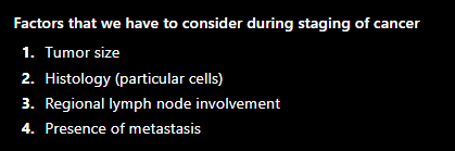
Localized primary tumor
Can still be removed.
stage 2
Cancer Staging and Progression (4)
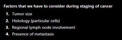
Cancer cells release MMP
Invasion of primary tumor through epithelium and into blood vessels
stage 3
Cancer Staging and Progression (4)
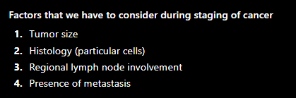
Migration of tumor into regional lymph nodes
stage 4
Cancer Staging and Progression (4)
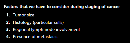
Metastasis and invasion of tumor to distant tissues
tumor markers
Produced directly by the tumor or as an effect of the tumor on the host
Note: these are not automatically associated with malignancy, it can be present in certain benign tumors, and can also be present in normal physiological processes.
types of tumor markers
serum proteins (β2-microglobulin)
oncofetal antigens (AFP, CEA)
Genes that are developed during pregnancy, malignancy in non-pregnancy.
hormones (calcitonin, β-hCG)
carbohydrate epitopes/cancer antigens (CA-125)
receptors (estrogen receptors, Her-2/neu)
enzymes (ALP)
PSA
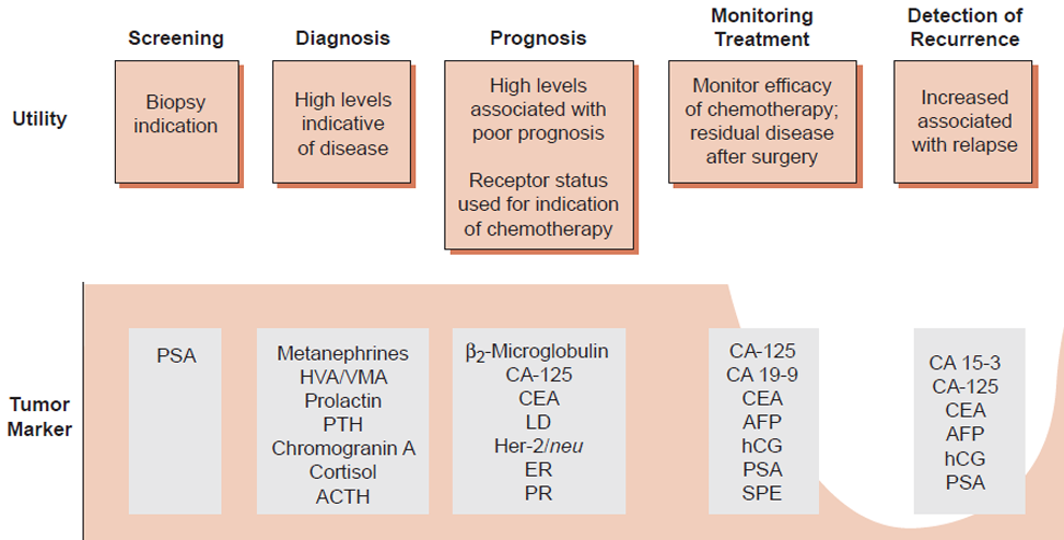
Applications of Tumor Marker Detection
Screening
Biopsy Indication
HPLC
Best method of choice for catecholamine tumor markers like HVA, 5HIAA, metanephrines and VMA?
commonly used for detection of small molecules (e.g. endocrine antibodies)
catecholamines and metabolites: carcinoid tumors, pheochromocytoma and neuroblastoma
IHC
Best method of choice for Receptor tumor markers like estrogen, progesterone receptor etc. It uses Biopsy as specimen.
immunoassay
Laboratory Consideration
Most commonly used method to measure tumor markers
But they require some considerations;
1. Since there is a wide range of values for out tumor markers
2. Lack of standardization
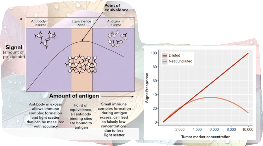
linearity
Immunoassay
Determined by analyzing specimens spanning the reportable range
“Linear range” span of analyte concentrations over which a linear relationship exist between the analyte and signal
hook effect
Immunoassay
Excessively high tumor marker concentration can result in falsely low measurements
“antigen excess:”
Note: There will be a hook effect of your graph if there is no ZOE or undiluted.
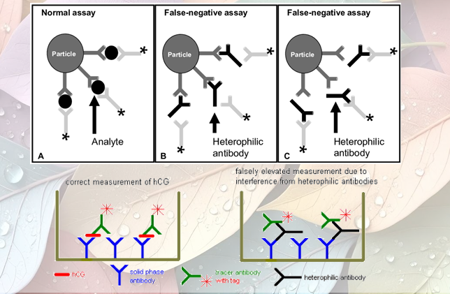
heterophile antibodies
Immunoassay
Human anti-mouse Abs (HAMAs)
Note: They are given to patients with auto-immune diseases
This Abs has to be tested first if the patient develops cancer. That is why it is also important to note for the px history
Because if the px was given these HAMAs before, these Abs binds to the latex beads = FALSE NEGATIVE.
analytical interferences
Immunoassay
Icterus
Lipemia
Hemolysis
Antibody cross reactivity
Neuroblastoma