Anatomy Lab Midterm 2
1/196
There's no tags or description
Looks like no tags are added yet.
Name | Mastery | Learn | Test | Matching | Spaced | Call with Kai |
|---|
No analytics yet
Send a link to your students to track their progress
197 Terms
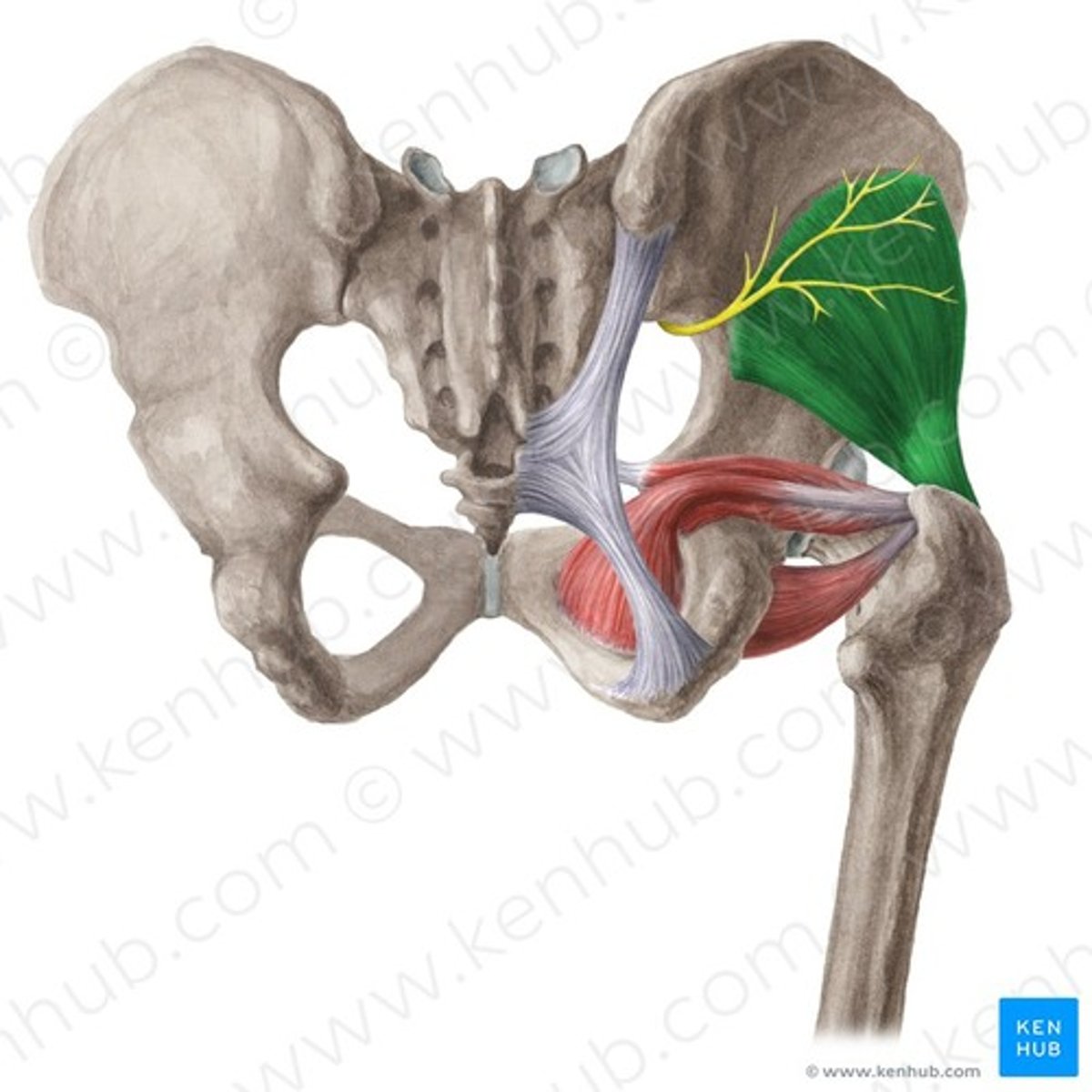
gluteus maximus
extends thigh
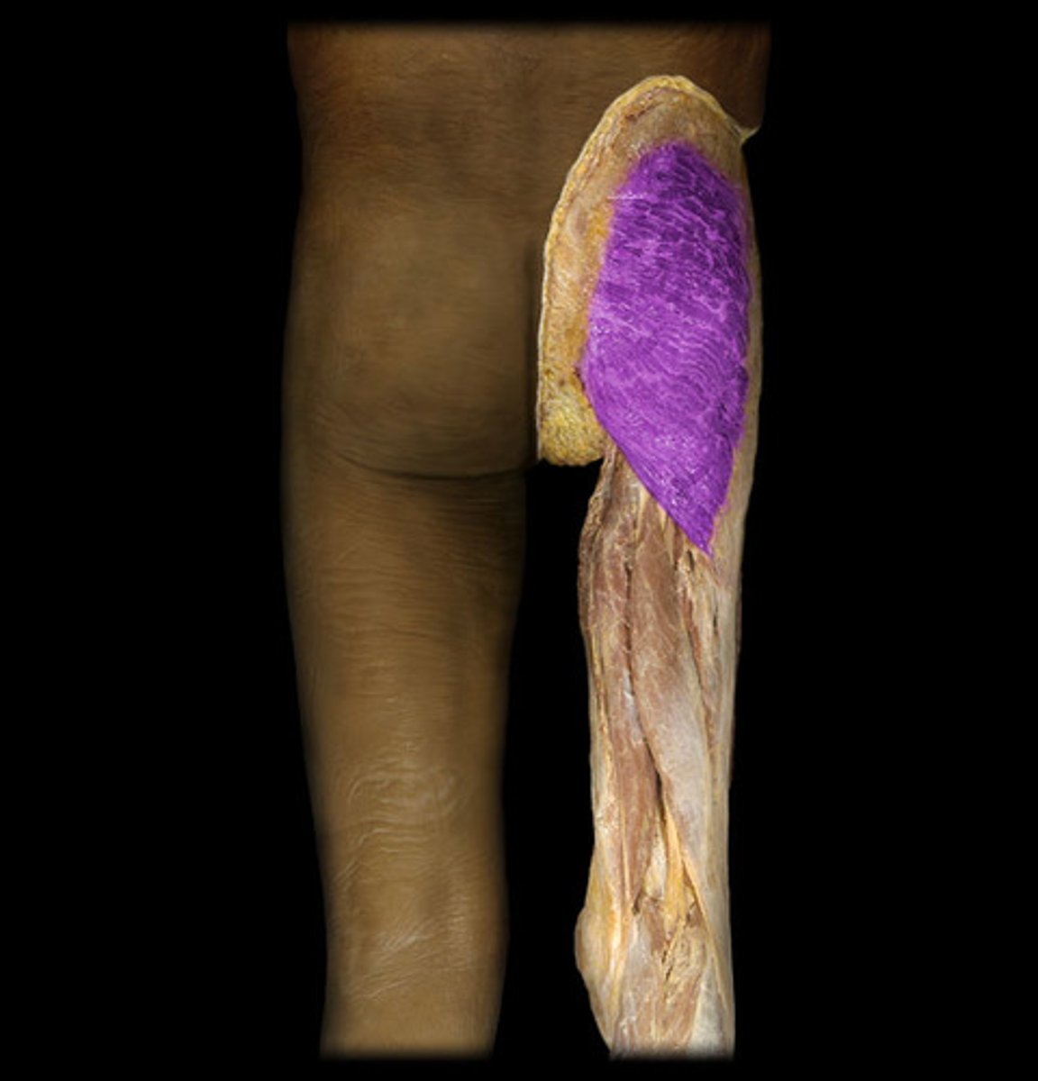
IA of Gluteus Maximus
Gluteal Line
Gluteal Tuberosity
Iliotibial Band
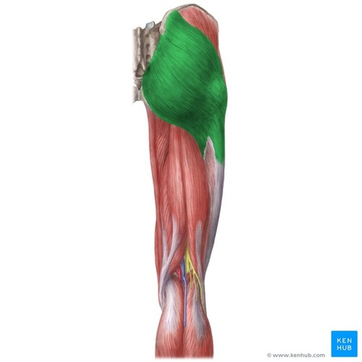
Gluteus Medius
abducts and medially rotates thigh (directly under maximus)
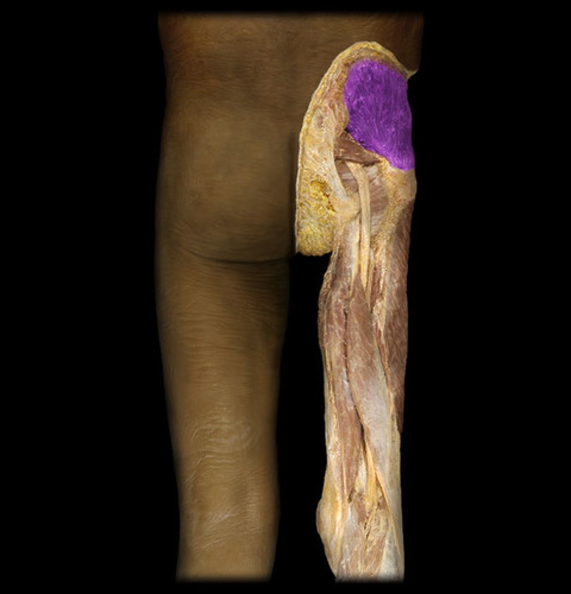
SA of Gluteus Medius
Ilium
IA of Gluteus Medius
Greater Trochanter of the femur

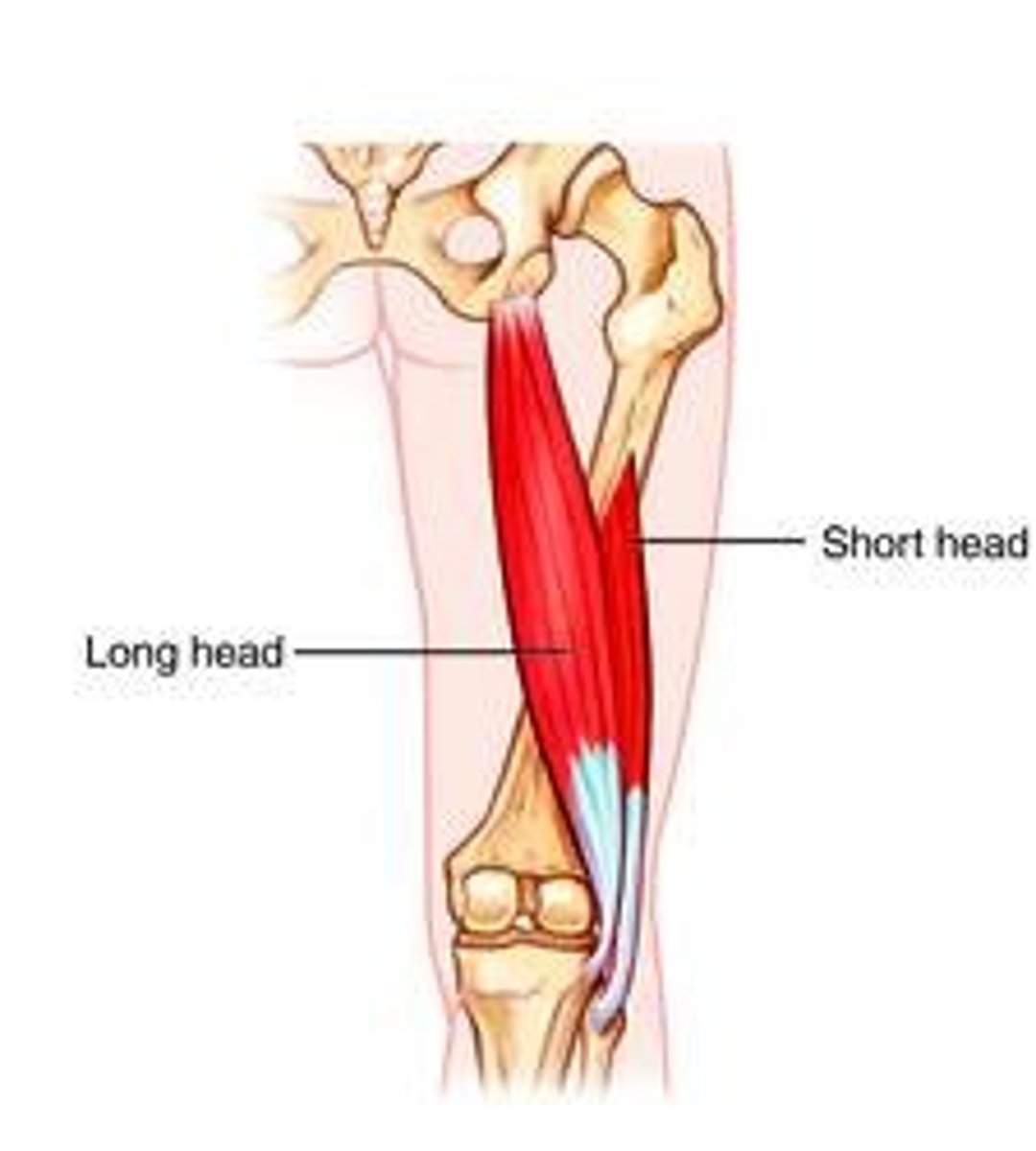
Biceps Femoris
extends thigh and flexes leg (2 heads)
SA of Biceps Femoris
Long Head: Ischial Tuberosity
Short Head: Linea Aspera
IA of Biceps Femoris
Fibular heads (both heads)

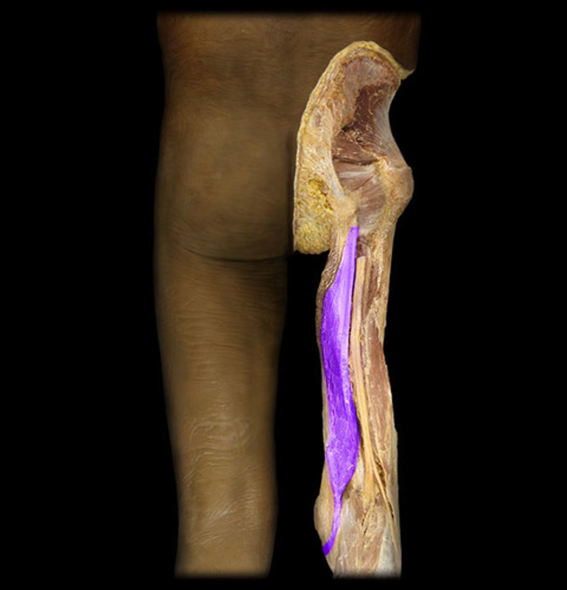
Semitendinosus
Flexes leg at the knee and extends thigh at the hip; belongs to the hamstring group
Opposite Biceps Femoris, more superficial
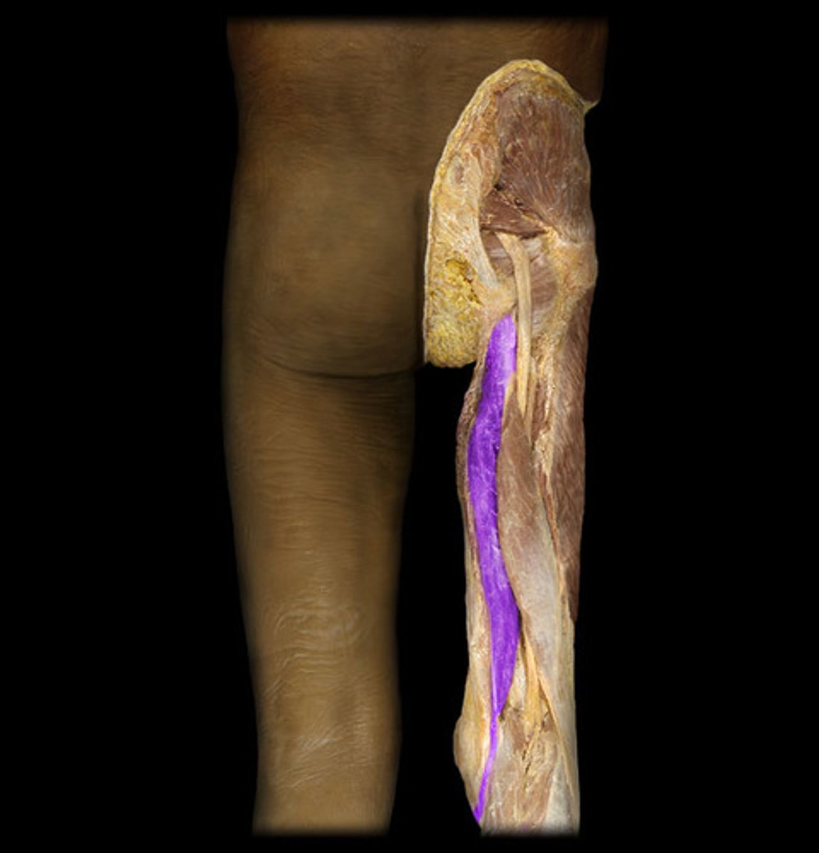
SA of Semiteninosus
Ischial Tuberosity
IA of Semitendinosus
Medial surface of the tibia
Semimembranosus
Flexes leg at the knee and extends thigh at the hip; belongs to the hamstring group
Deep to Semitendinosus, take up similar space
SA of Semimembranosus
Ischial Tuberosity
IA of Semimembranosus
Medial Condyle of the tibia
Fibularis Longus
plantar flexes and everts foot
Anterior, most lateral muscle, on Fibula
SA of Fibularis Longus
Proximal shaft of the fibula
IA of Fibularis Longus
Metatarsal I, Medial Cuneiform
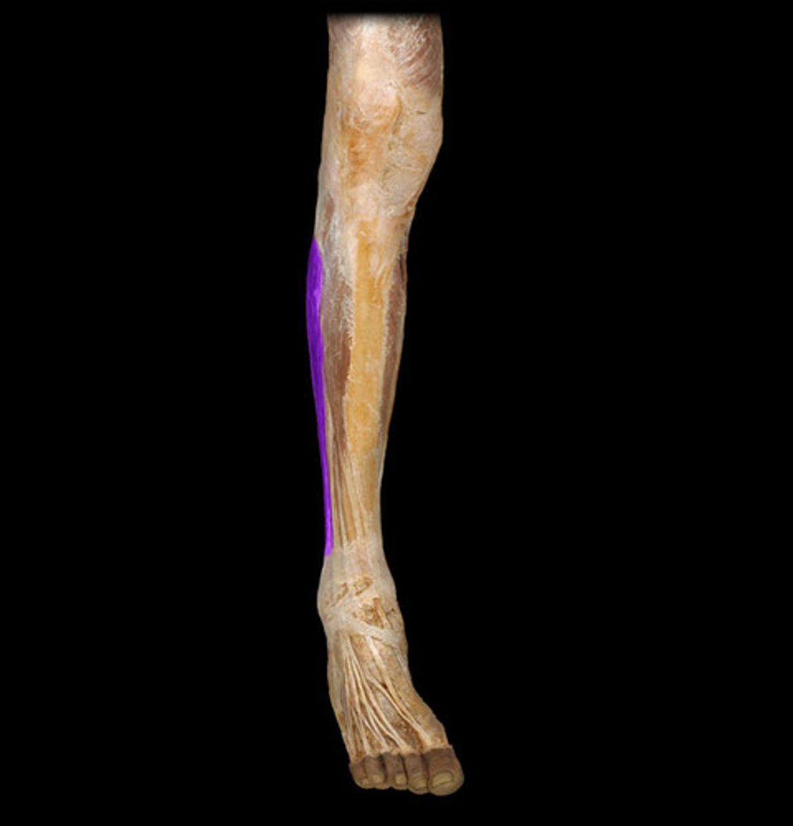
fibularis brevis
plantar flexes and everts foot
Lower, closer to heel than longus
SA of Fibularis Brevis
Distal shaft of fibula
IA of Fibularis Brevis
Metatarsal V (pinky)
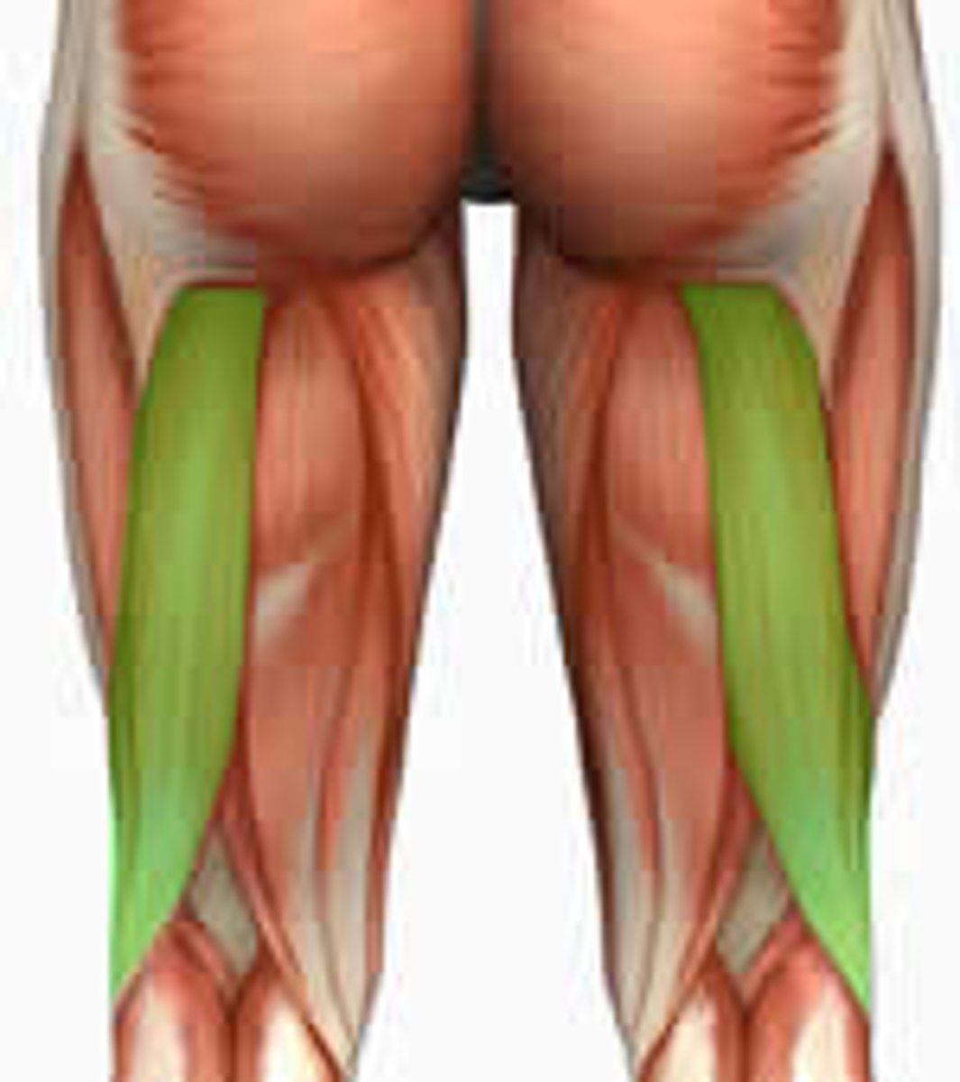
Gastrocnemius
Calf muscle
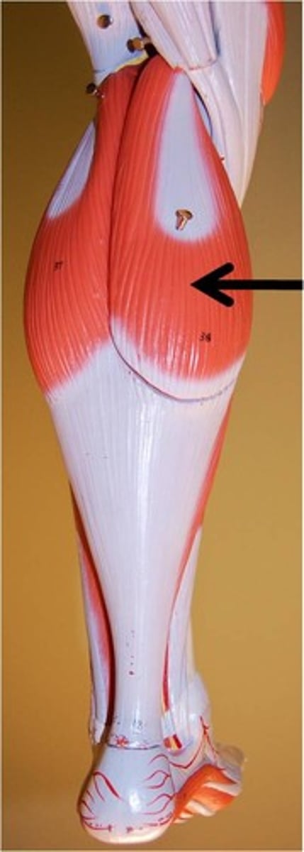
SA of Gastrocnemius
Medial Belly: Femur, Medial Condyle
Lateral Belly: Femur, Lateral Condyle
IA of Gastrocnemus
Calcaneal Tendon
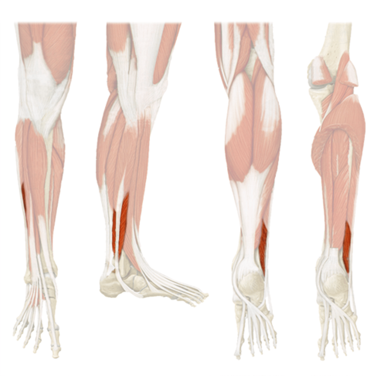
Soleus
plantar flexes foot, directly under Gastrocnemus
SA of Soleus
Fibula, Tibia (Proximal posterior shaft)
IA of Soleus
Calcaneal tendon (Achilles tendon)
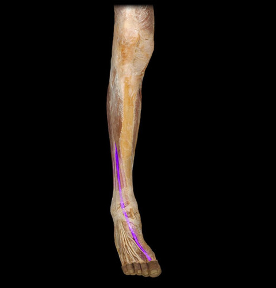
flexor digitorum longus
flexes toes, medial on posterior of leg
SA of Flexor Digitorum Longus
Middle posterior shaft of tibia
IA of Flexor Digitorum Longus
Distal phalanges of toes 2-5
Flexor hallucis longus
flexes great toe
Posterior side, goes to base of big toe
SA of Flexor Hallucis Longus
Fibula, Interosseus Membrane
IA of Flexor Hallucis Longus
Distal Phalanx of big toe
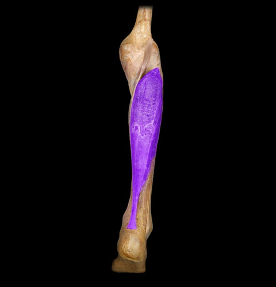
tibialis anterior
dorsiflexes and inverts foot
Lateral, anterior, fusiform muscle
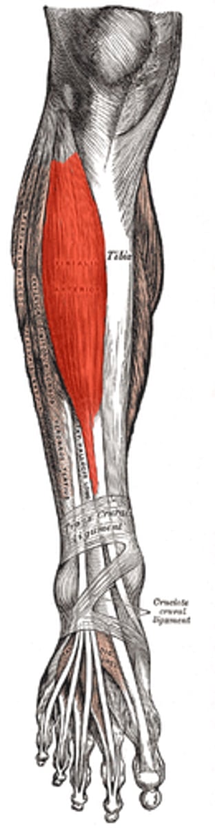
SA of Tibialis Anterior
Tibial, Lateral condyle and shaft, Interosseous Membrane
IA of Tibialis Anterior
Metatarsal I (big toe)
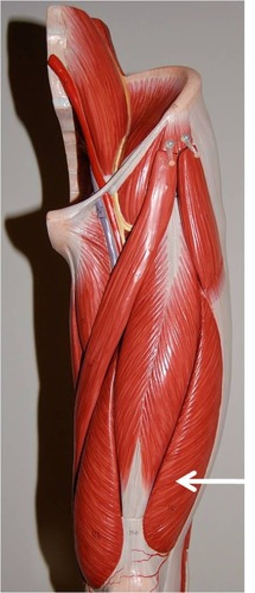
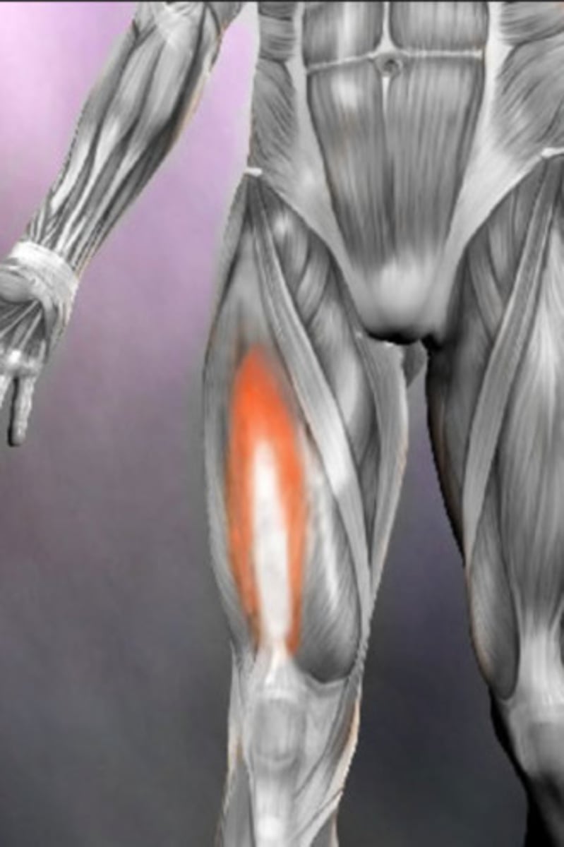
rectus femoris
extends leg at knee
Large, medial muscle upper knee, unipennate
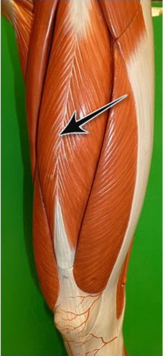
SA of Rectus Femoris
Anterior Inferior Iliac Spine (AIIS)
IA of Rectus Femoris
Proximal border of Patella
Vastus Medialis
extends knee
Muscle to medial side of Rectus Femoris
SA of Vastus Medialis
Linea Aspera
IA of Vastus Medialis
Patella (via common quadriceps tendon)
Vastus Lateralis
extends knee
Muscle to lateral side of Rectus Femoris
SA of Vastus Lateralis
Linea Aspera
IA of Vastus Lateralis
Patella (via common quadriceps tendon)
Vastus Intermedius
extends knee
Deep to Rectus Femoris, underneath and between Vastus lateralis and medialis
SA of Vastus Intermedius
Anterolateral surface of femur shaft
IA of Vastus Intermedius
Patella
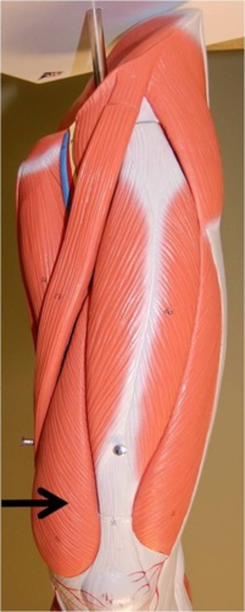
Sartorius
Flexes, abducts, and laterally rotates thigh at the hip; flexes knee
Band of muscle that runs from later to medial on anterior upper thigh
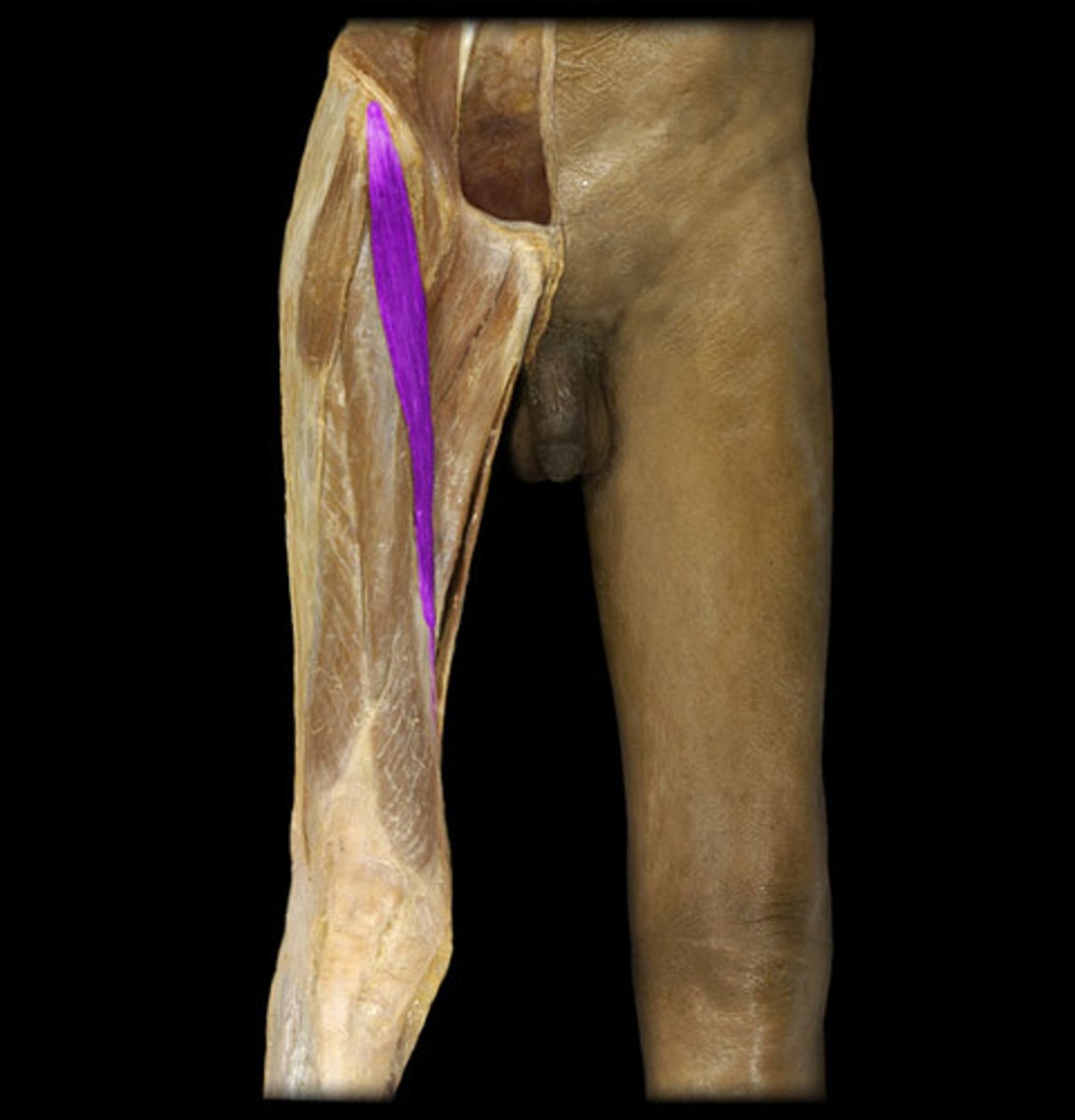
SA of Sartorius
ASIS (Anterior Superior Iliac Spine)
Inferior Attachment of Sartorius
Tibia
Tensor Fascia Latae (TFL)
abducts hip, flexes hip
Most medial muscle on anterior upper thigh, often alone and not near other muscles
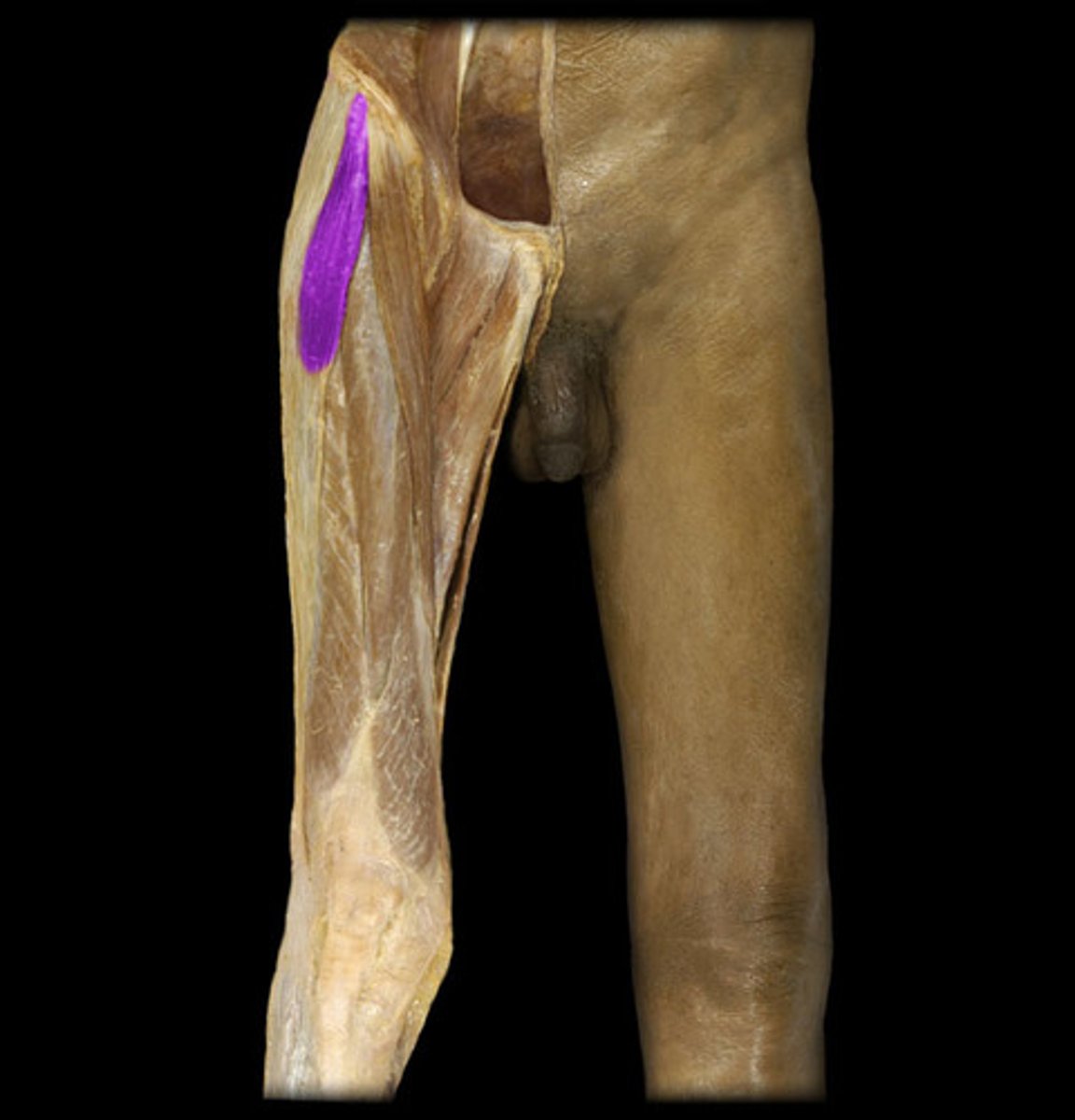
SA of TFL
Iliac crest, ASIS
IA of TFL
Iliotibial Band
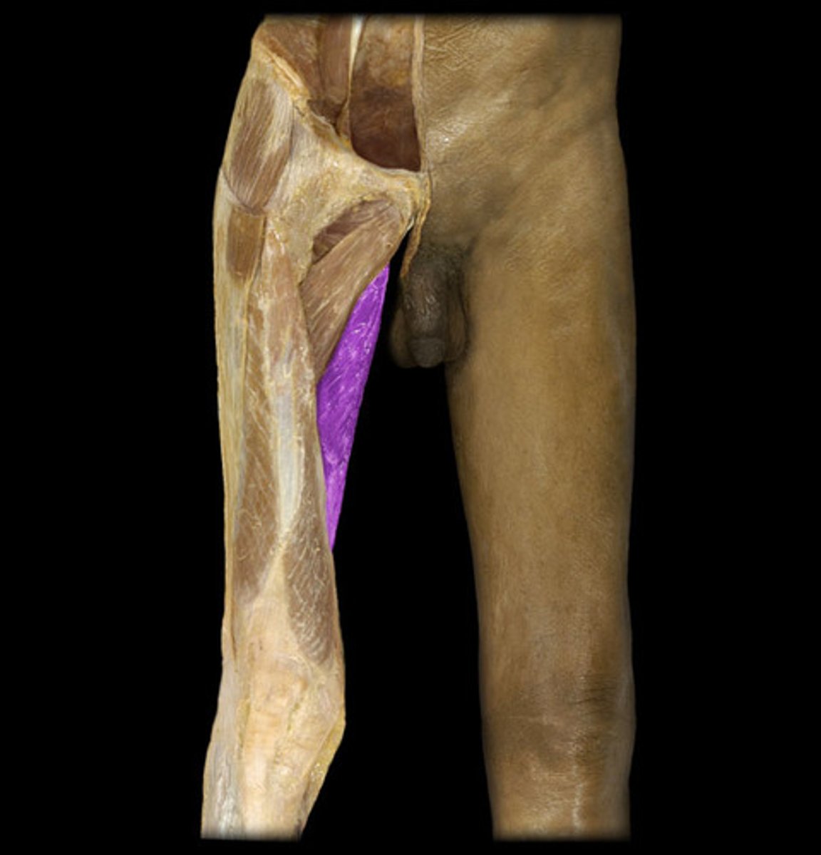
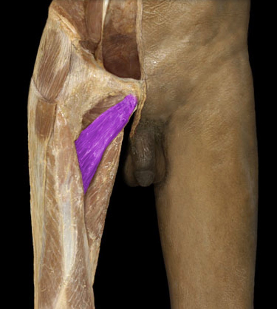
Gracialis
adducts thigh
Thin muscle on the most medial part of anterior upper thigh
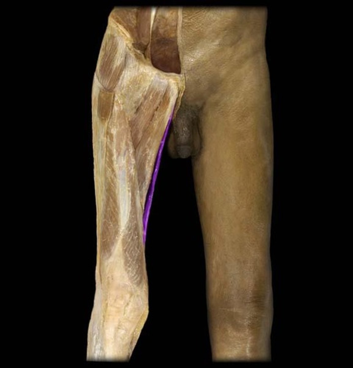
SA of Gracilis
Pubis
IA of Gracialis
Tibia
Adductor Longus
adducts, flexes, and medially rotates thigh
Longest and LOWEST of the adductors, most medial
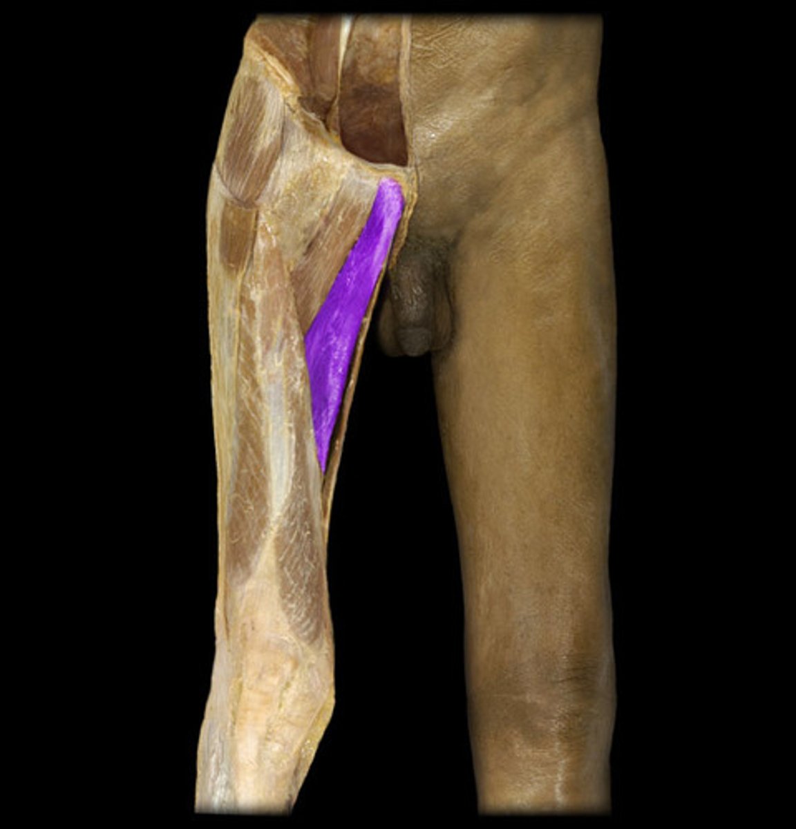
SA of Adductor Longus
Pubis
IA of Adductor Longus
Linea Aspera of femur
Adductor Magnus
adducts thigh
DEEP to Adductor Longus
SA of Adductor Magnus
Pubis
IA of Adductor Magnus
Gluteal tuberosity
Adductor Tubercle
Adductor Brevis
adducts thigh
Above Longus, superficial to Magnus
SA of Adductor Brevis
Pubis
IA Adductor Brevis
Linea Aspera
iliotibial band
Helps to maintain knee extension, especially while standing
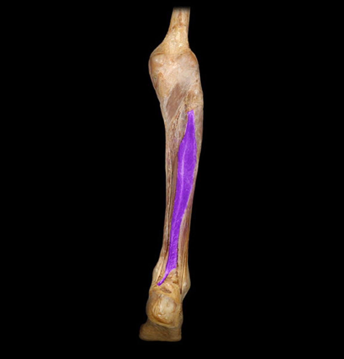
extensor digitorum longus
extends toes
Anterior side, connects to toes
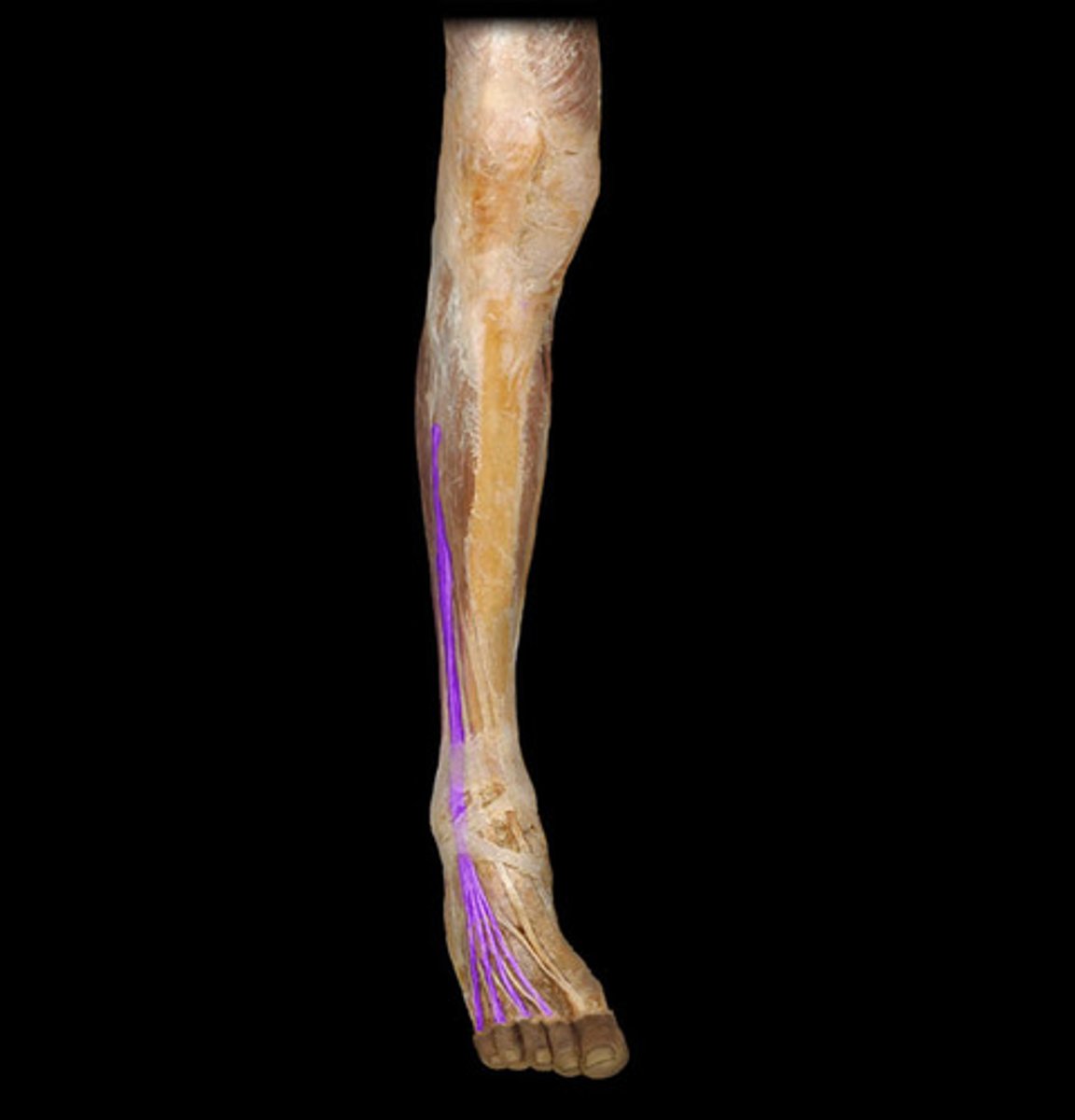
SA of Extensor Digitorum Longus
Lateral condyle of tibia
Shaft of fibula
Interosseous Membrane
IA of Extensor Digitorum Longus
Middle and distal phalanges II-V
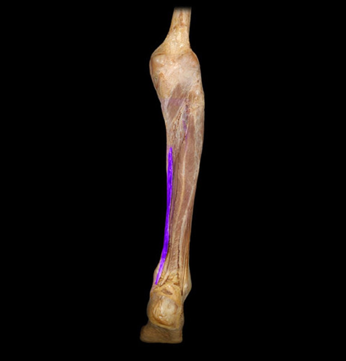
Extensor Hallucis Longus
extends great toe
Anterior, connects to big toe
SA of Extensor Hallucis Longus
Anterior surface of mid fibula, Interosseous membrane
IA of Extensor Hallucis Longus
Distal phalanx of big toe
Pectoralis Minor
Stabilizes, protracts, rotates scapula
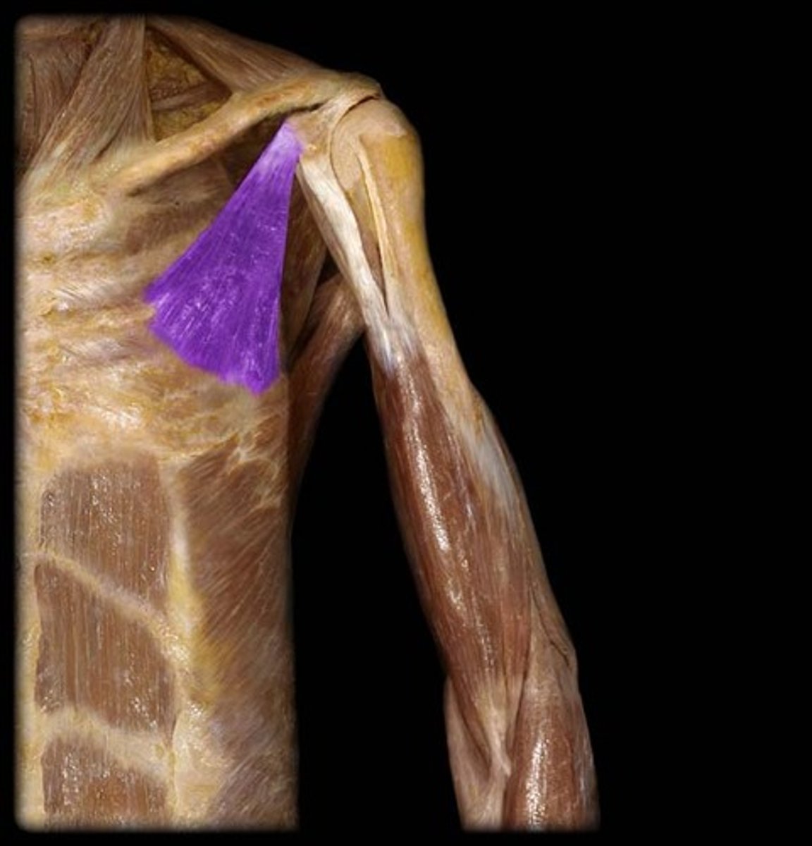
MA of Pectoralis Minor
Ribs 3-5
LA of Pectoralis Minor
Coracoid process of scapula
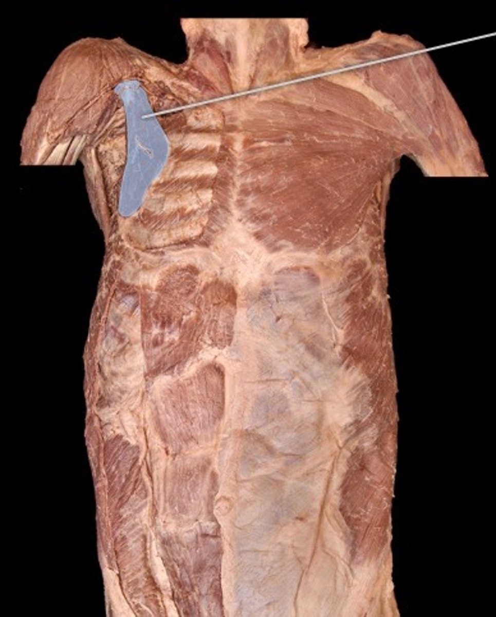
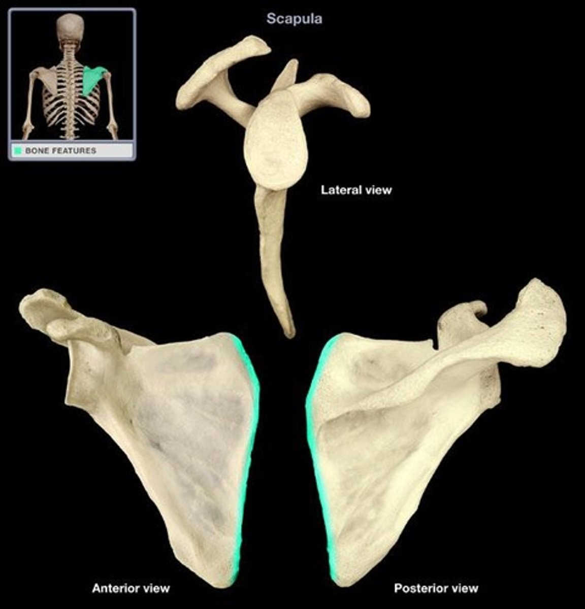
serratus anterior
Stabilizes, protracts, rotates scapula (Hard to see on cadaver!)
Deep to subscapularis, almost like fingers covering the ribs

Anterior Attachment (AA) of Serratus Antierior
Ribs 1-9
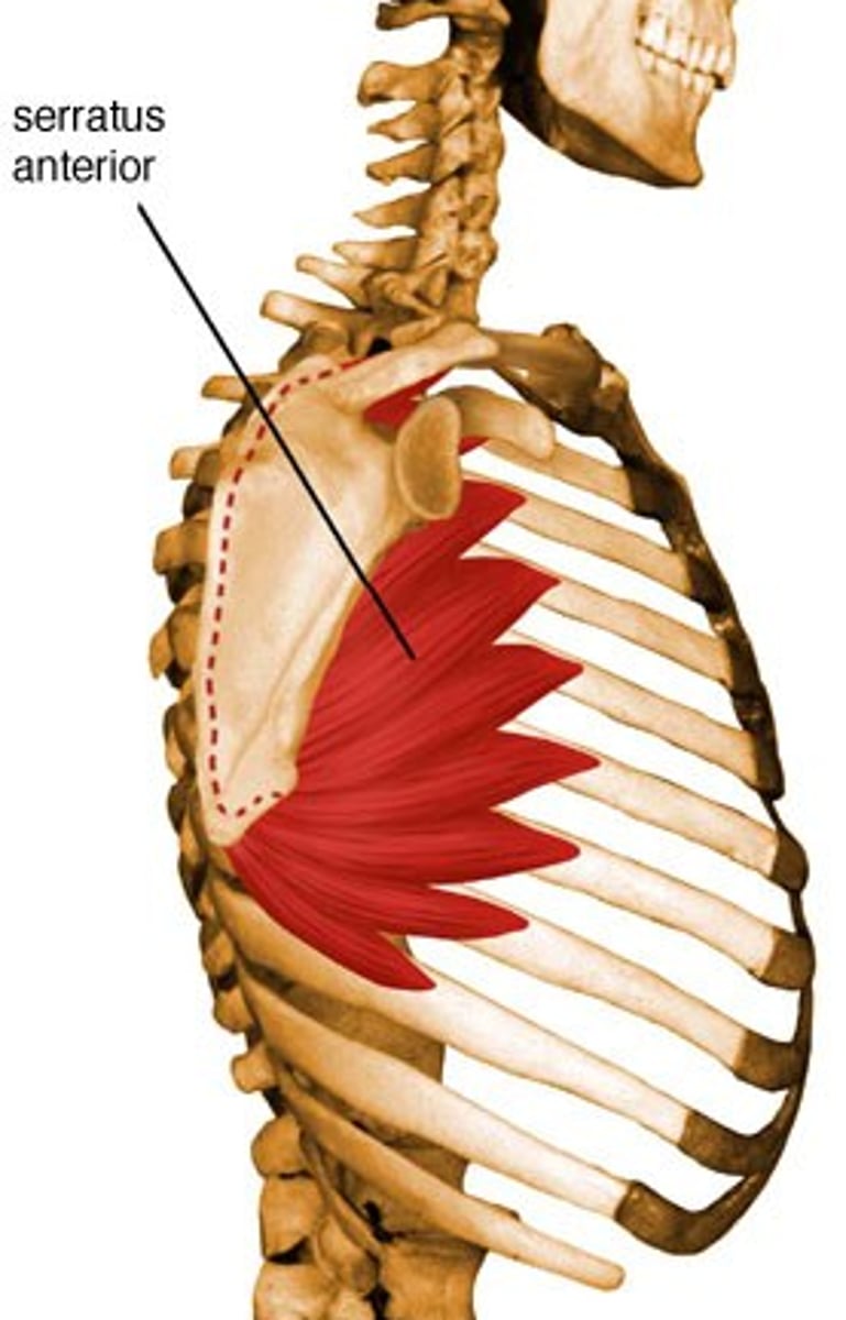
PA of Serratus Anterior
Medial border of scapula
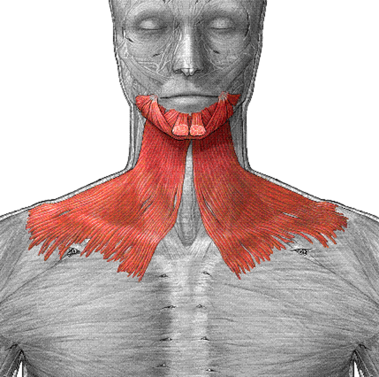
Sternocleidomastoid
Rotates head, base of muscle attached to anterior side of clavicle
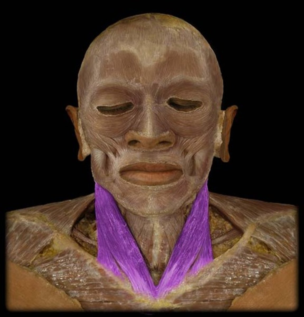
Superior Attachment of Sternocleidomastoid
Mastoid process of temporal bone, superior nuchal line
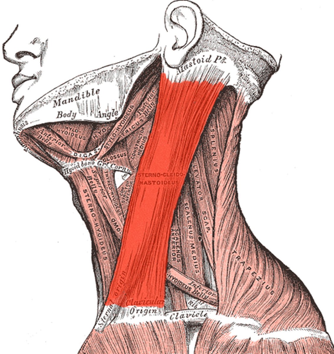
Inferior attachments of Sternocleidomastoid
Clavicle and Manubrium
Pectoralis Major
Flexion of arm, adduction of arm, medial rotation of arm
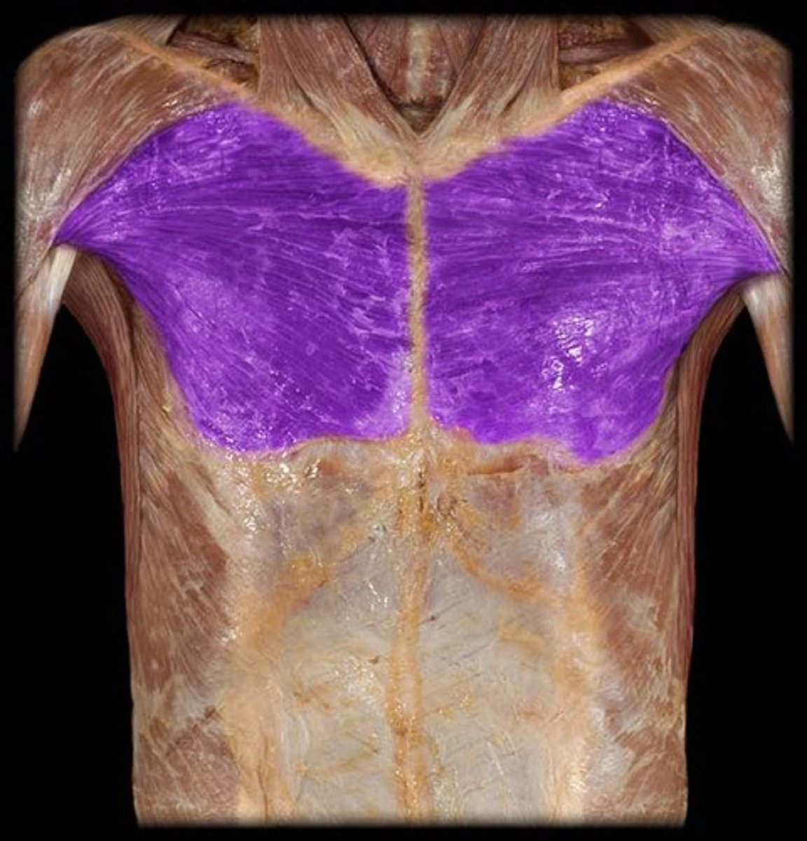
MA of Pectoralis Major
Clavicle (medial), Sternum, Costal Cartilage 1-6
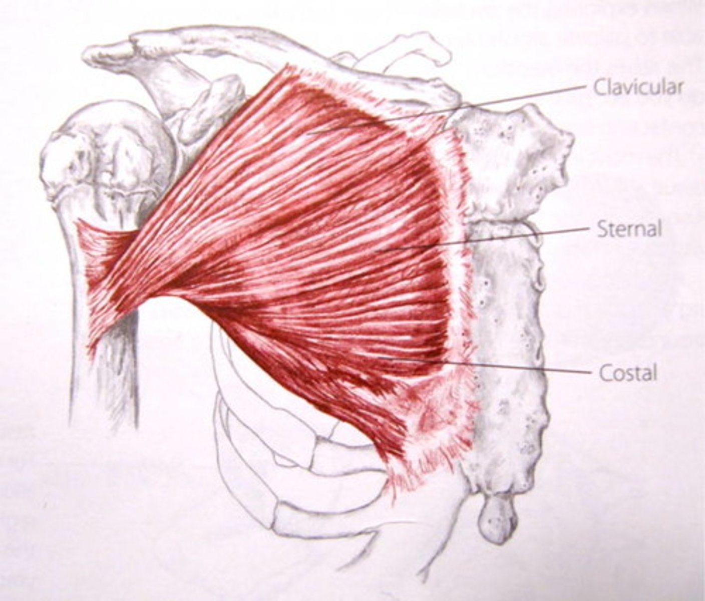
LA of Pectoralis Major
Lateral lip of intertubercular sulcus
Deltoid
Flexion and medial rotation, abduction of arm, extension and lateral rotation
3-headed muscle
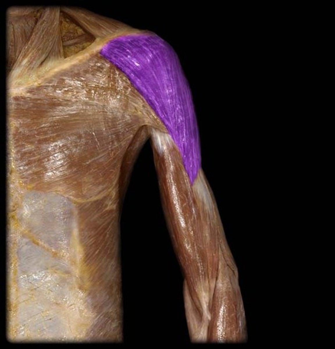
PA of Deltoid
Clavicle (anterior)
Acromion process of scapula (lateral)
Scapular spine (posterior)
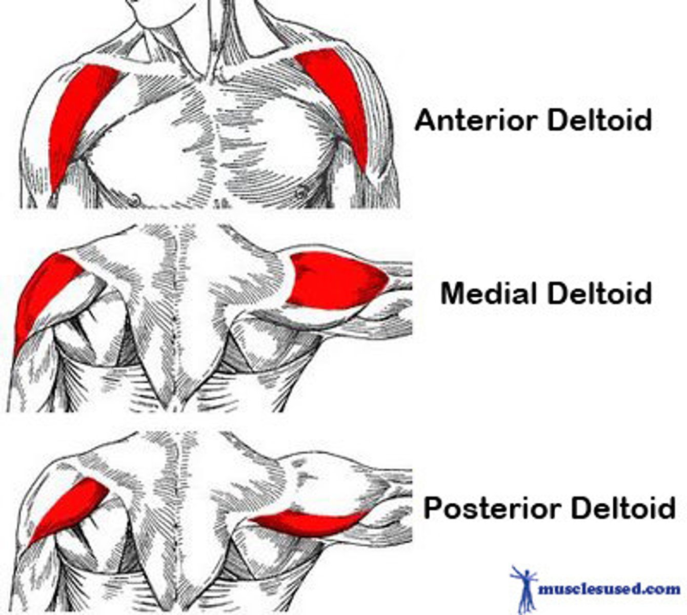
DA of Deltoid
deltoid tuberosity of humerus
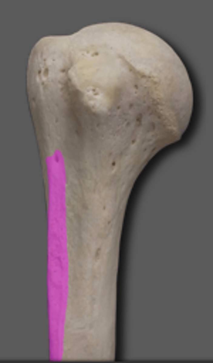
Biceps Brachii
Adduction of shoulder, supination of forearm, flexion and extension
Two-headed
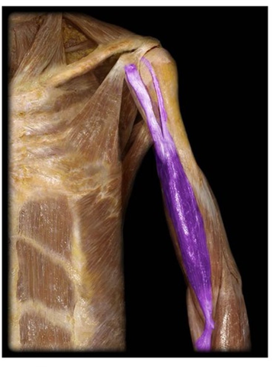
PA of Biceps Brachii
Long head - Supraglenoid tubercle, lateral
Short head - Coracoid process of scapula, medial
DA of biceps Brachii
Radial tuberosity
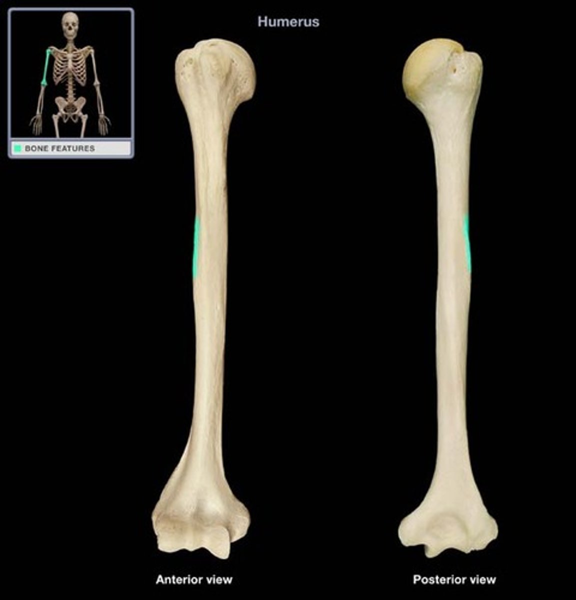
Brachialis
Flexion of forearm
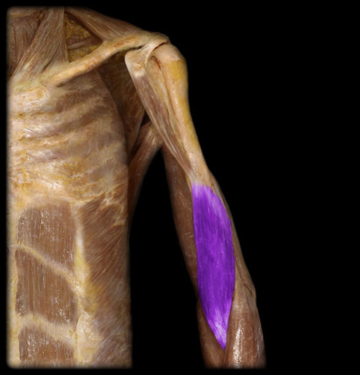
PA of Brachialis
Anterior distal shaft of Humerus
DA of Bachialis
Tuberosity and Coronoid process of Ulna
Brachioradialis
Flexes forearm, assists in pronation and supination
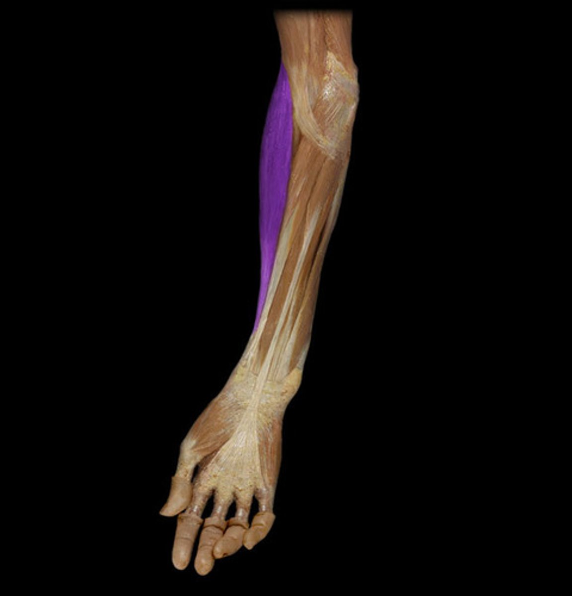
PA of Brachioradialis
Lateral distal shaft of Humerus
(Far down the humerus)
DA of Bracioradialis
Styloid process of Radius
External Obliques
compress abdomen, flex vertebral column, rotate trunk, lateral flexion of trunk
Fibers go from outside -> down
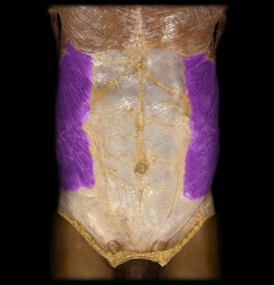
Superior attachements of External Obliques
Ribs and costal cartilages 5-12
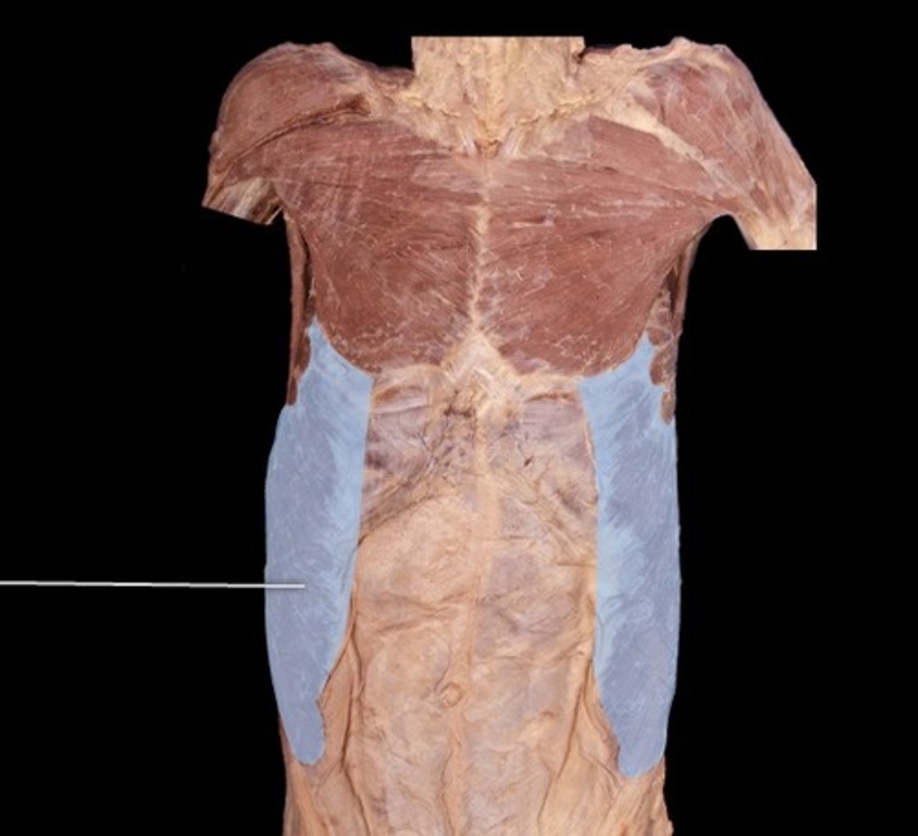
Inferomedial attachments of External obliques
Linea alba
Iliac crest
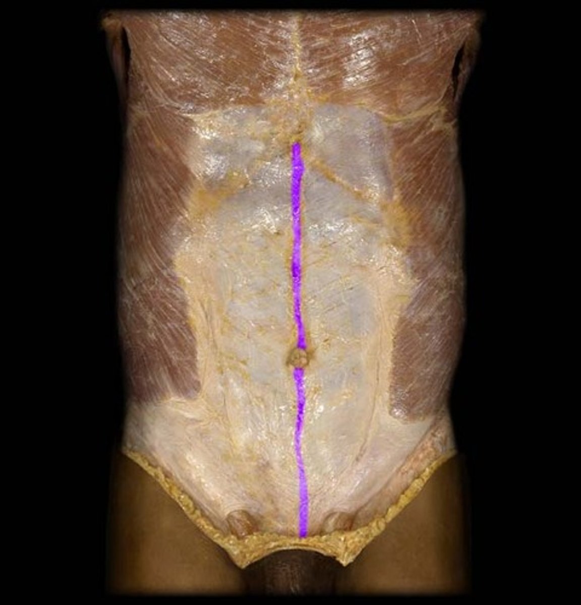
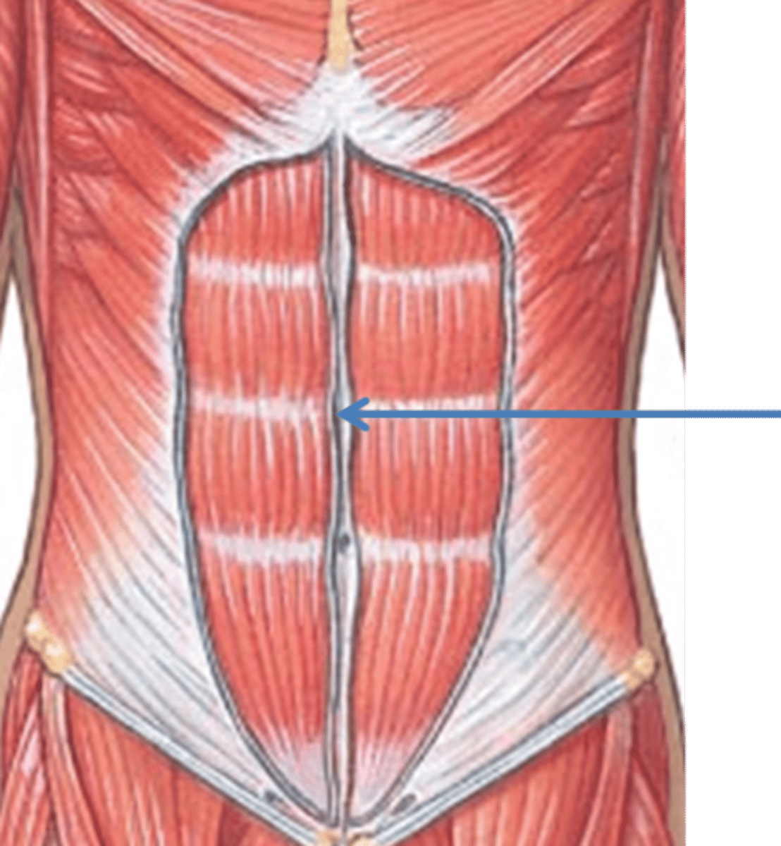
Linea alba
midline tendinous seam joining the abdominal muscles