Lab
1/1334
There's no tags or description
Looks like no tags are added yet.
Name | Mastery | Learn | Test | Matching | Spaced | Call with Kai |
|---|
No analytics yet
Send a link to your students to track their progress
1335 Terms
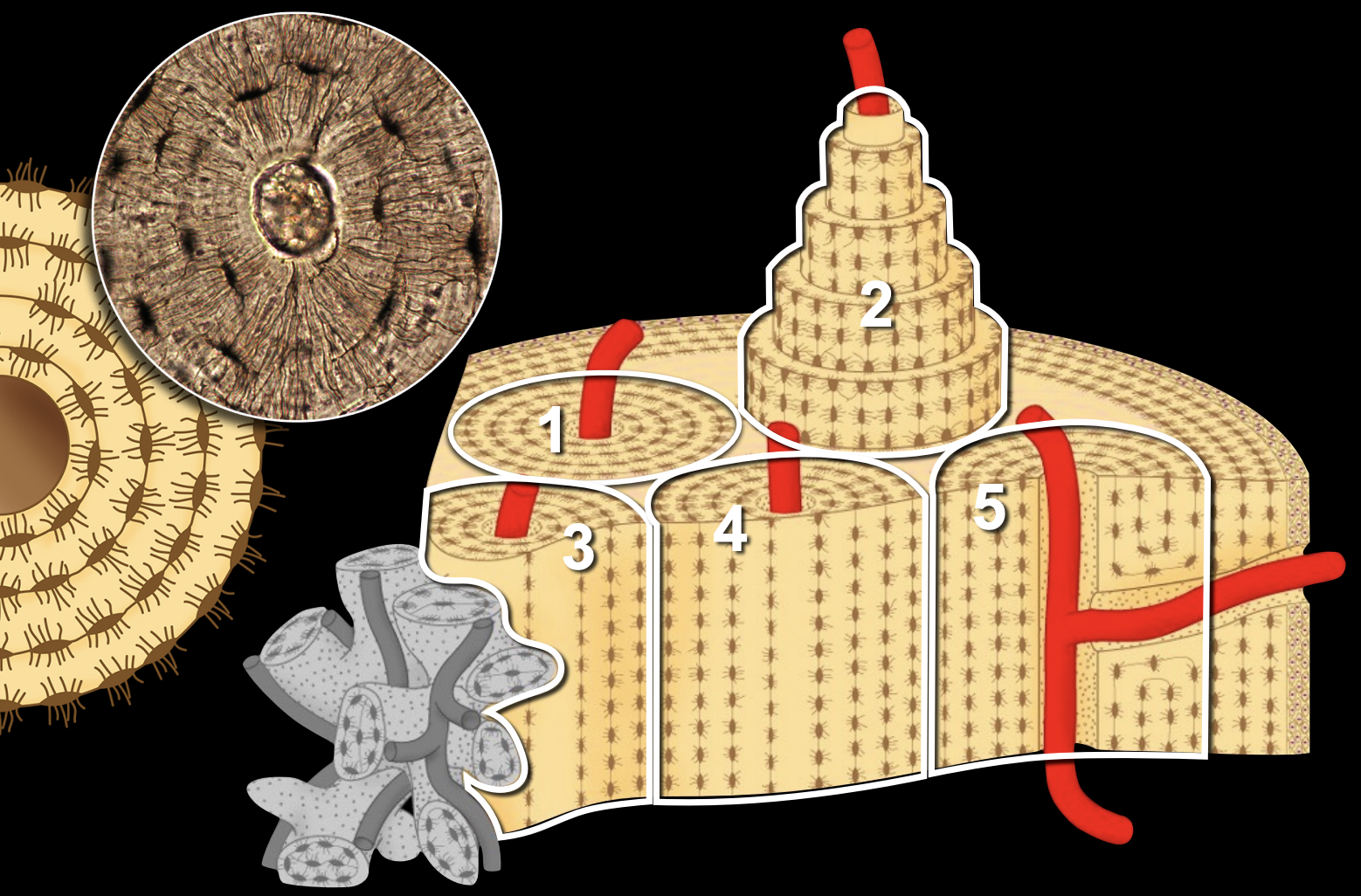
Osteon
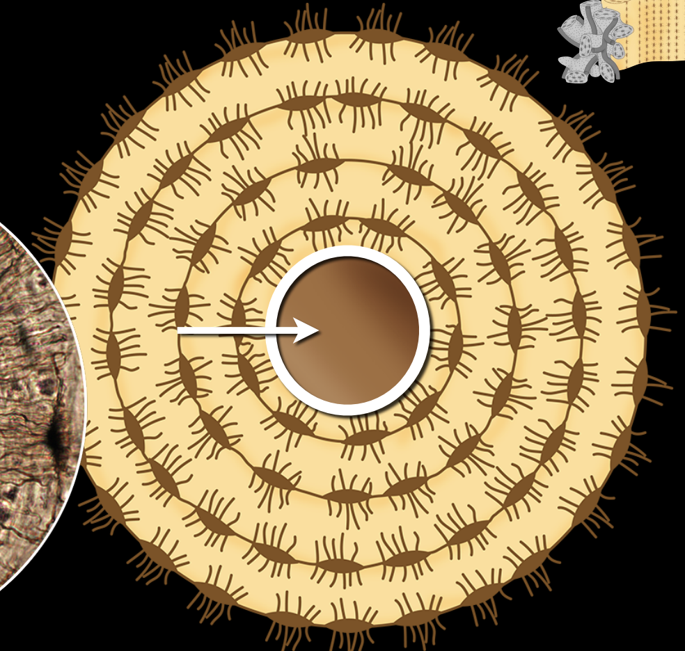
Osteonic canal

Lacuna
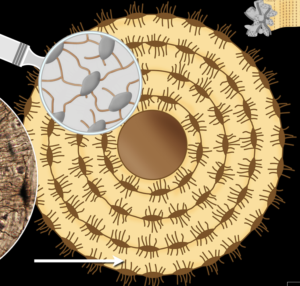
Canaliculi
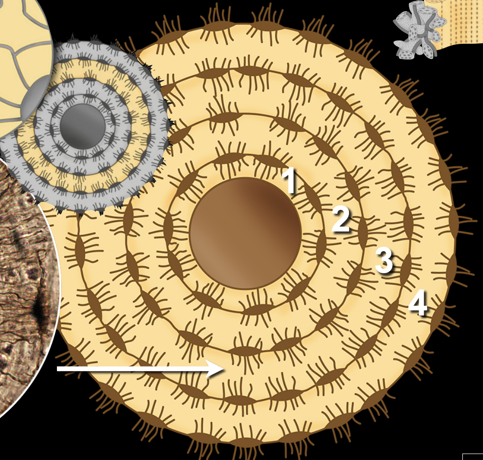
Lamella
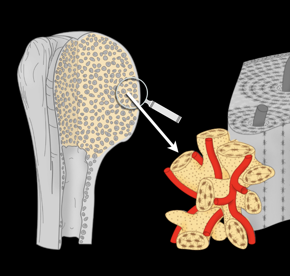
Spongy bone
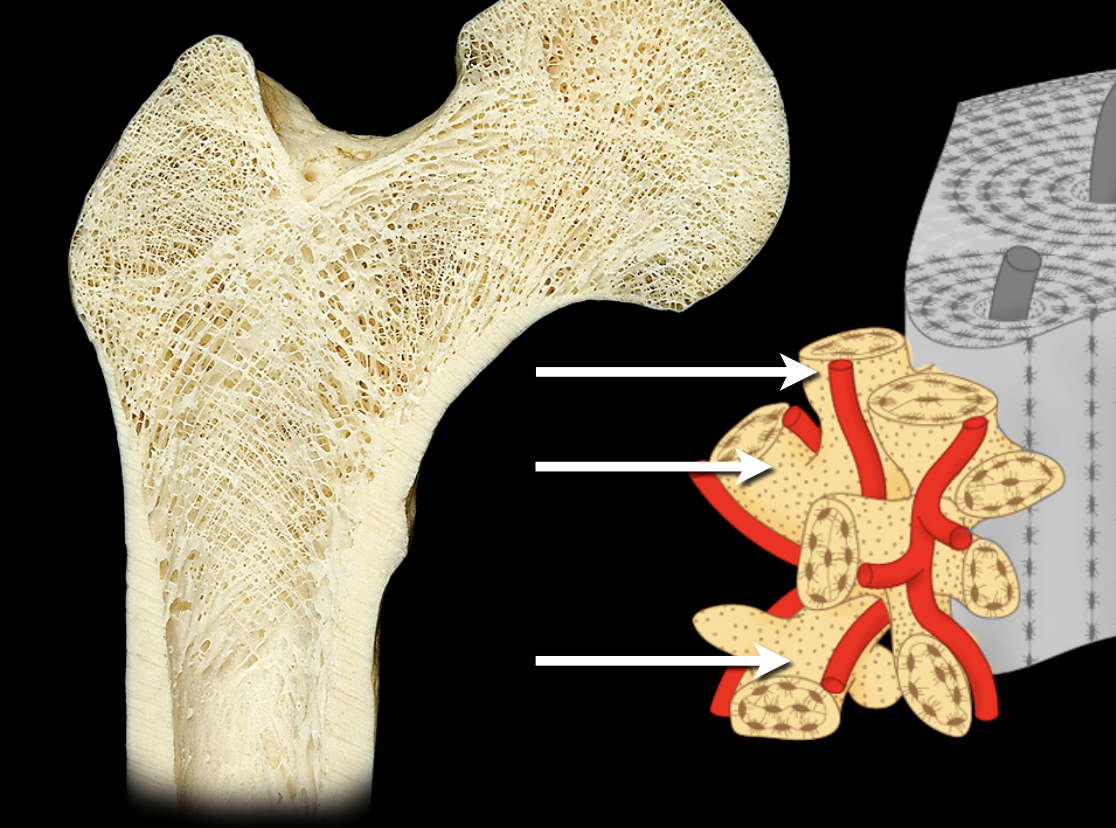
Bone trabecula
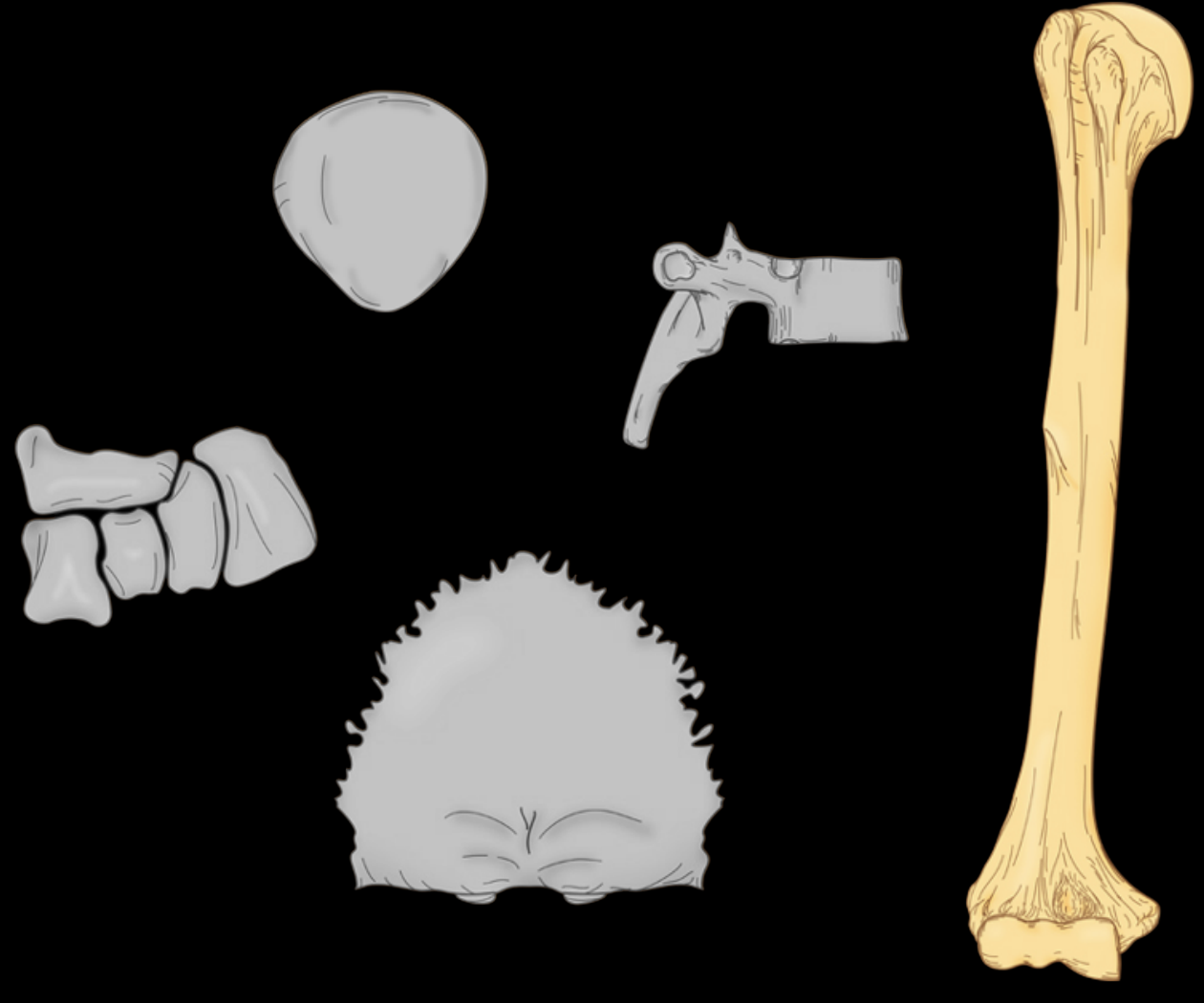
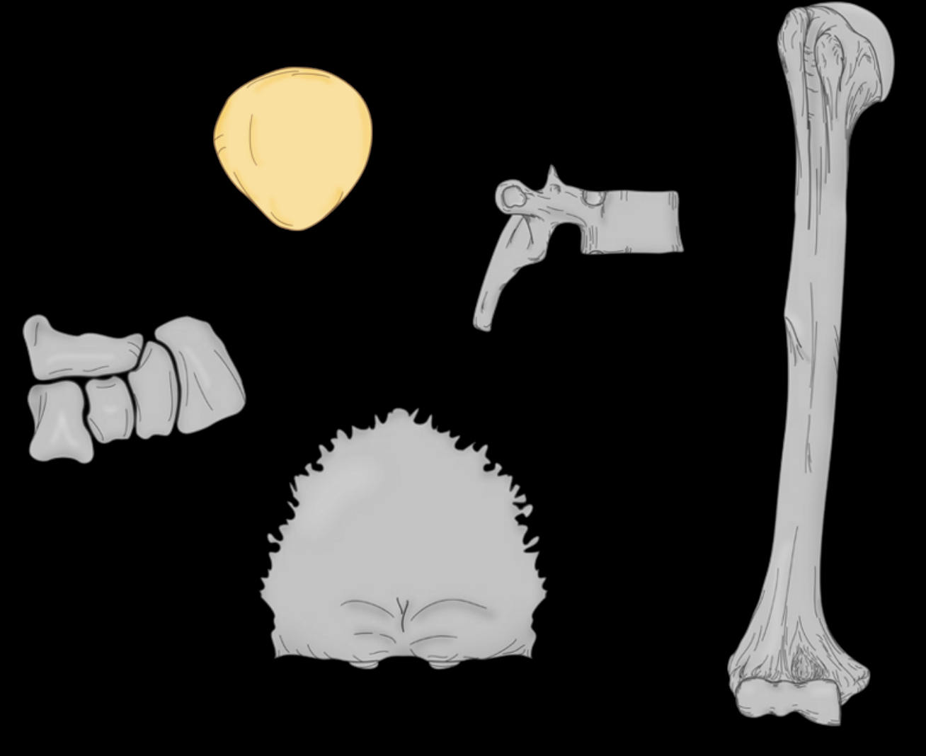
Long bone
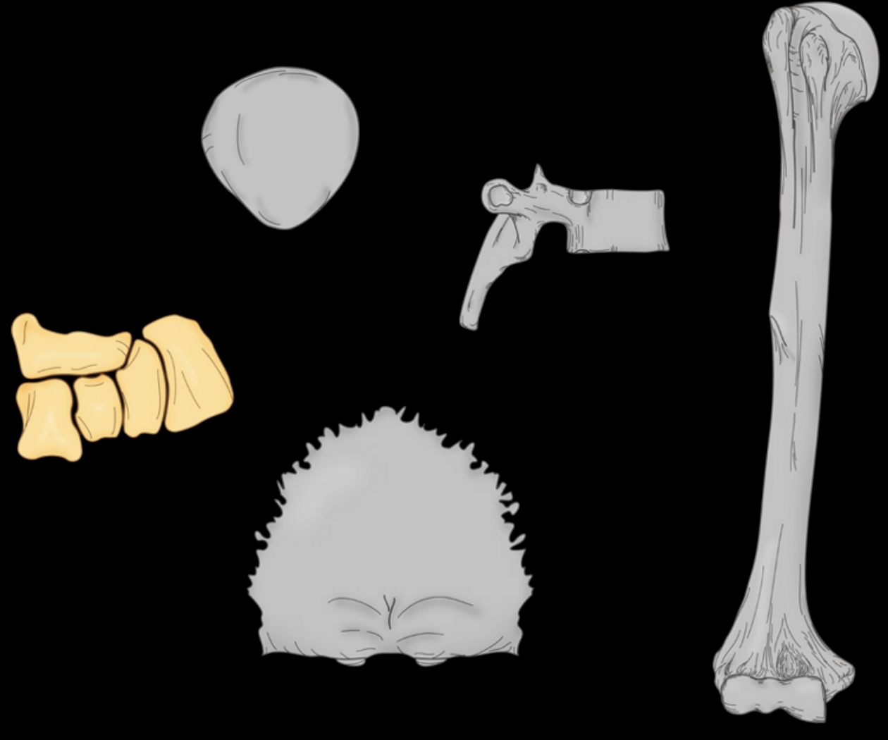
Short bone
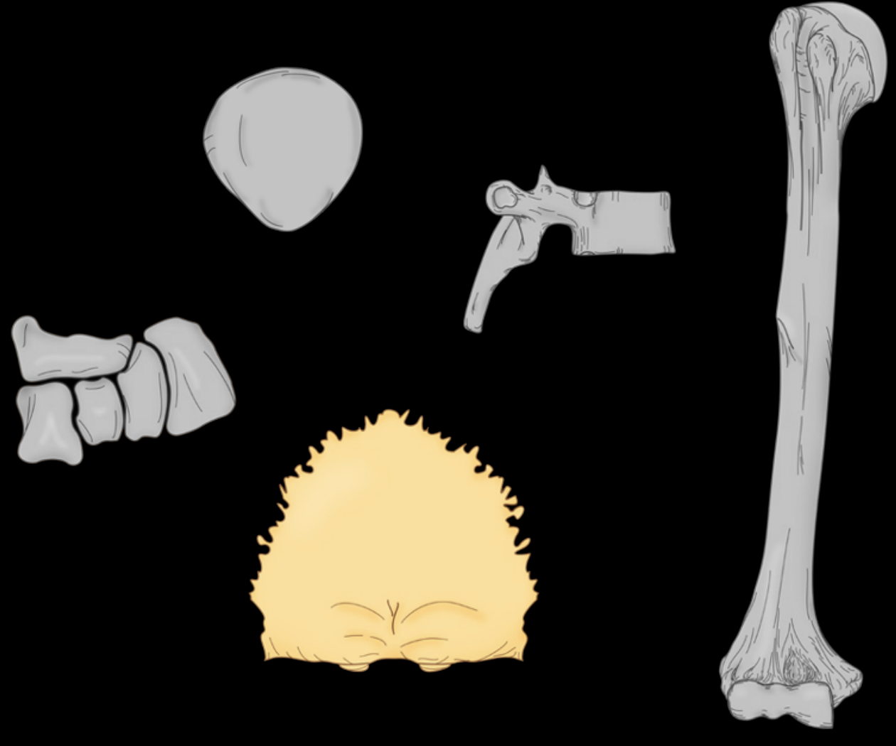
Flat bone
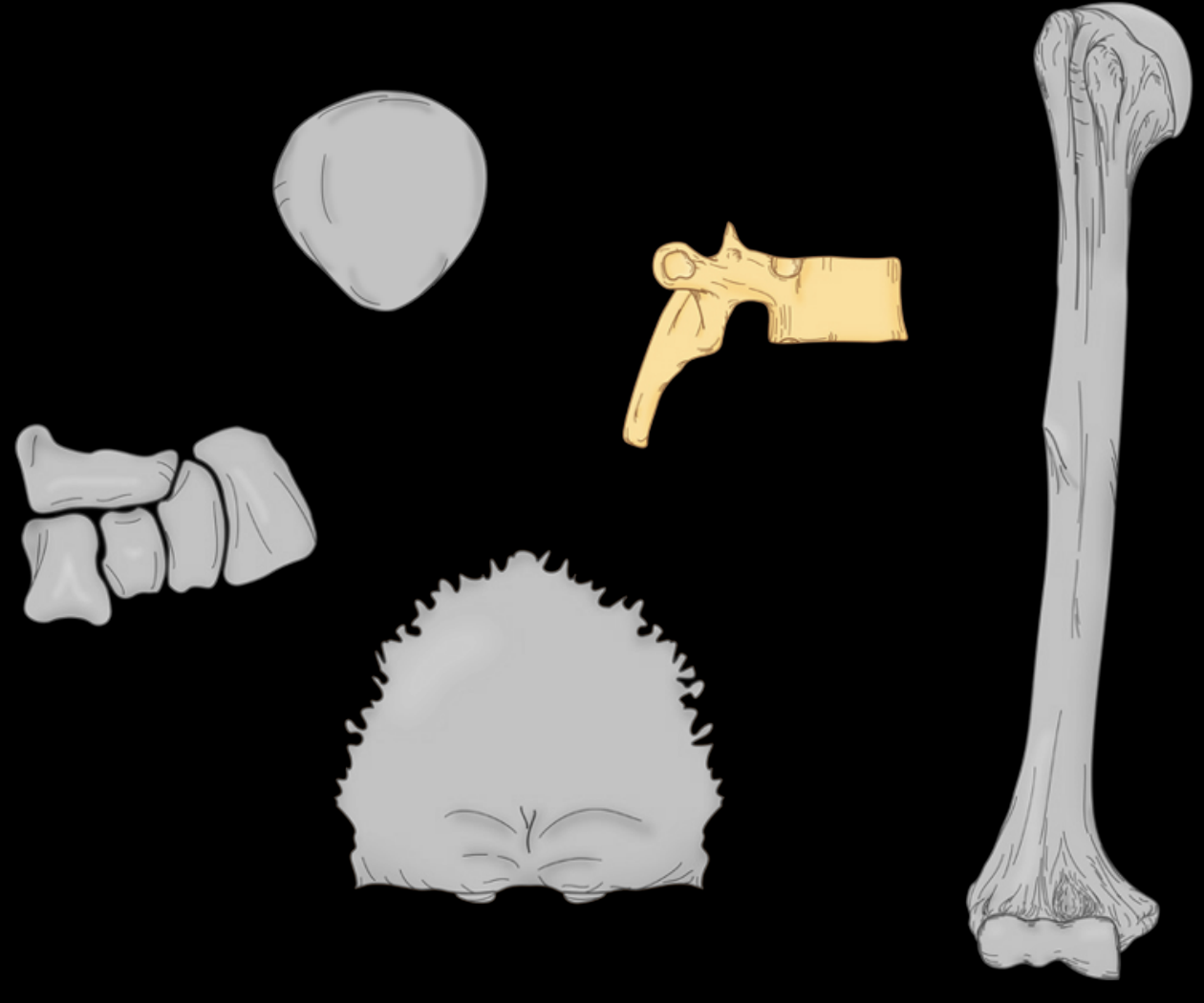
Irregular bone
Pneumatized bone
Sesamoid bone
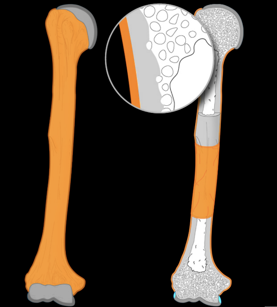
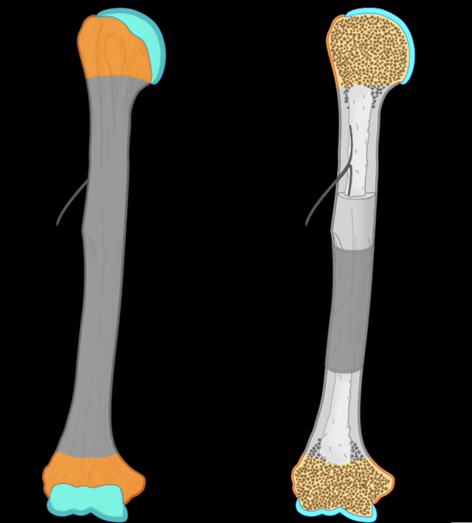
Peristeoum
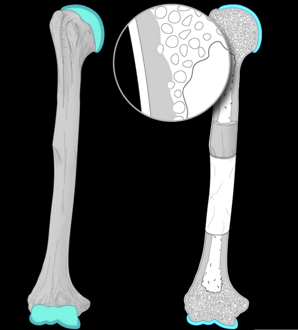
Arcticular cartilage
What type of cartilige is Articular cartilige
Hyaline cartilage
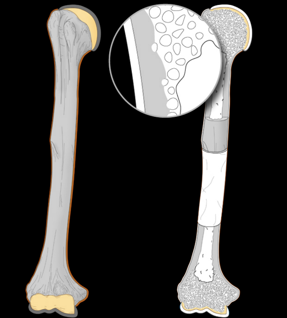
Subchondral bone region
The specialized, vascularized bone layer located directly beneath the articular cartilage and its underlying calcified cartilage layer
Subchondral bone
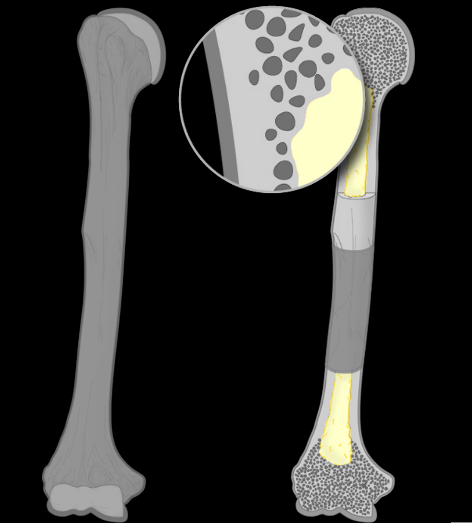
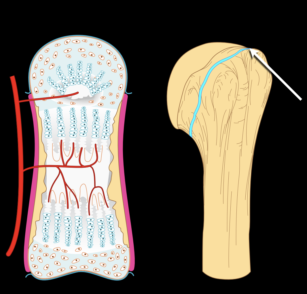
Medullary cavity
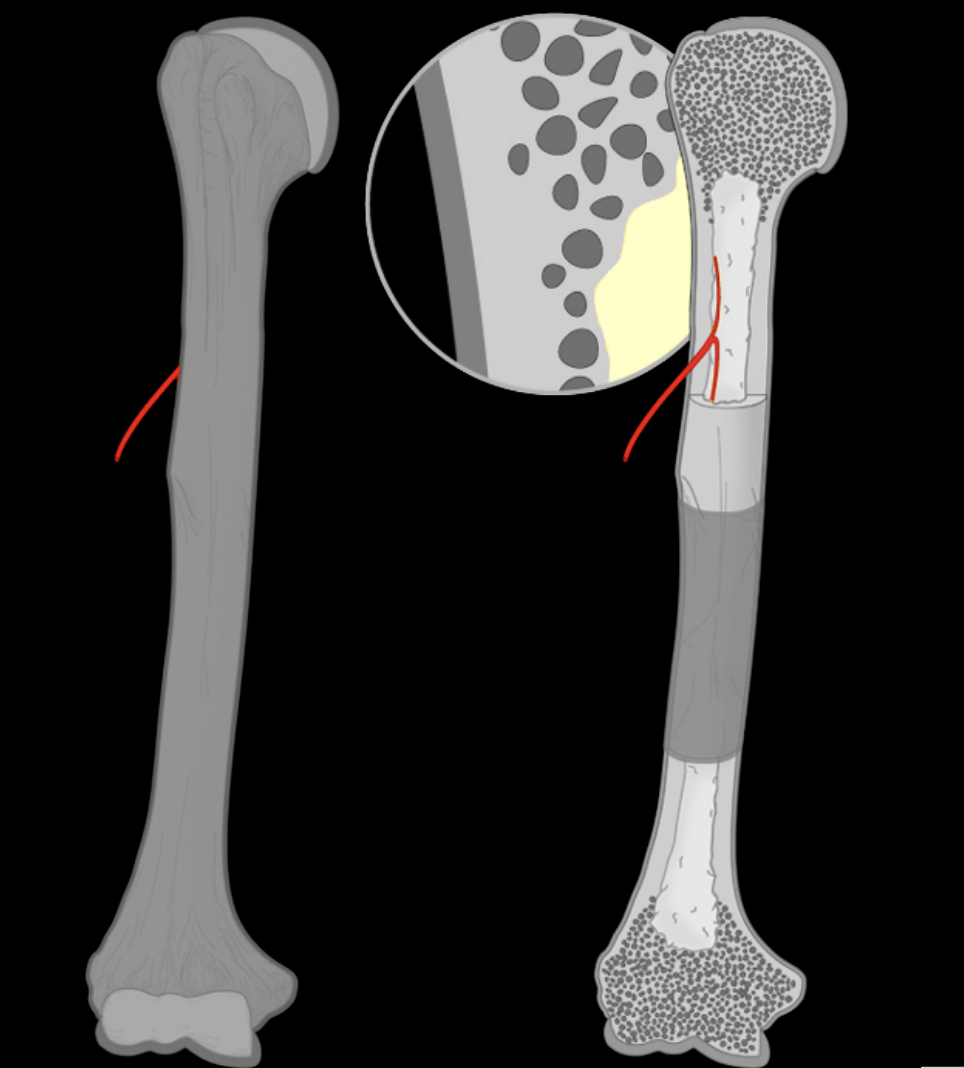
Nutrient foramen and canal
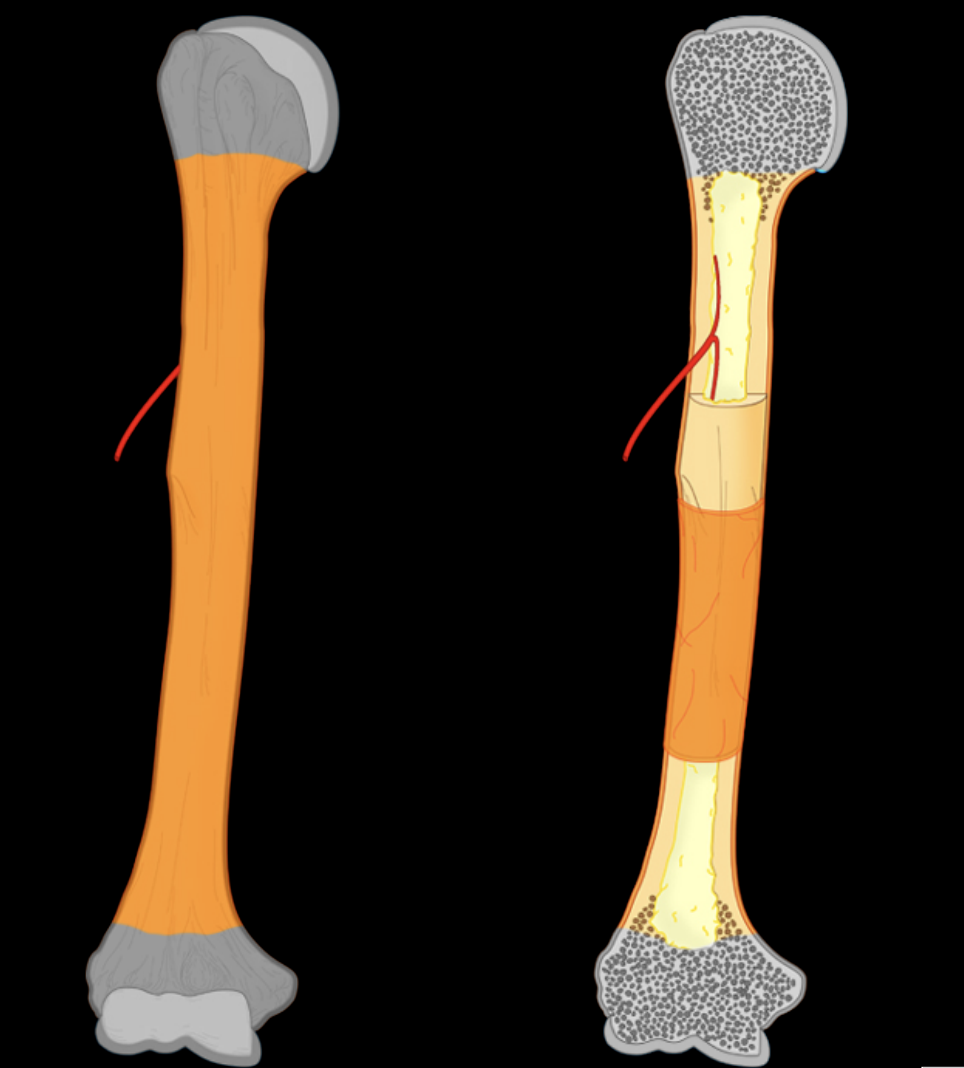
Diaphysis
Epiphysis
Mature, less active cells derived from chondroblasts that maintain the bone matrix.
Chondrocytes
Immature, active progenitor cells that secrete the extracellular matrix (ECM) to form cartilage
Chrondroblasts
Fibers present in the extracellular matrix - cartilage matrix
Collagen and elastic fibers
Fibrous joint
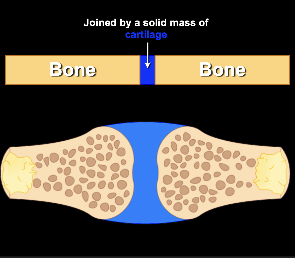
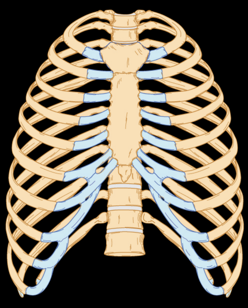
Cartilaginous joint
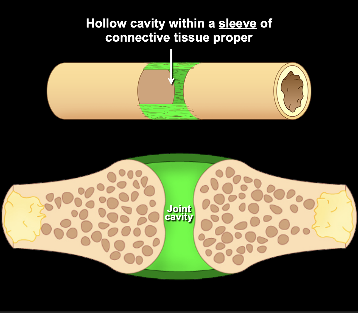
Synovial joint
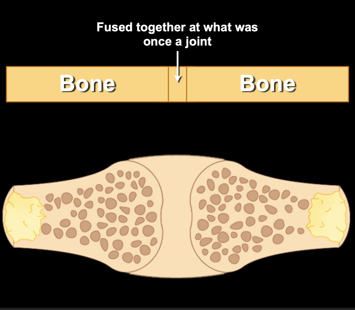
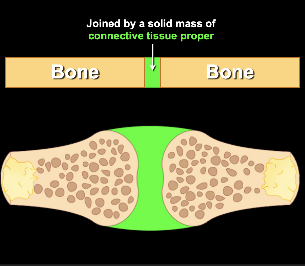
Syntosis
What do you call it when 2 bones are joined together overtime
Syntosis
A fibrous joint characterized by two adjacent bones connected by ligamentous structures
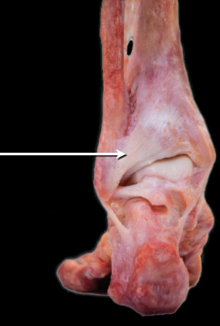
Syndesmosis
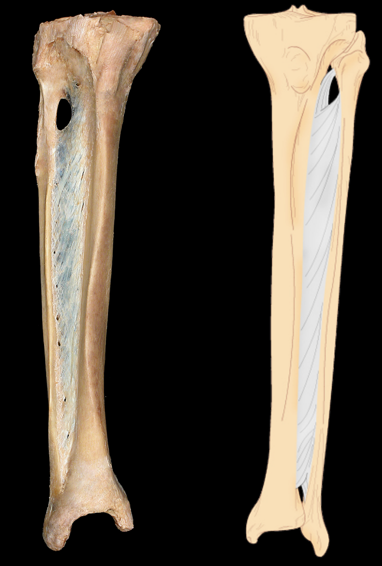
What type of fibrous joint and subtype
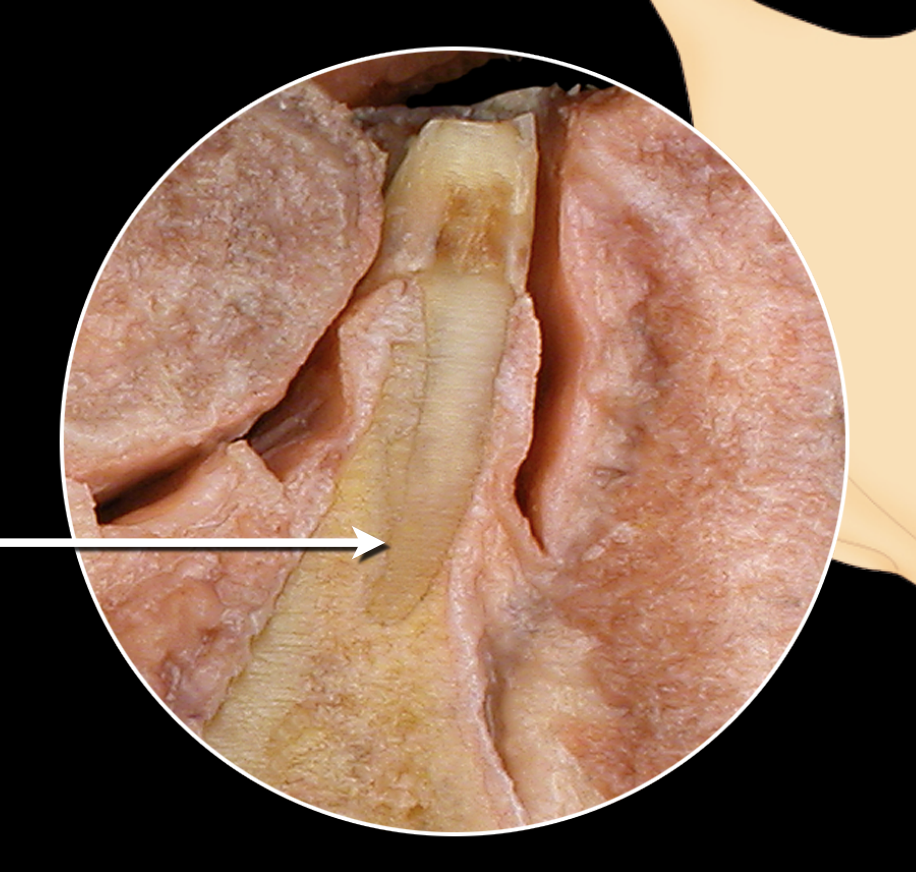
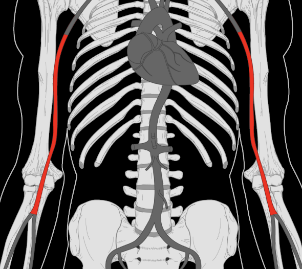
Syndesmosis and interosseous membrane
What type of fibrous joint and subtype
Syndesmosis and interosseous ligament
What type of fibrous joint and subtype
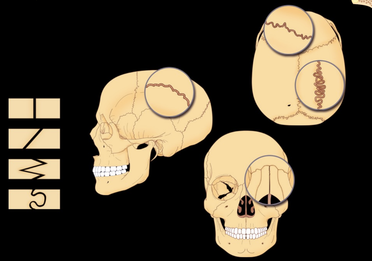
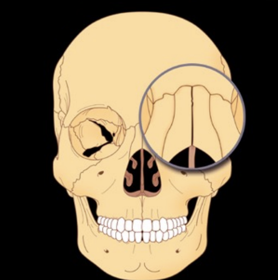
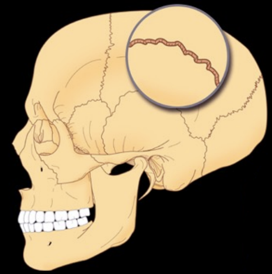
Syndesmosis and sutures
What type of fibrous joint and subtype
Syndesmosis and gomphosis
What type of suture
Plane
What type of suture
Limbous
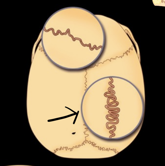
What type of suture
Denticulate
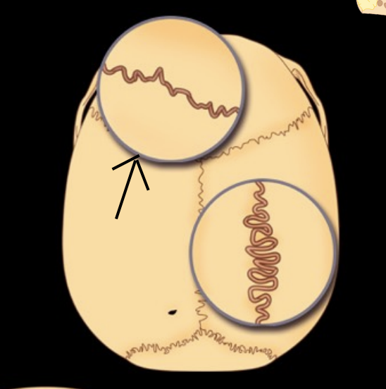
What type of suture
Serrate
What type of joint and subtype
Cartilaginous and synchondroses
What type of joint and subtype
Cartilaginous and epiphysial cartilage

What type of joint and subtype
Cartilaginous and epiphysial cartilage
What type of joint and subtype
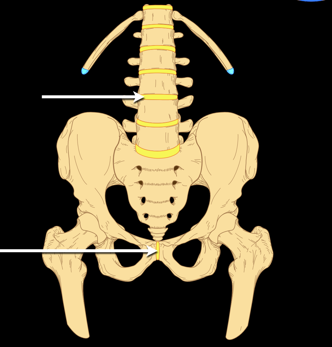
Cartilaginous and symphysis
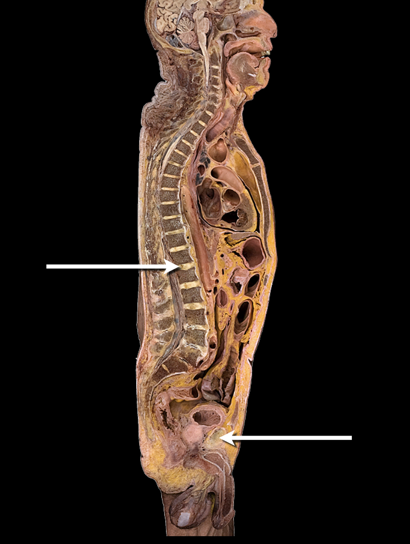
What type of joint and subtype
Cartilaginous and symphysis
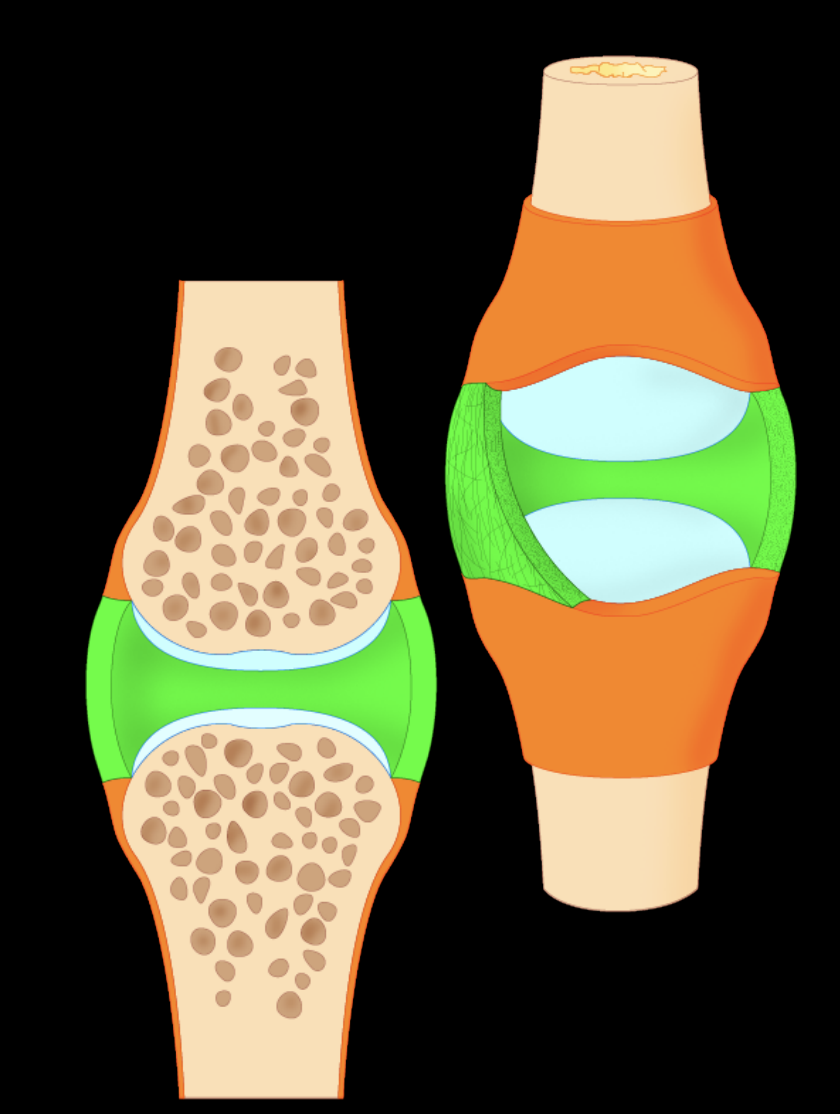
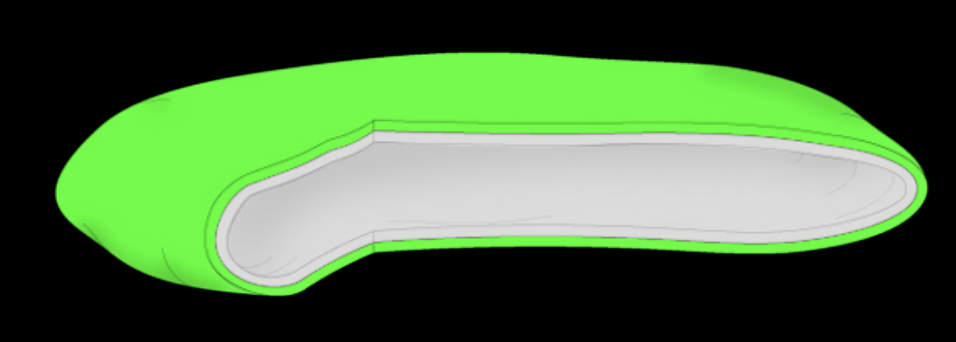
Capsular ligament or fibrous layer
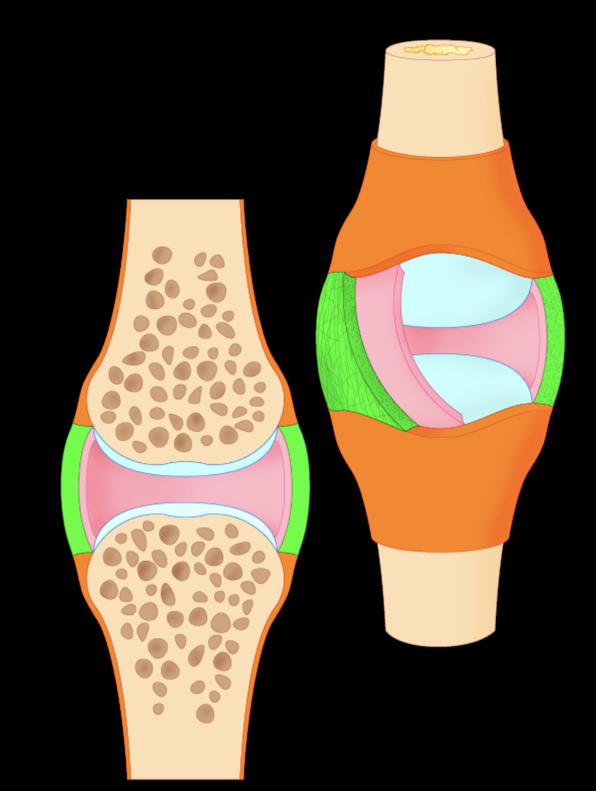
Synovial layer
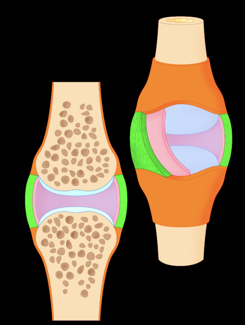
Synovial fluid
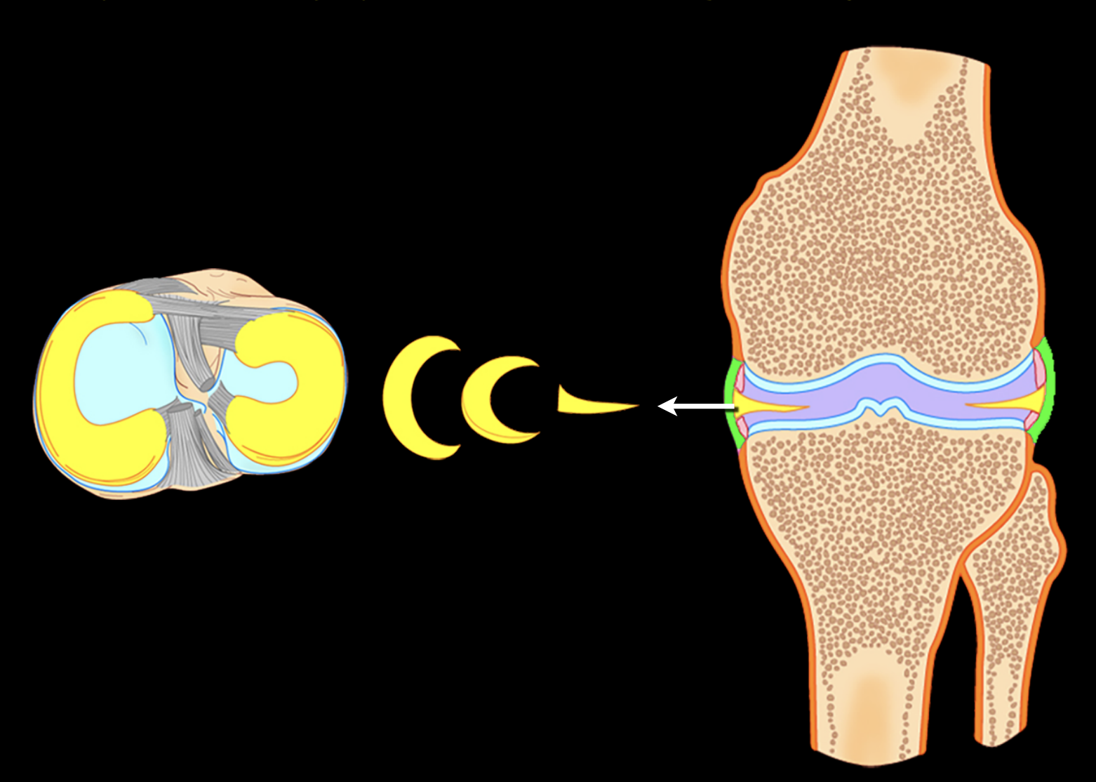
Meniscus
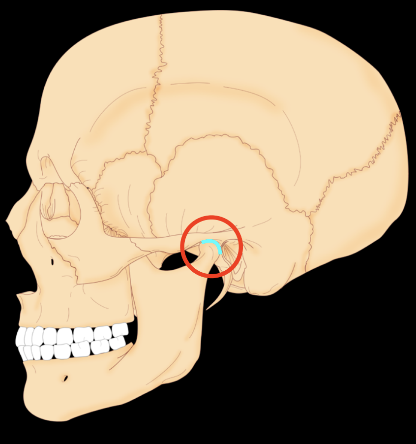
Articular disc
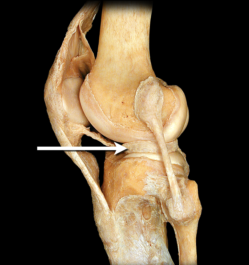
Meniscus
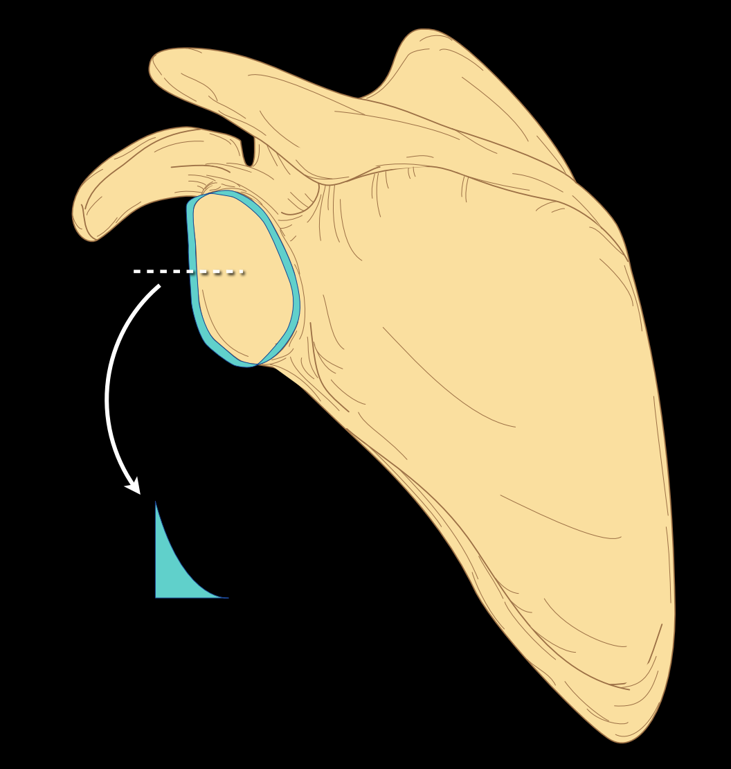
Labrum
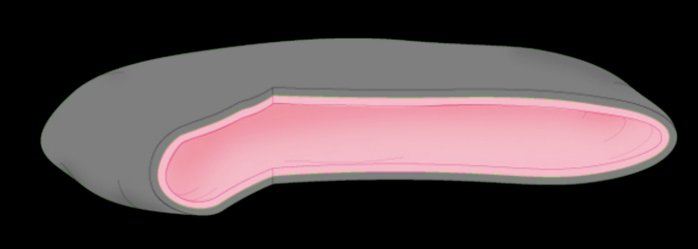
Synovial membrane
Fibrous membrane
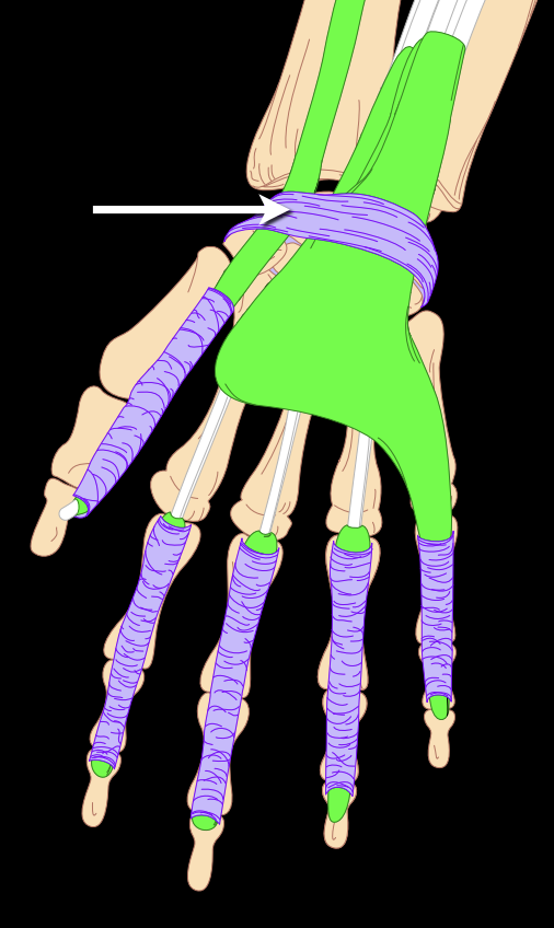
Retinaculum
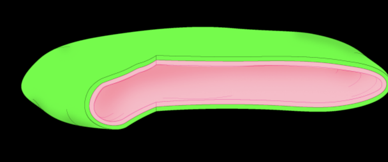
Synovial bursa
Large cells that contain up to 25 nuclei and are most abundant in the areas of bone undergoing resorption. It absorbs and digests the matrix components of the bone tissue.
Osteoclasts
Living within the canaliculi, they have long arm processes that extend to neighboring cells. These cells play an active role in the release of calcium from bone tissue to the blood. They are maintenance cells of the bone tissue.
Osteocytes
Cells that produce the bone matrix are prevalent at the advancing surfaces of developing tissue. They rearrange themselves in a single layer of cells, and they develop and remodel the edge of the bone. Essentially, they make bone.
Osteoblasts
Bone stem cells that originate from mesenchymal stem cells and are crucial for bone repair and growth. They form a population of stem cells that can differentiate into the more specialized bone-forming cells.
Osteoprogenitor cells
A place where two bones are closely joined, either forming an immovable joint or completely fused.
Symphysis
Cartilaginous joint where bones are joined together by hyaline cartilage, or where bone is united to hyaline cartilage.
Synchondrosis
A viscous, clear-to-pale-yellow fluid secreted by the synovial membrane into joint cavities, serving primarily to lubricate joints, reduce friction between articular cartilages, and provide nutrients to joint tissues.
Synovial fluid
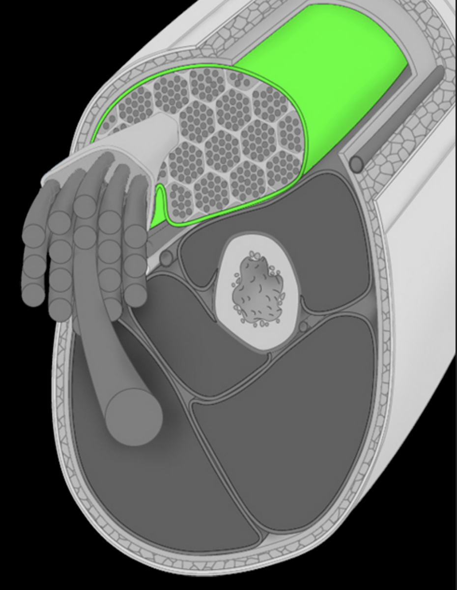
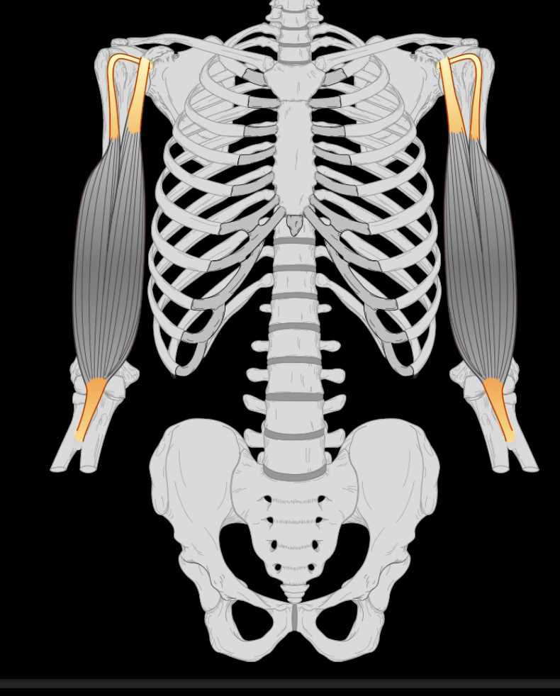
Epimysium
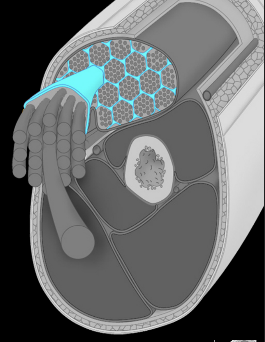
Perimysium
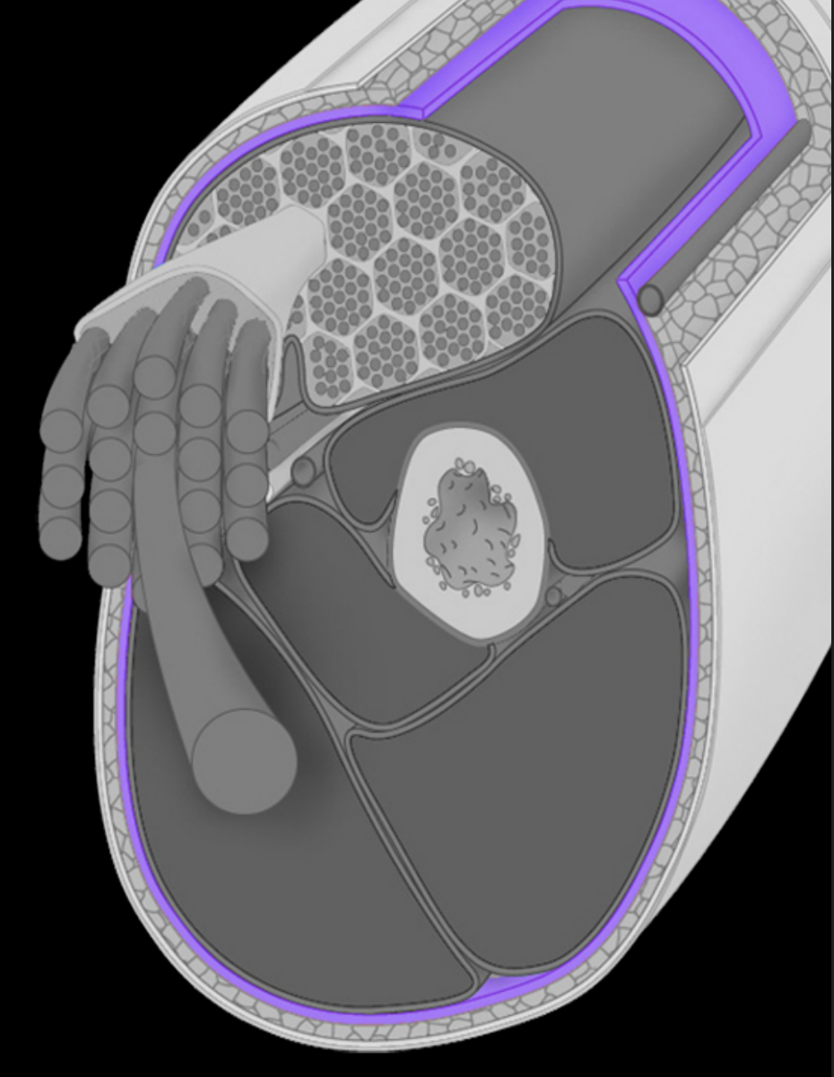
Fascia
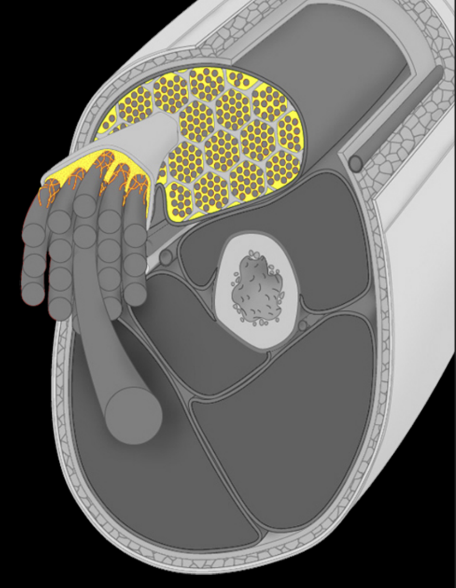
Endomysium
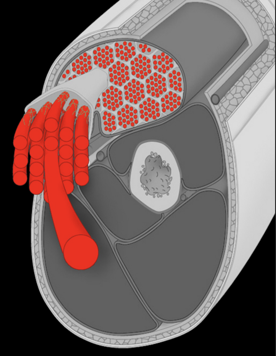
Muscle fiber
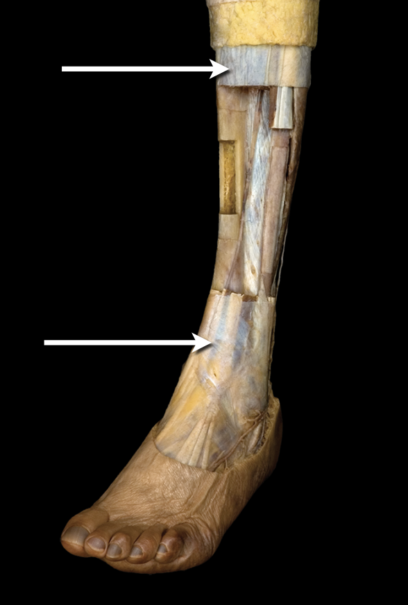
Fascia and retinaculum
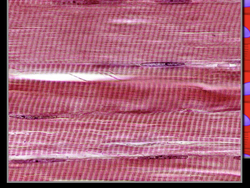
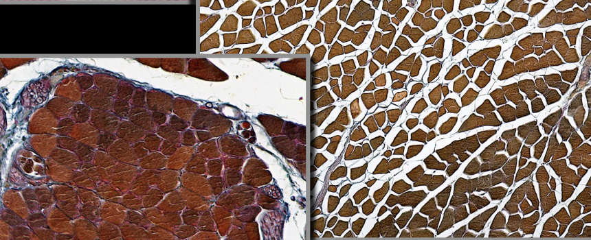
Skeletal striated muscle
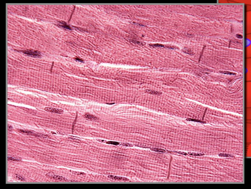
Cardiac striated muscle
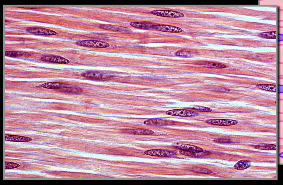
Smooth non striated muscle
Identify the type of muscle
Skeletal striated muscle
Identify the type of tisse
Dense regular connective tissue
What type of tissue makes tendones
Dense regular fibrous connective tissue
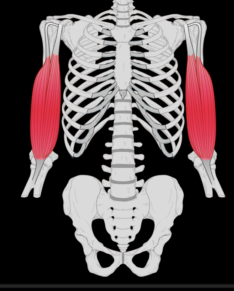
What is the only voluntary muscle
Skeletal muscle
Skeletal muscle fibers

Name structure 1.
Perimysium
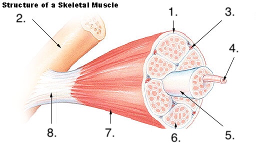
Name structure 2.
Bone

Name structure 3.
Blood vessel

Name structure 4.
Muscle fiber

Name structure 5.
Fascicle

Name structure 6.
Endomysium

Name structure 7.
Epimysium

Name structure 8.
Tendon
These are the largest high-pressure vessels nearest the heart, characterized by a thick tunica media about 1/10th of the vessel diameter.
Elastic artery
These are medium to small arteries that regulate blood flow to organs via significant smooth muscle, controlling vasoconstriction and dilation. The tunica media is about 1/4th of the vessel diameter and is the main distributing vessel.
Muscular artery
These have a much thinner diameter. They have smooth muscle wrapped around the vessel, allowing for rapid dilation and constriction. The muscle contraction can completely occlude the lumen of the vessel therefore having a sphincter function.
Arteriole
The body's smallest blood vessels, forming a network that connects arteries and veins, acting as the crucial site for exchanging oxygen, nutrients, and waste products between the blood and surrounding tissues
Capillary
Inner layer of blood vessels
Tunica initima
Middle layer of blood vessels
Tunica media
Outside layer of blood vessels
Adventitia
What is the internal layer of the blood vessels made out of
Simple squamous epithelium and loose connective tissue.
What is the middle layer of the blood vessels made out of
Smooth muscle and connective tissue
What is the outermost layer of the blood vessels made out of
Dense irregular connective tissue
Blood vessels that carry deoxygenated blood from the body's tissues back to the heart. They have thin walls and often contain one-way valves to prevent blood from flowing backward, especially against gravity, relying on muscle contractions to help pump blood upwards.
Vein
very small blood vessels that connect your capillaries with your veins throughout your body. They move blood that contains waste and lacks oxygen from your capillaries to your veins.
Venule
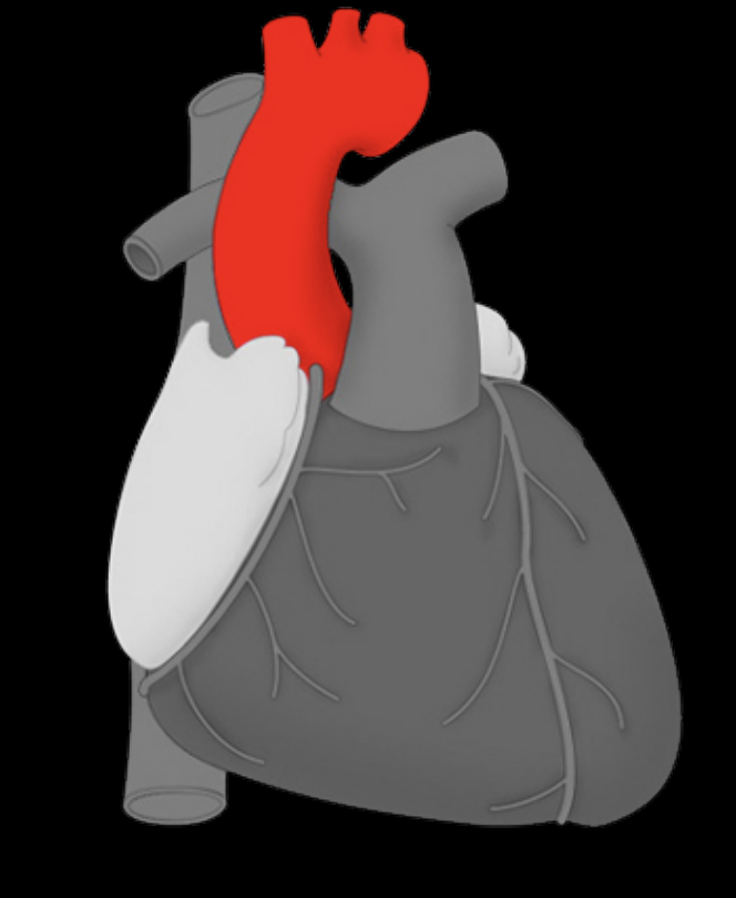
Aorta
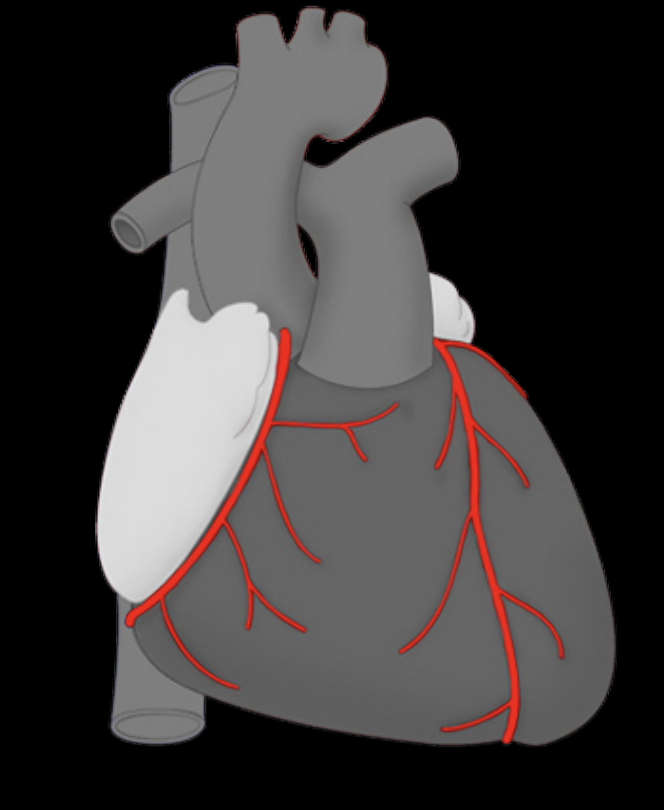
Coronary arteries
Brachial artery
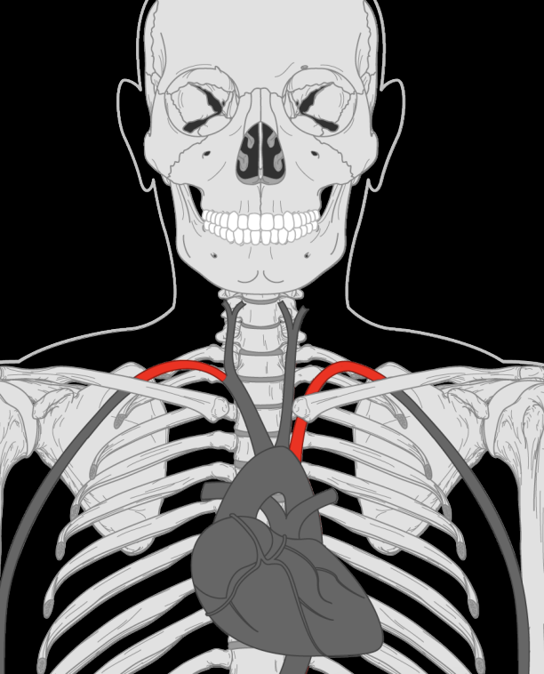
Subclavian artery