EXAM 2 - Regional Neck Anatomy
1/94
There's no tags or description
Looks like no tags are added yet.
Name | Mastery | Learn | Test | Matching | Spaced | Call with Kai |
|---|
No analytics yet
Send a link to your students to track their progress
95 Terms
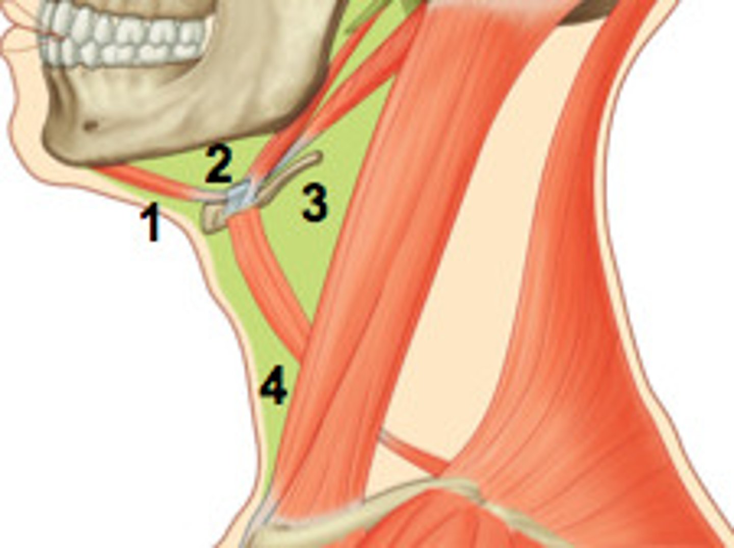
Anterior cervical triangle borders
1. anterior border of Sternocleidomastoid
2. inferior margin of mandible
3. Anteromedial line of neck
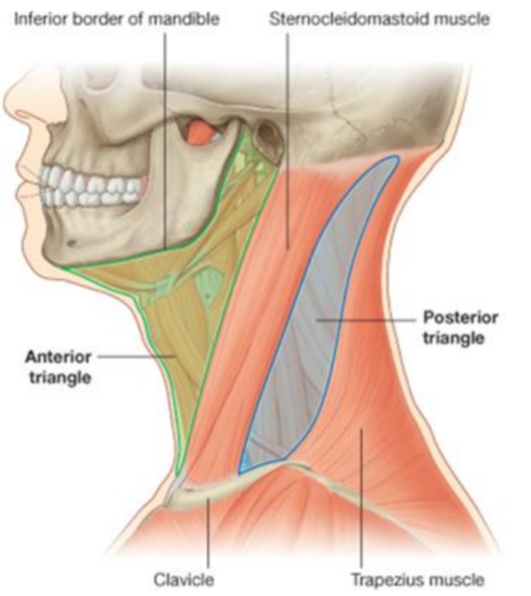
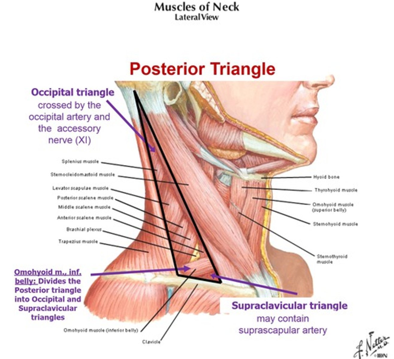
posterior cervical triangle borders
1. posterior border of SCM
2. anterior border of traps
3. middle 1/3 of clavicle
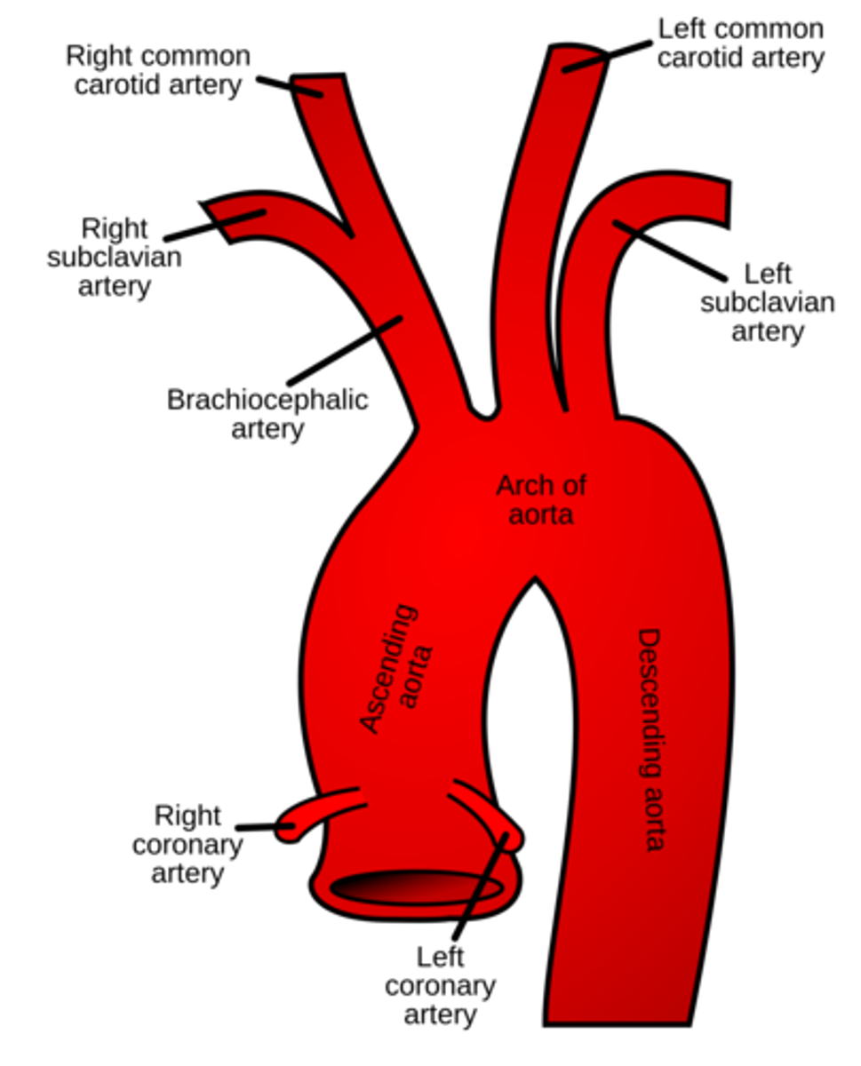
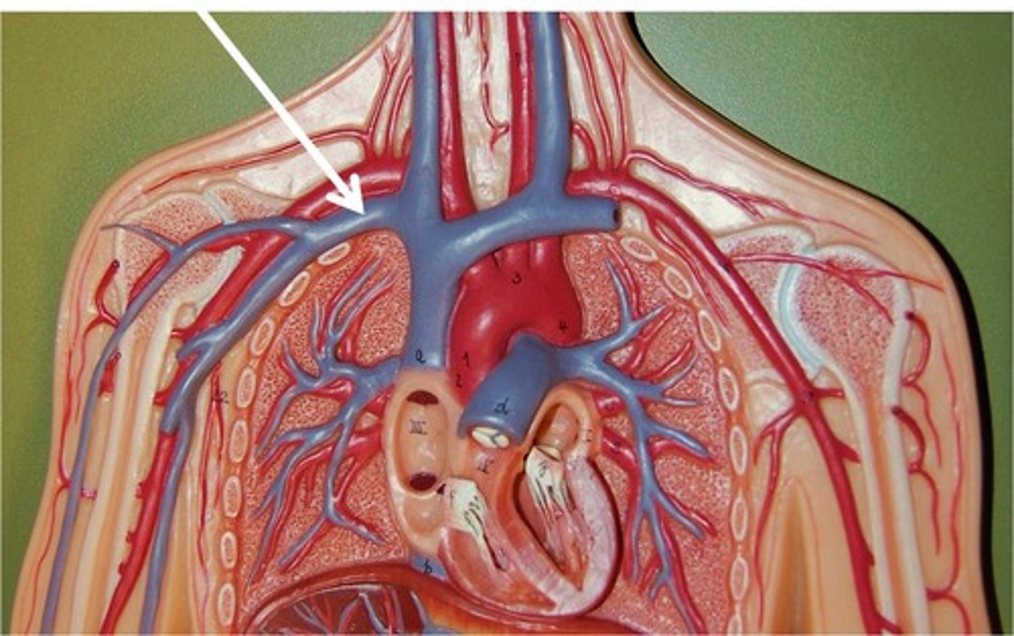
Major Vessels of the Neck
► Arterial
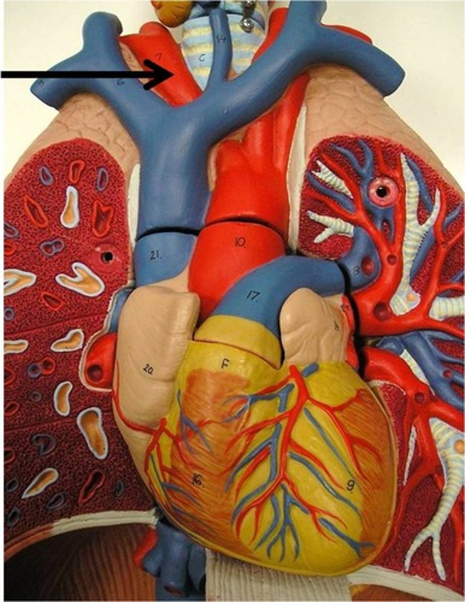
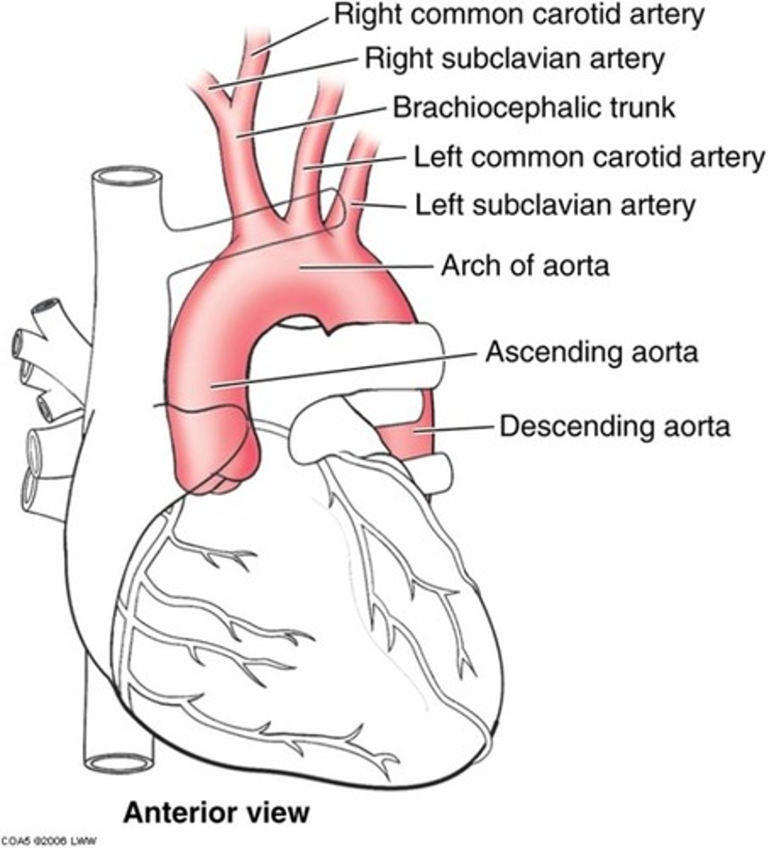
◦ Brachiocephalic a (not paired)
◦ L/R Common carotid aa (CCA)
◦ External carotid aa
◦ Internal carotid aa
◦ L/R Subclavian aa
Major Vessels of the Neck
► Venous
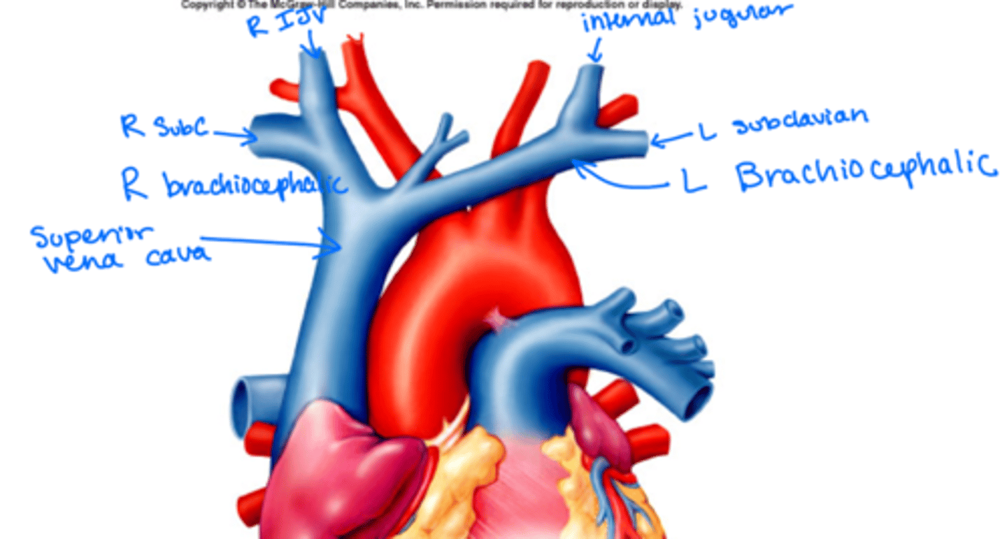
◦ L & R subclavian vv
◦ External Jugular vv (EJV)
◦ Internal Jugular vv (IJV)
◦ L & R brachiocephalic vv
Brachiocephalic a
► Arises directly off of the aortic arch (1st branch)
► A single vessel that gives rise to:
◦ 1. R subclavian artery
◦ 2. R CCA
Common Carotids (CCA)
► Right CCA
◦ Arises from the brachiocephalic artery
► Left CCA
◦ Arises from the aortic arch - the second branch
Both CCAs run cranially in neck through
__________________ just deep to ant border of
______
the carotid sheath
SCM
carotid triangle borders
superior belly of omohyoid muscle
posterior belly of digastric muscle
anterior border of SCM
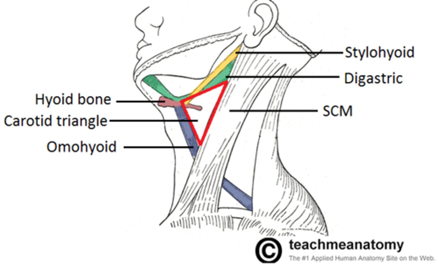
Carotid Triangle - contents
common carotid arteries and branches
internal and external carotid arteries
internal jugular veins and tributaries
vagus nerve (in the middle)
hypoglossal nerve
spinal accessory n
thyroid gland
larynx
deep cervical lymph nodes
branches of the cervical plexus
Carotid Sheath - contents (must know all 7)
1. CCAs (L&R)
2. internal carotid arteries
3. internal jugular veins
4. vagus n (shiny and pull and the head will come up...extremely strong)
5. cervical lymph nodes
6. carotid sinus
7. sympathetic n fibers
Common Carotids Continued
► Above upper border of thyroid cartilage they
divide into
1. external carotid a (C)
2. internal carotid a (B)
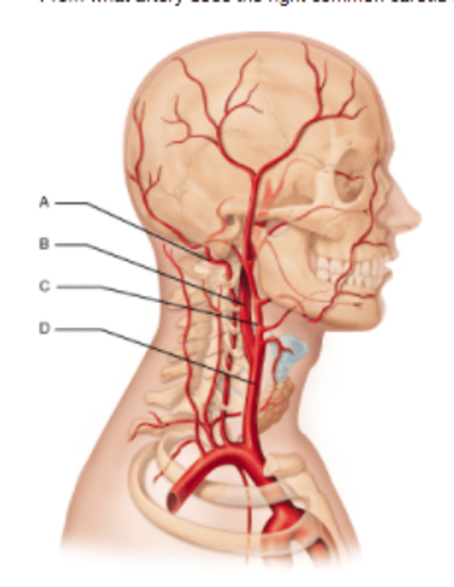
External Carotid aa - supplies
outside of the face and skull
External Carotid aa - branches
1. superficial thyroid
2. ascending pharyngeal
3. lingual
4. facial
5. occipital
6. posterior auricular
7. superficial temporal
8. maxillary
External Carotid aa - path
upper boarder of thyroid cartilage - parotid gland behind the mandible - emerges at anterior border of SCM (with the carotid triangle)
can you palpate the External Carotid aa
yeah - its not within the carotid sheath
Internal Carotid aa - branches
NO BRANCHES IN THE NECK
Branches in the head:
1. ophthalmic
2. posterior communicating
3. anterior cerebral artery
4. middle cerebral artery
Internal Carotid aa - path
upper border thyroid cartilage throughout the neck
- enters the cranial cavity via the carotid canal (temporal bone) - terminates into anterior and middl cerebral arteries
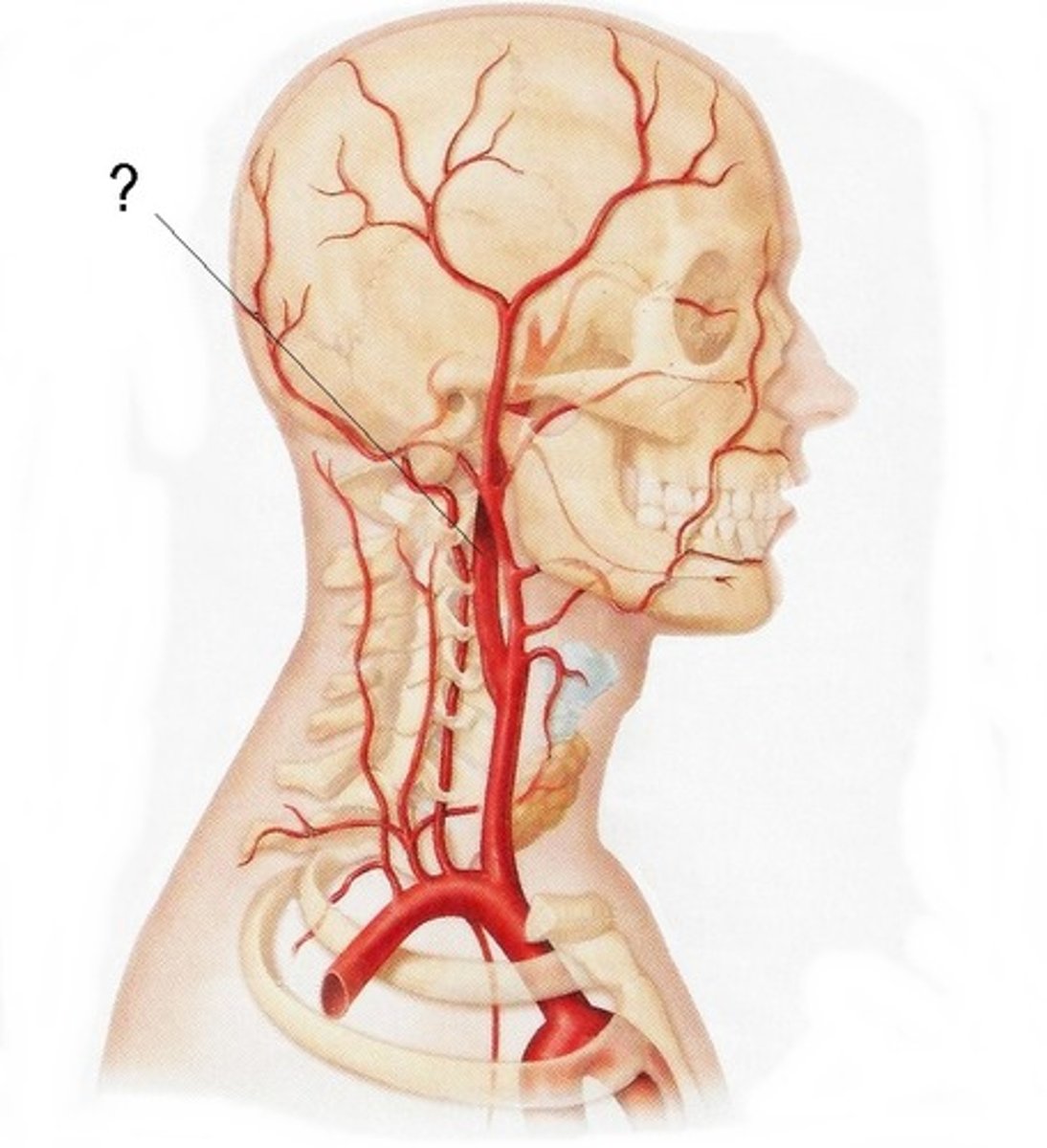
R Subclavian aa arises
from the brachiocephalic artery
arches up and laterally over the lung pleura and runs between two muscles
what muscles does the R Subclavian aa run though
anterior scalene and the middle scalene
where does the R Subclavian aa become the axillary a
level of the first rib
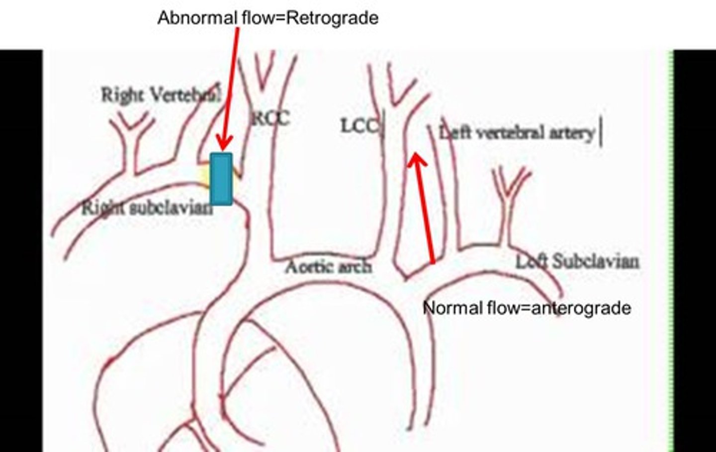
Subclavian steal syndrome
Causes someone to pass out when they raise their right arm above their head
Occurs due to retrograde flow in the vertebral artery
occurs when the subclavian artery is occluded - between the right common carotid and vertebral artery - , and blood flow is diminished, or obstructed, to the head and upper extremities.
Left subclavian artery arises
directly from the aortic arch (3rd branch)
Subclavian vv
Continuation of axillary vv at level of 1st rib
path of the subclavian vv
runs through a groove (along with the subclavian a) on the superior surface of rib 1
- anterior groove is for the subclavian vein
- posterior groove is for the subclavian artery
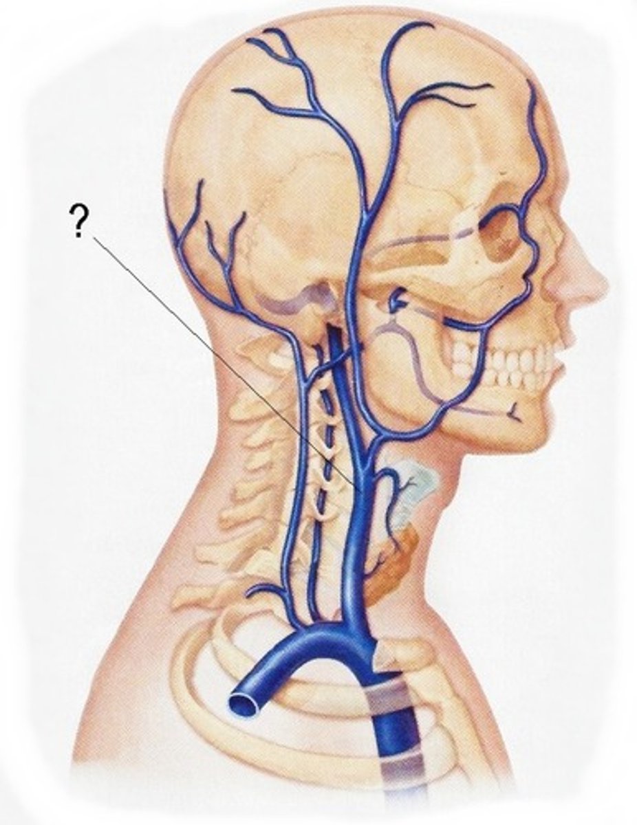
joins with the internal jugular vein to form the brachiocephalic veins
what does the subclavian vv drain
1. External jugular vein
2. left thoracic duct
3. right lymphatic duct
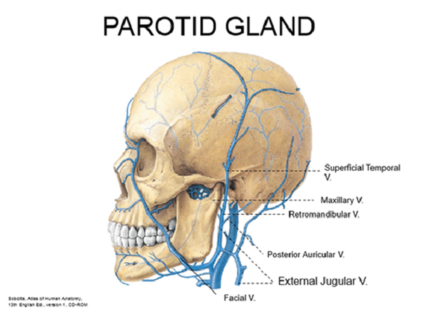
External Jugular vv (EJV) - Formed
behind the angle of the mandible by the union of the posterior auricular vein and the retromandibular vein
clinical application of EJV
you can visually see the venous pressure above the clavicle in the EJV and IJV
Internal Jugular vv (IJV) - drains
brain!!, face, scalp, and neck
Internal Jugular vv (IJV) is a continuation of
sigmoid venous sinus in brain
Internal Jugular vv (IJV) - leaves the skull through what foramen
jugular foramen
Internal Jugular vv (IJV) - path
descends through the neck within the carotid sheath lateral to the vagus n and internal common carotid artery
-joins the subclavian vv behind the medial end of the clavicle to form the brachiocephalic vv
Internal Jugular vv (IJV) - major tributaries
1. facial vein
2. inferior petrosal sinus
Brachiocephalic vv (R & L) - major tributaries
IJV
Subclavian v
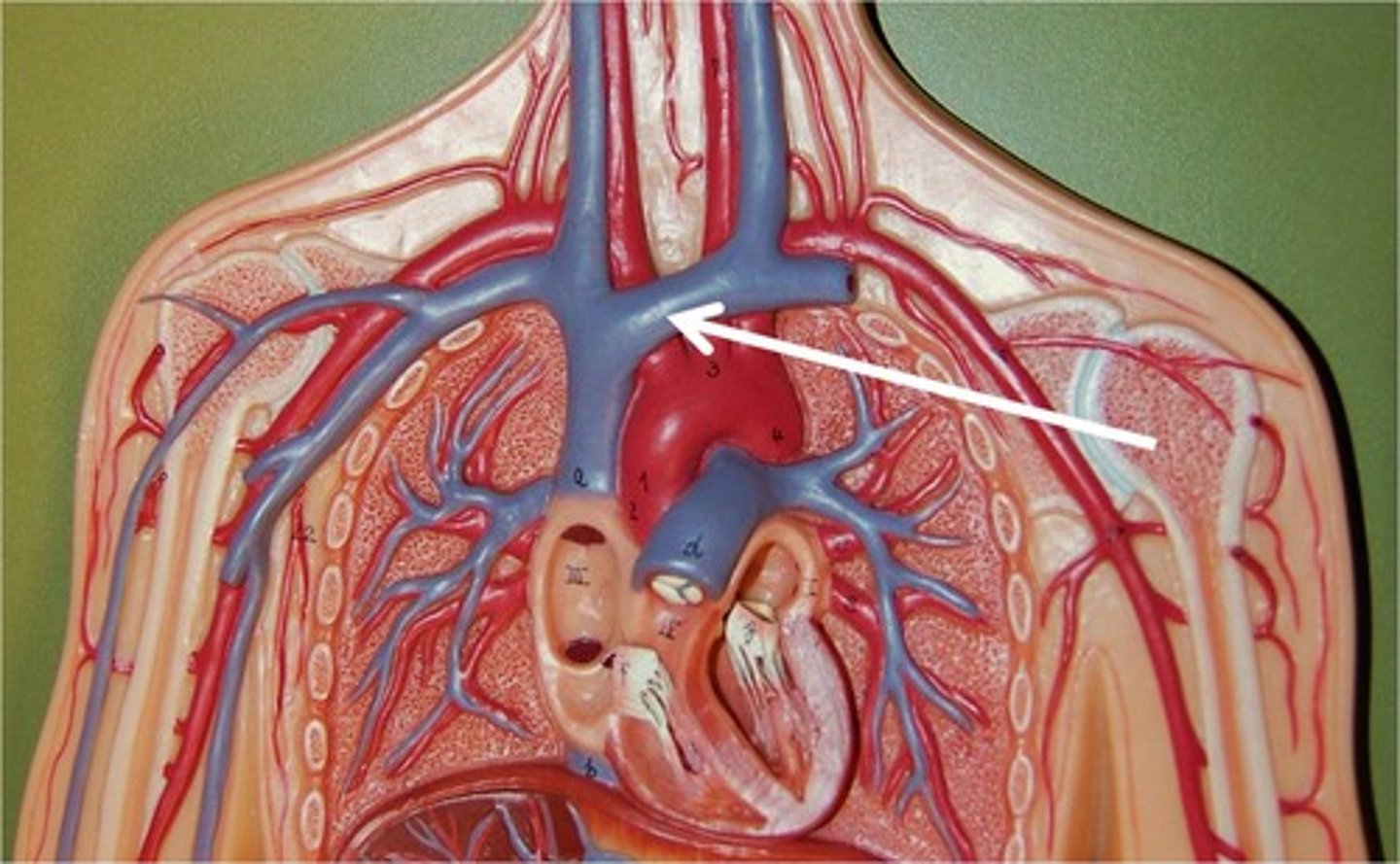
where does the Brachiocephalic vv begin
at the medial end of the clavicle
Platysma (O, I, A, innervation)
-Superficial & Large
► O: fascia of upper chest
► I: fascia of chin and jaw
► A: depress and draw lower lip laterally & draw up skin of chest
► N: facial n - cervical branch
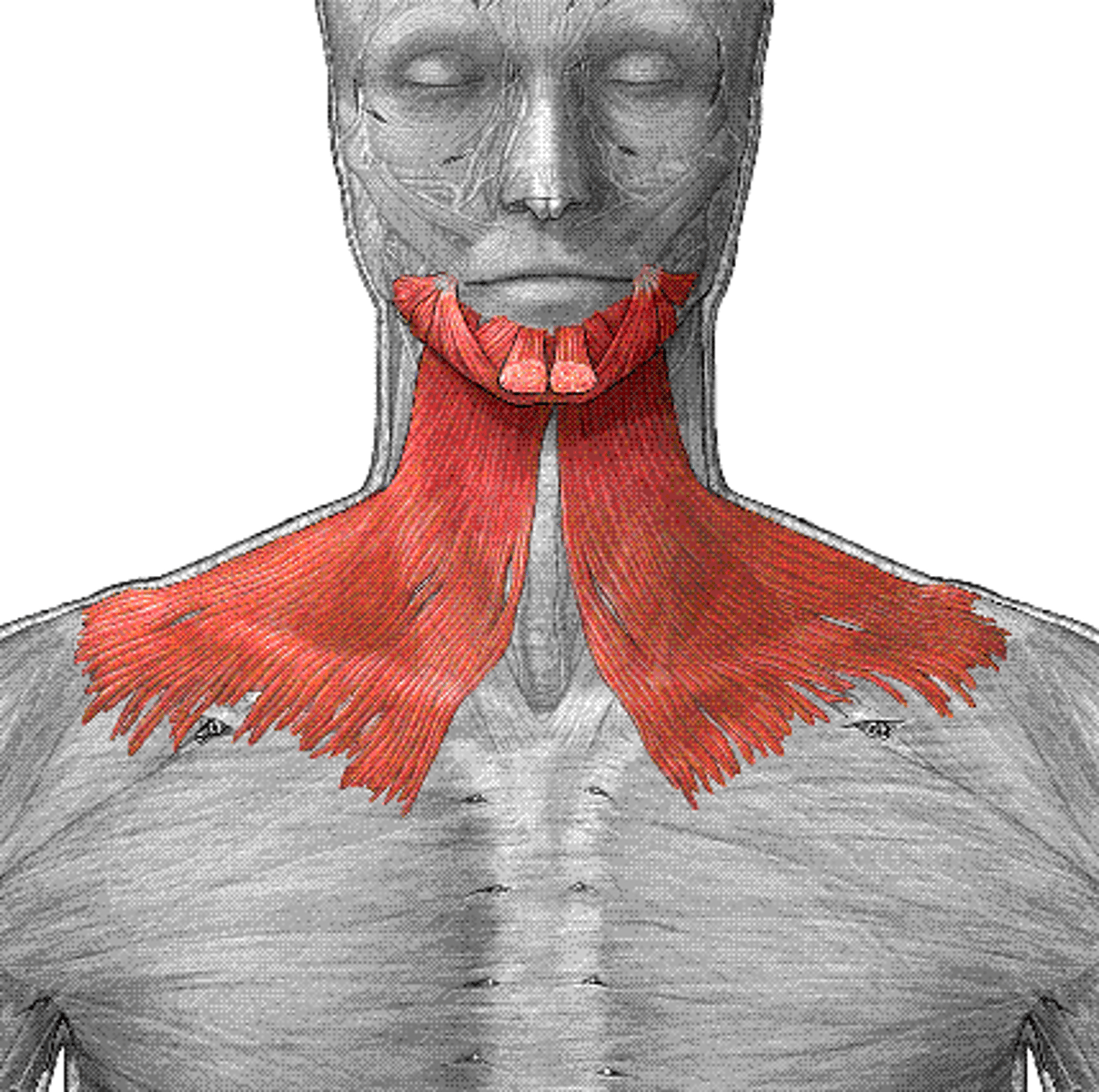
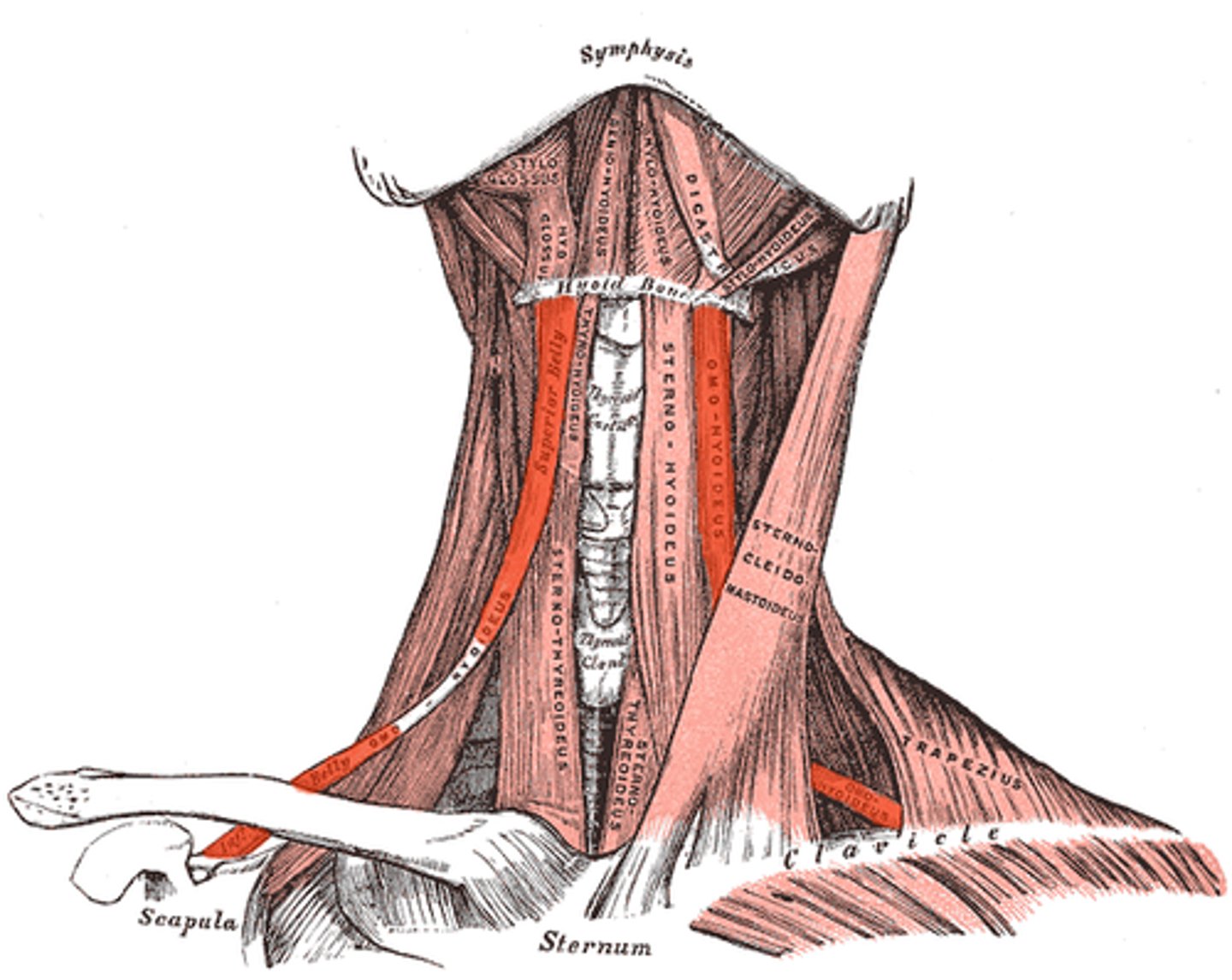
Muscles of the Hyoid/Larynx - general groups and actions
► Suprahyoid Group
◦ General action - elevate
► Infrahyoid Group
◦ General action - depress
Suprahyoid Group
digastric, geniohyoid, mylohyoid, stylohyoid
Digastric (anterior)
O I A INN
O: inferior border of mandible
I: tendon attached to hyoid
A: elevate hyoid and lower mandible
INN: V3 - trigeminal N

Digastric (posterior)
O I A INN
O: mastoid
I: tendon attached to hyoid
A: move hyoid posteriorly
INN: facial N - Cervical branch
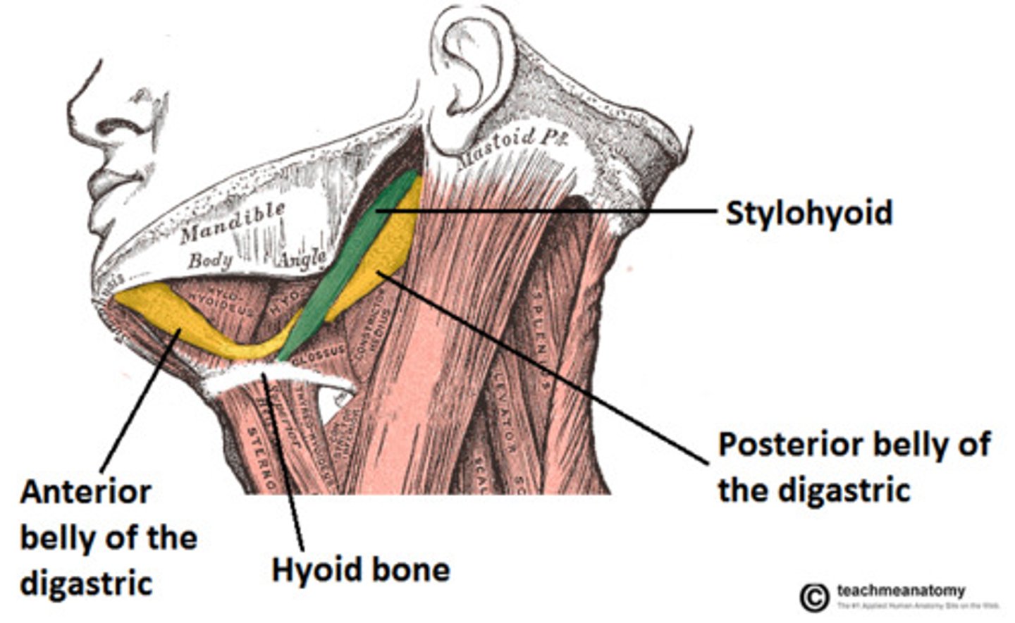
Stylohyoid - A & N
A: move hyoid backward and elevate tongue
N: Facial N
mylohyoid - A & N
A: elevate hyoid and raise the floor of the mouth and tongue
N: V3 - trigeminal
geniohyoid A&N
A: protrude hyoid and tongue
N: hypoglossal n (CN XII) and C1 nerve
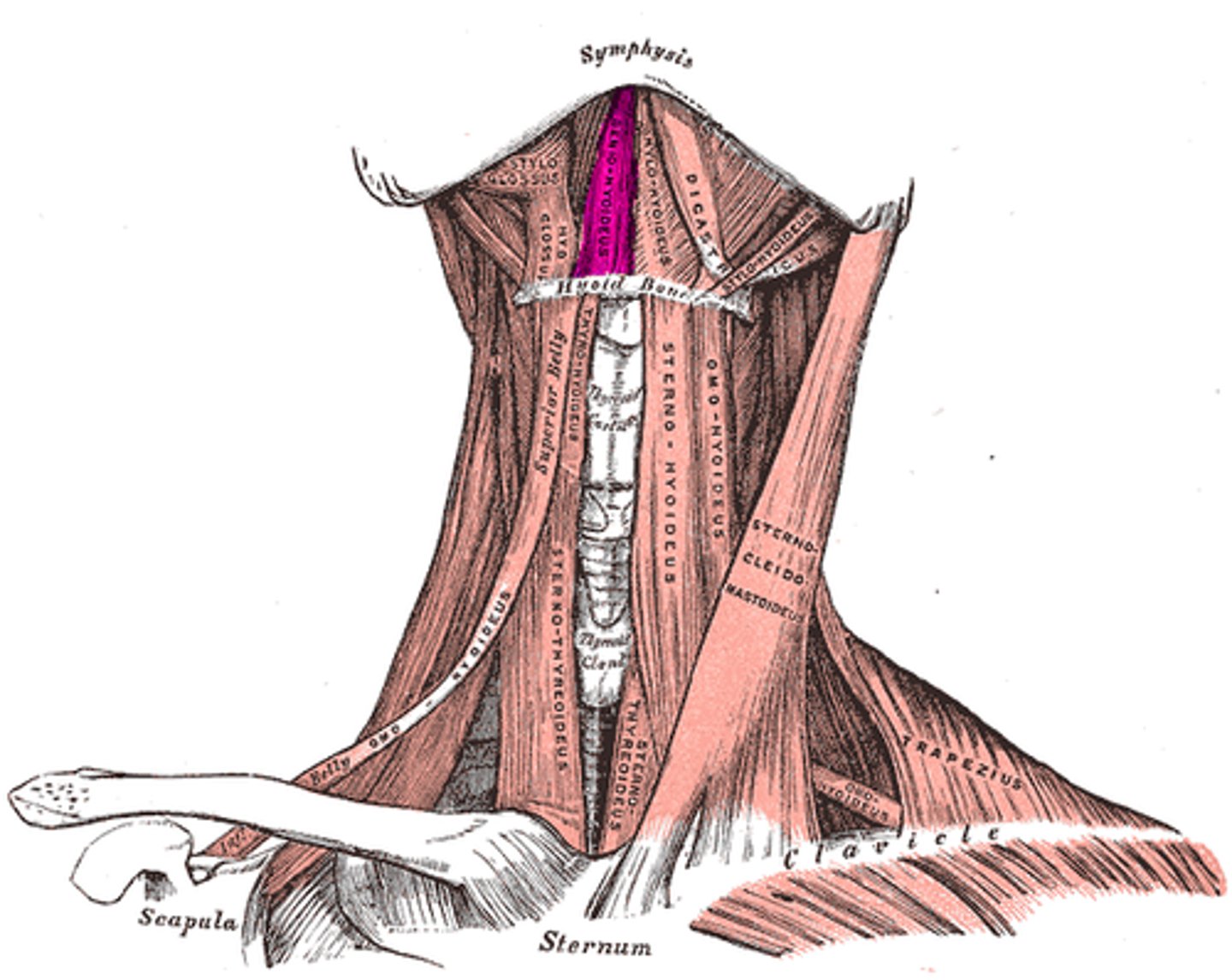
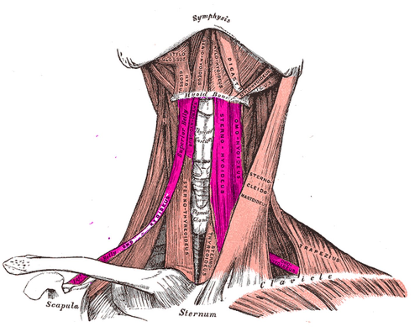
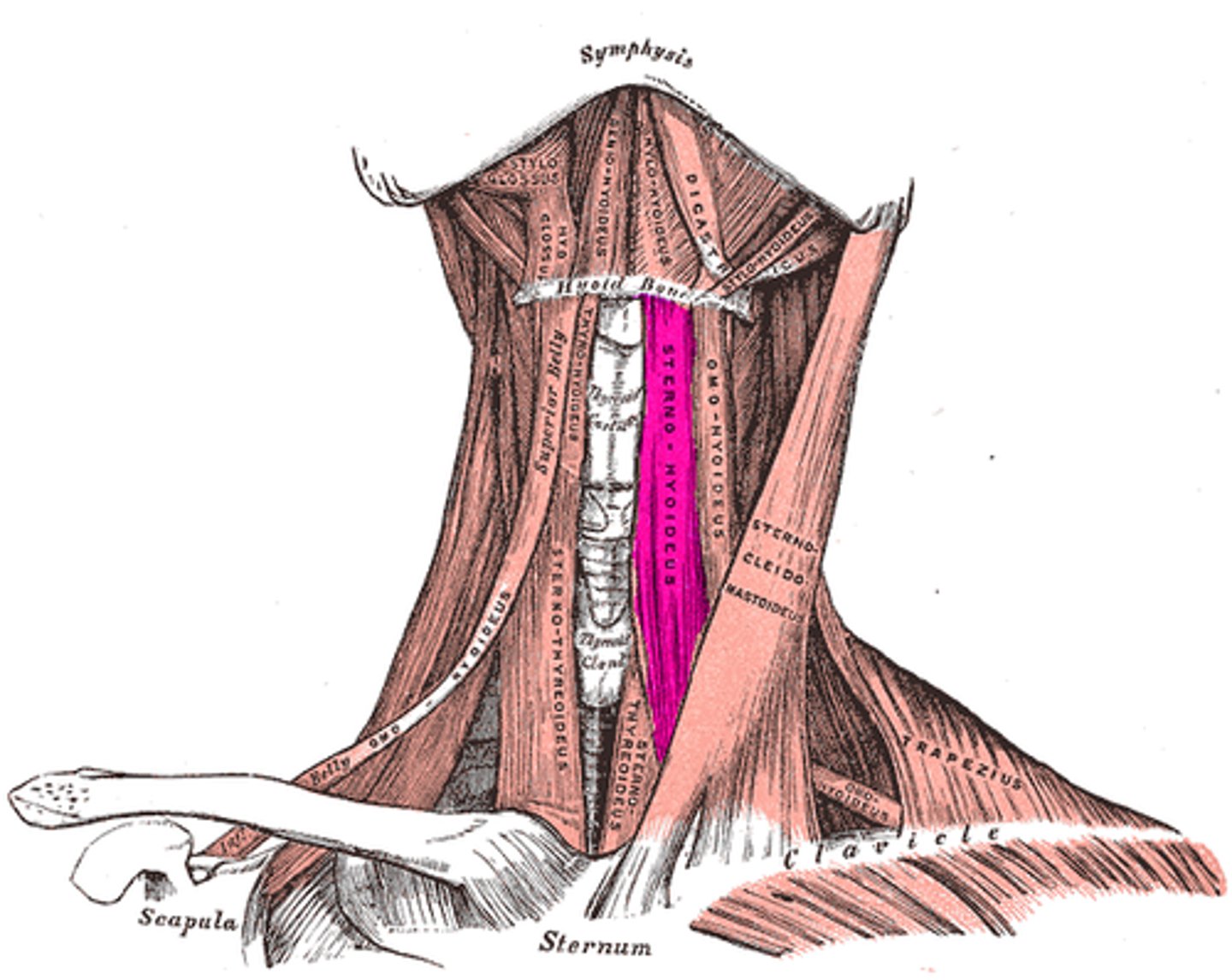
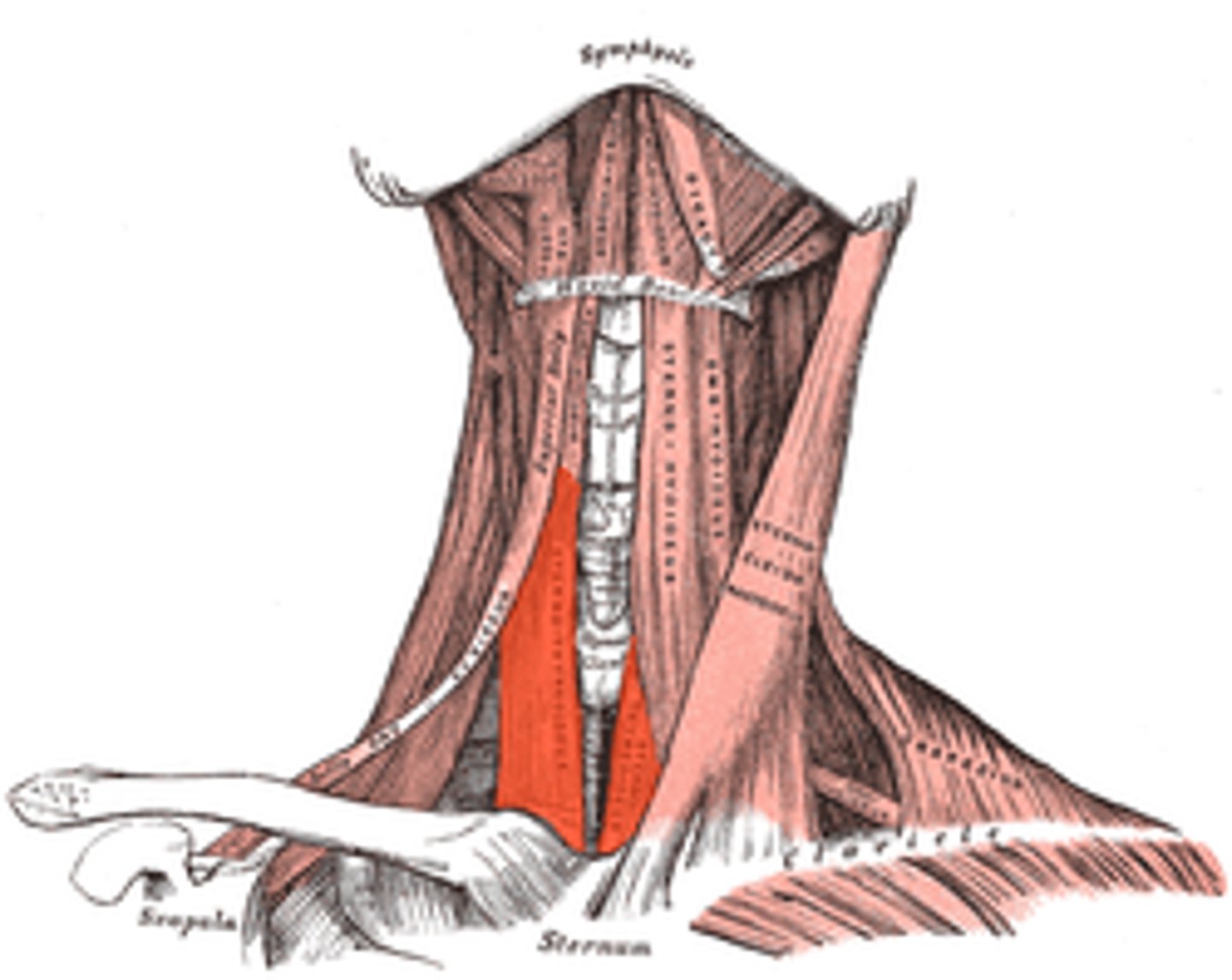
Infrahyoid Group consists of
sternohyoid, sternothyroid, omohyoid, thyrohyoid
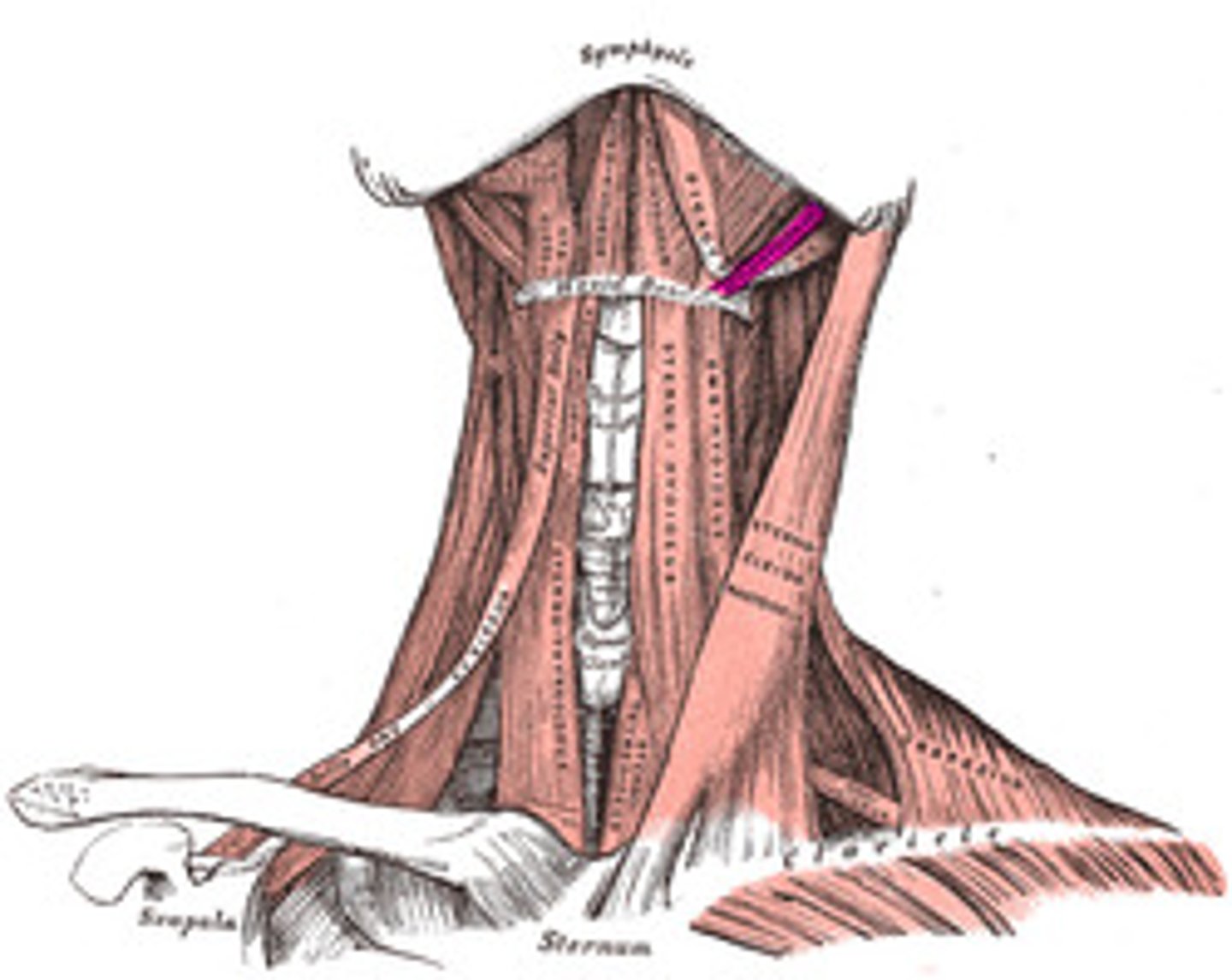
sternohyoid A&N
A: depress hyoid
N: C1-C3
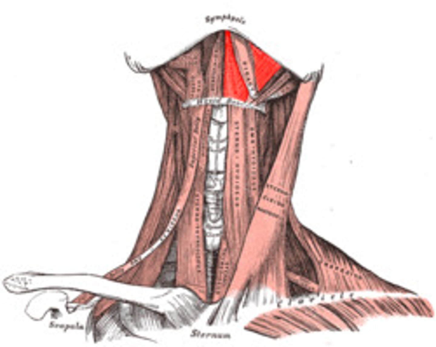
sternothyroid A&N
A: depress thyroid cartilage
N: C1-C3
Omohyoid A&N
A: depress hyoid
N: C2-C3
There is an superior and inferior belly
thyrohyoid A&N
A: depress hyoid and elevate thyroid cartilage
N: hypoglossal n and C1

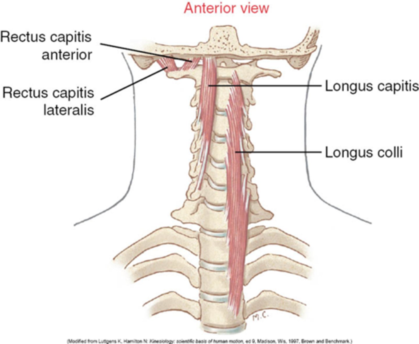
Anterior Cervical Vertebral Group
longus colli
longus capitis
rectus capitis anterior
rectus capitis lateralis
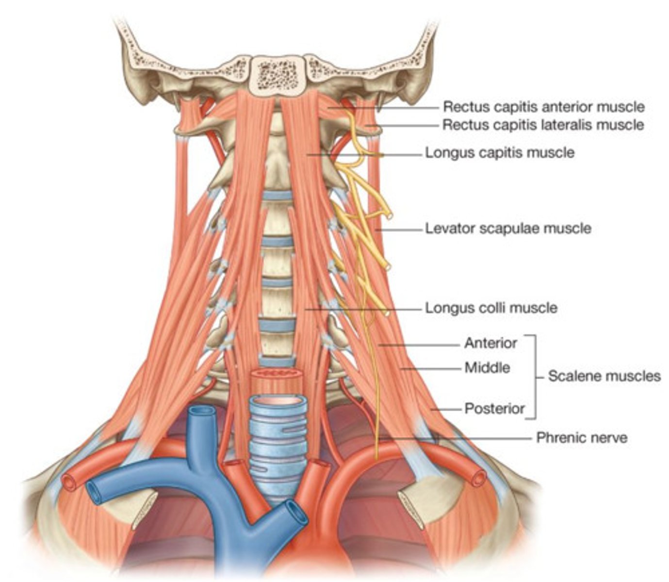
Longus Colli A&N
A: flex vertebral column
N: C2-C7
longus capitis OIAN
O: TP of C3-C6
I: occipital bone
A: flex vertebral column/neck
N: C1-C3
Rectus Capitis Anterior OIAN
O: C1 (atlas)
I: occipital bone
A: flex vertebral column/neck
N: C2-C3

rectus capitis lateralis OIAN
O: C1 (atlas)
I: occipital bone
A: flex vertebral column laterally
N: C2-C3
Lateral Cervical Vertebral Group
anterior, medius, and posterior scalene
scalenus anterior OIAN
O: TP of C3-C6
I: First rib
A: elevate rib 1 (inhale)
N: C2-C3
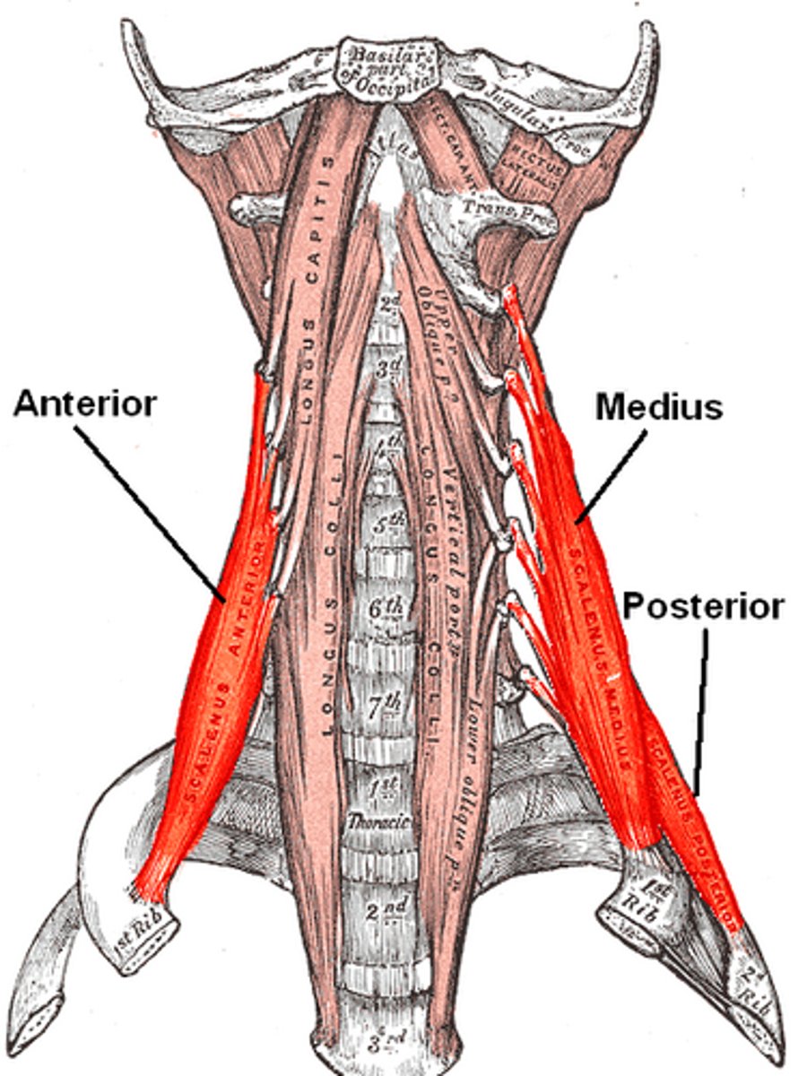
scalenus medius OIAN
O: TP of C2-C7
I: First rib
A: elevate rib 1 (inhale)
N: C2-C3

scalenus posterior OIAN
O: TP of C5-C7
I: second rib
A: elevate rib 2 (inhale)
N: C3-C5

Additional Actions of anterior and lateral cervical vertebrae
Bilateral: flexion of the vertebral column (flexion in cervical vert/neck)
Unilateral: ipsilateral neck flexion and slight rotation
Superficial Group of neck
sternocleidomastoid
SCM - OIAN
Sternal head O: manubrium of sternum
Clavicular head O: head of clavicle
Common I: mastoid process
Bilateral A: flex neck, elevate chin, draw head ventrally and sternum superiorly
Unilateral A: lateral flexion of neck, rotation of head contralaterally
Common N: C2-C3
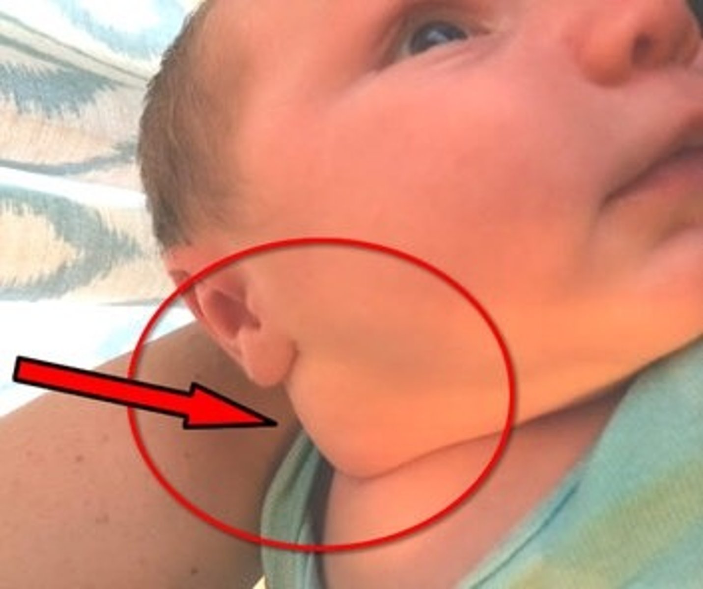
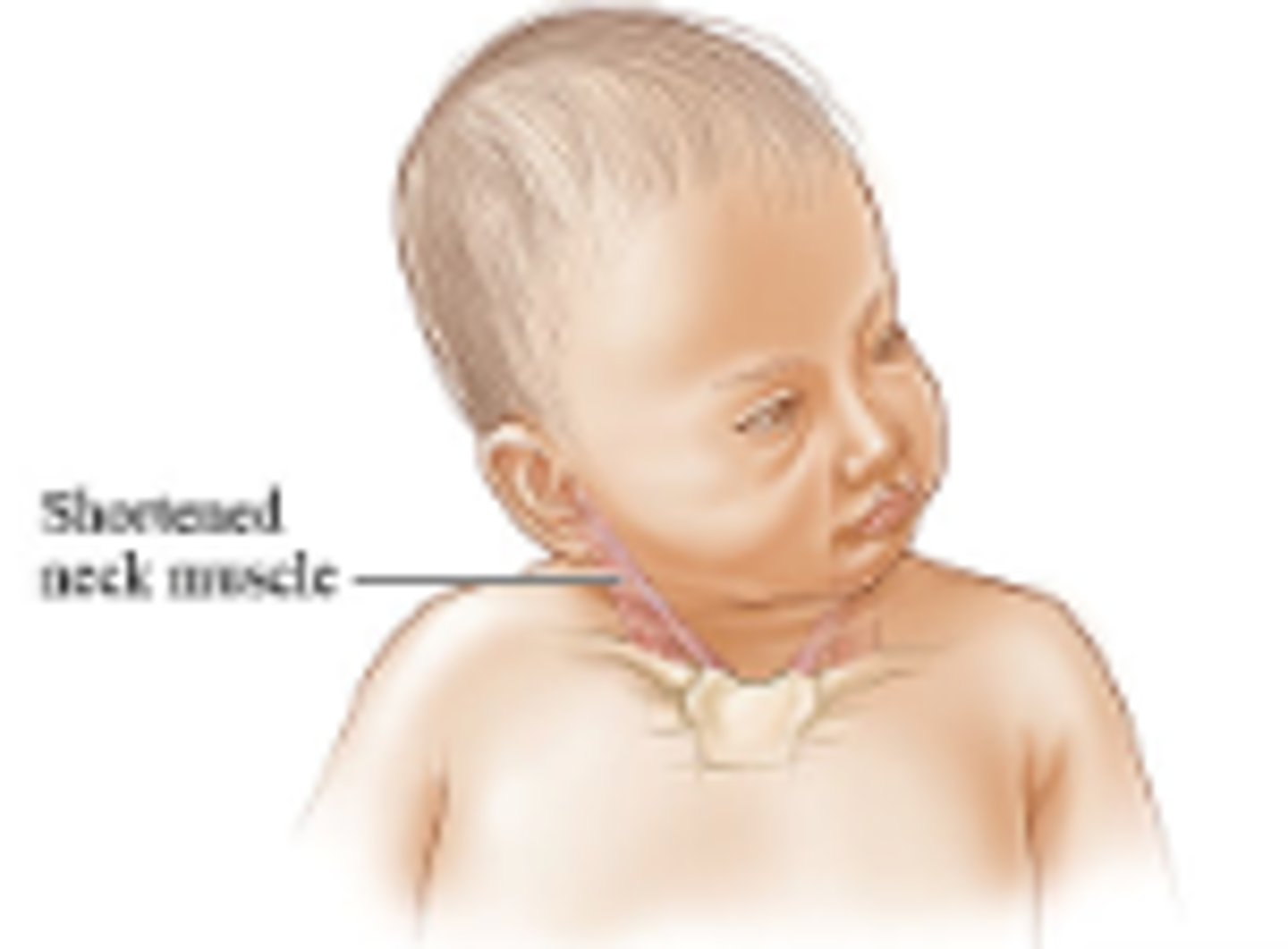
Congenital Torticollis & Types
twisted neck - present at birth
3 major types
- Fibromatosis colli
- muscular torticollis
- spasmodic torticollis
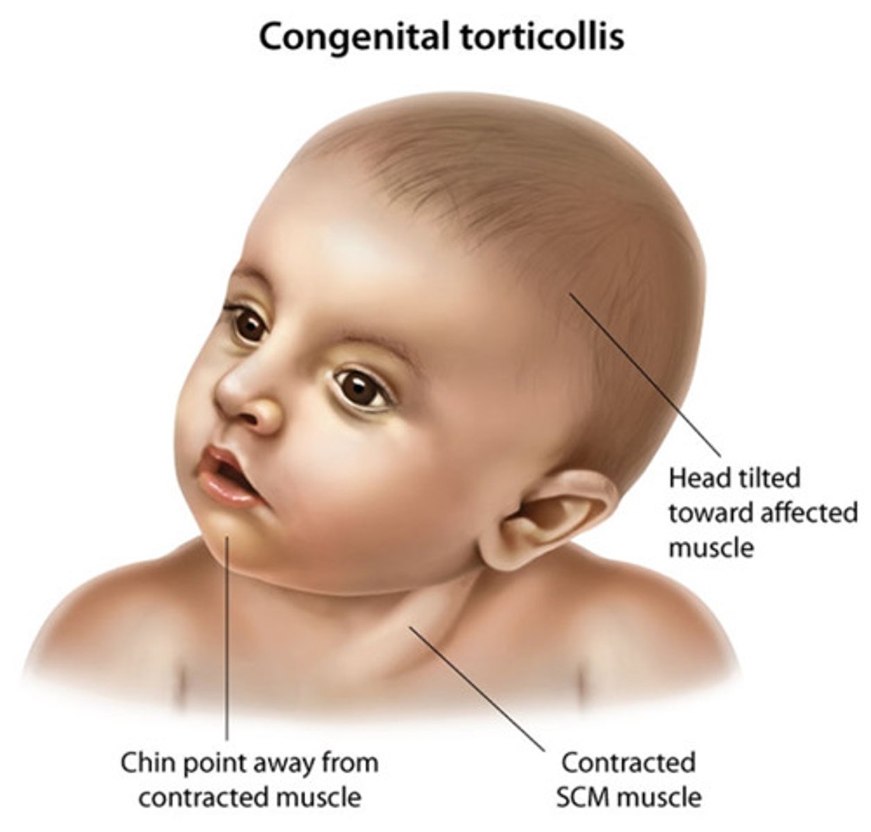
Fibromatosis colli
- most common
- develop in SCM
- prenatal - generally occurs in a breached delivery
- results in a twisted neck from unilateral action of SCM
(this means that if the right SCM is contracted, the neck is flexed towards the right but contralaterally flex head towards the left side)
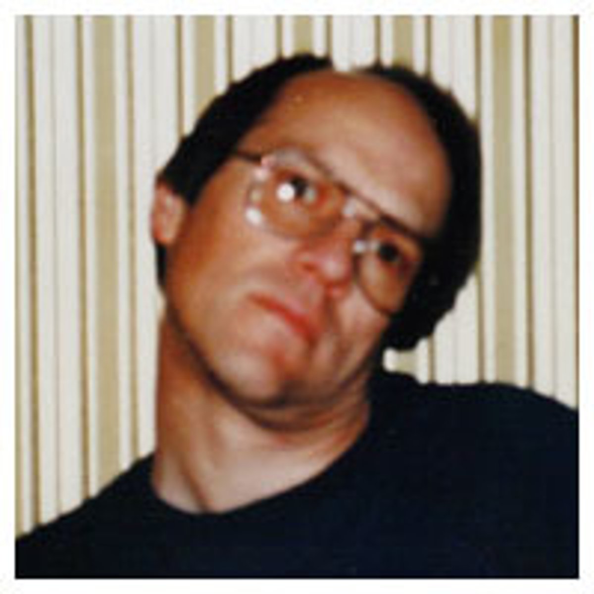
muscular torticollis
result of difficulty in delivery of baby - use of forceps usually
- treatment with a surgical release of the SCM from the origin and repair
spasmodic torticollis
- can be adult onset (not congenital)
- treat with botox
- sustained muscle spasms/contraction in the SCM - causing flexion on the impaired side and turn head contralaterally towards other side
Thyroid Gland - location
anterior aspect C5 - T1
Thyroid Gland - comprised of
- right and left lobe
- isthmus - connection between lobes
sometimes a pyramidal lobe on top of isthmus
Thyroid Gland - arterial supply
superior and inferior thyroid arteries
-unpaired thyroid ima artery (only in 10% of population)
Thyroid Gland - venous drainage
superior, middle, inferior thyroid veins
Thyroid Gland - lymphatic drainage
Prelaryngeal node, pretrachial node, and paratrachial node
Thyroid Gland -innervation
superior, middle, and inferior cervical sympathetic ganglia
- vasomotor: constriction of blood vessels to inhibit the blood flow (this is innervation that controls blood flow to organ by constricts blood vessels that supply it)
Parathyroid Glands - location
embedded in posterior surface of the thyroid gland
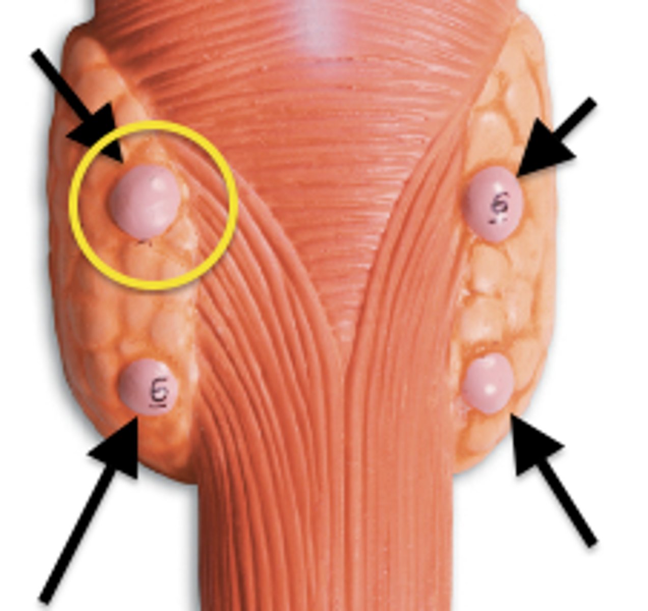
Parathyroid Glands - comprised of
2 superior and 2 inferior glands
can have more=ectopic glands as well
Parathyroid Glands - arterial supply
thyroid arteries
Parathyroid Glands - venous supply
thyroid veins
Parathyroid Glands - lymphatic drainage
cervical and paratrachial nodes
Parathyroid Glands - innervation
superior, middle, and inferior cervical sympathetic ganglia
- vasomotor: constriction of blood vessels to inhibit the blood flow
important in surgery of a thyroidectomy to leave the parathyroid glands behind and the nerves and blood supply
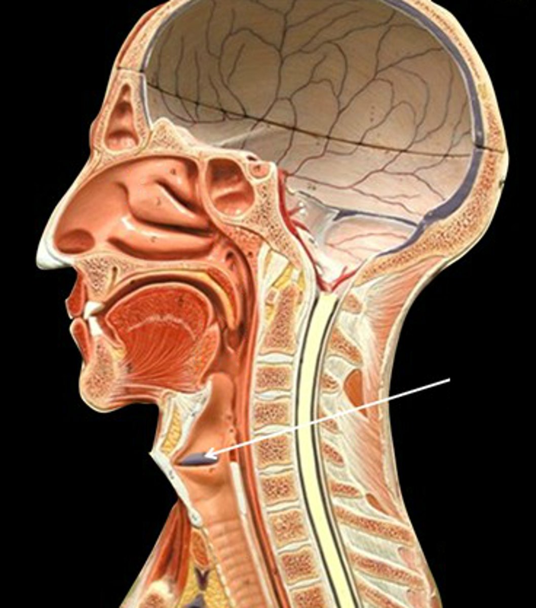
Larynx - functions
► Cartilagenous chamber about 4cm
► Primary fxn - keeping things out of airway (solids and liquids)
► Secondary fxn - phonation
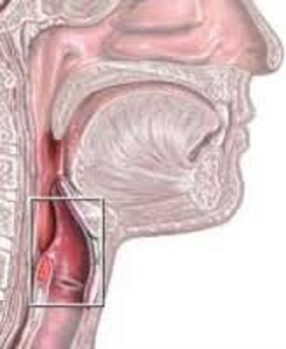
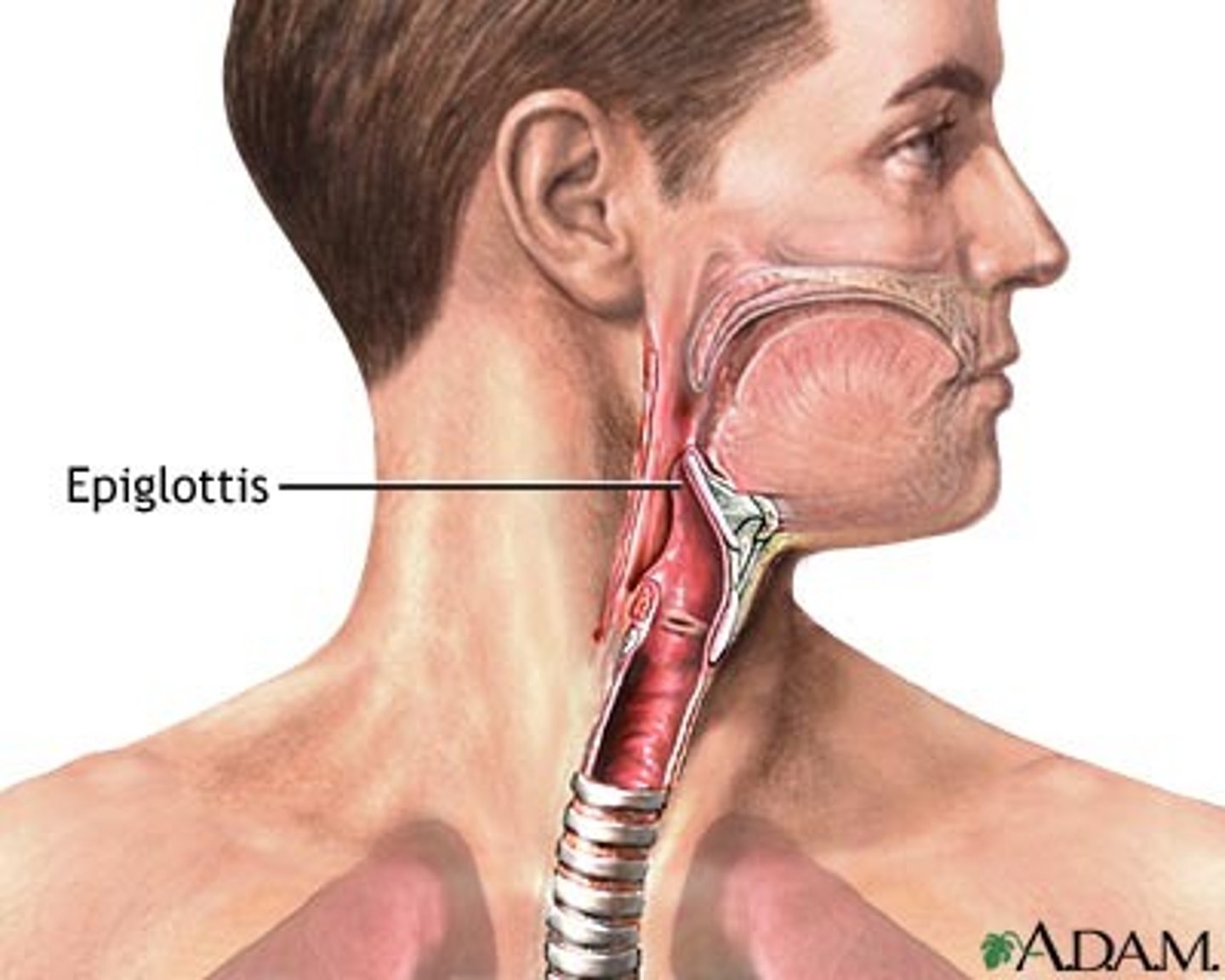
Epiglottis
A cartilaginous flap of tissue that guards opening of larynx
vestibular folds
false vocal cords - also prevent food and water from entering airway
Laryngeal Muscles - Extrinsic m
-originate outside larynx and insert on larynx
Move larynx as a whole
◦ 2 groups
● 1. Depressors
● A. Examples: infrahyoids
● 2. Elevators
● A. Examples: suprahyoids
Laryngeal Muscles - intrinsic m
-originate and insert on larynx
Move individual larynx parts
● 1. alter length & tension - of vocal cords
● 2. size & shape - rim glottidis
Walls of the Larynx - 2 folds
interior wall has 2 folds on each side
- superior and inferior vestibular folds
superior vestibular folds
false vocal cords
close the larynx off during swallowing
play no role in speech
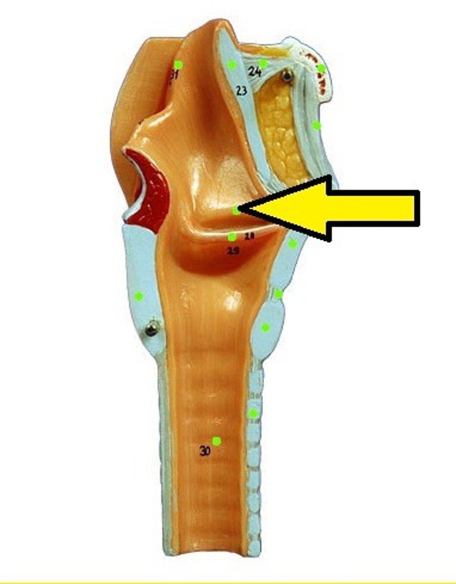
inferior vestibular folds
true vocal cords
produce the sound when air is force through/between them
contains the vocal ligaments
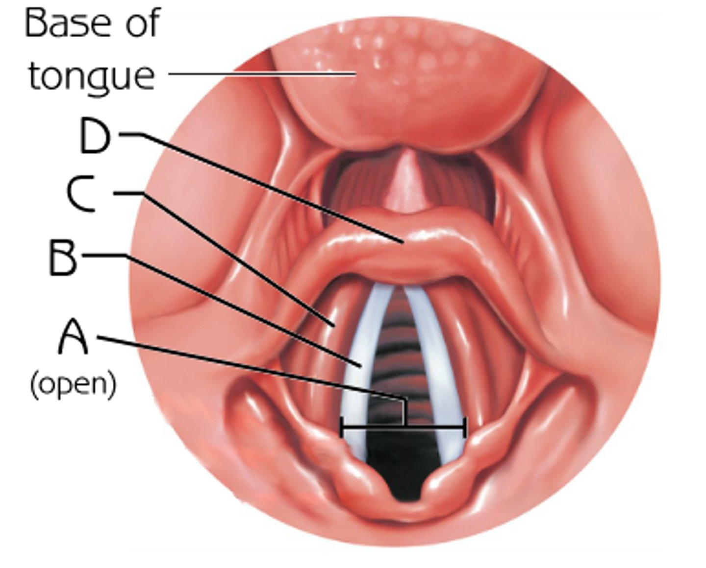
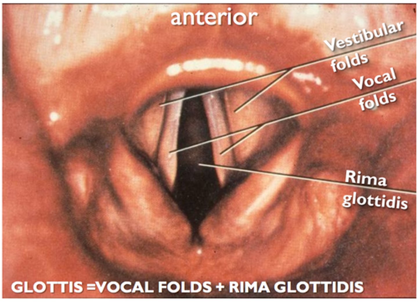
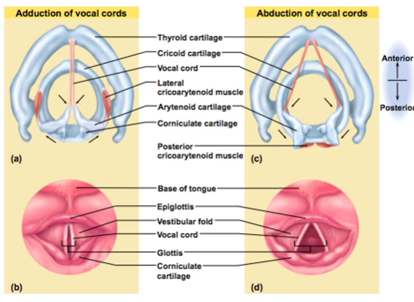
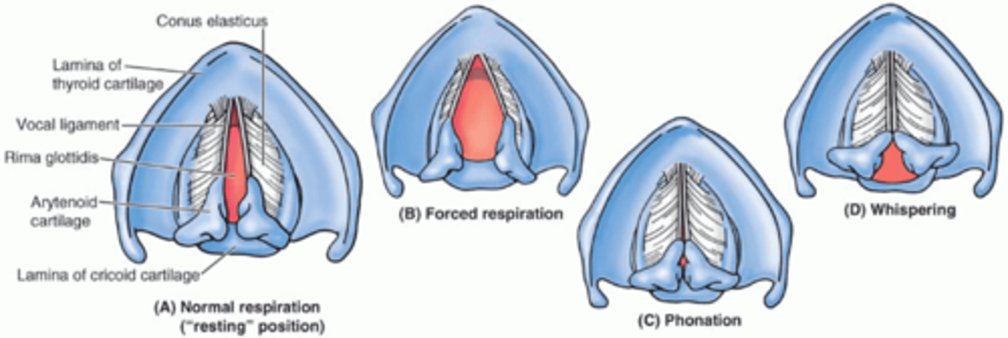
glottis
the vocal apparatus of the larynx - opening of larynx
-your vocal cords and the opening between them, the rima glottidis
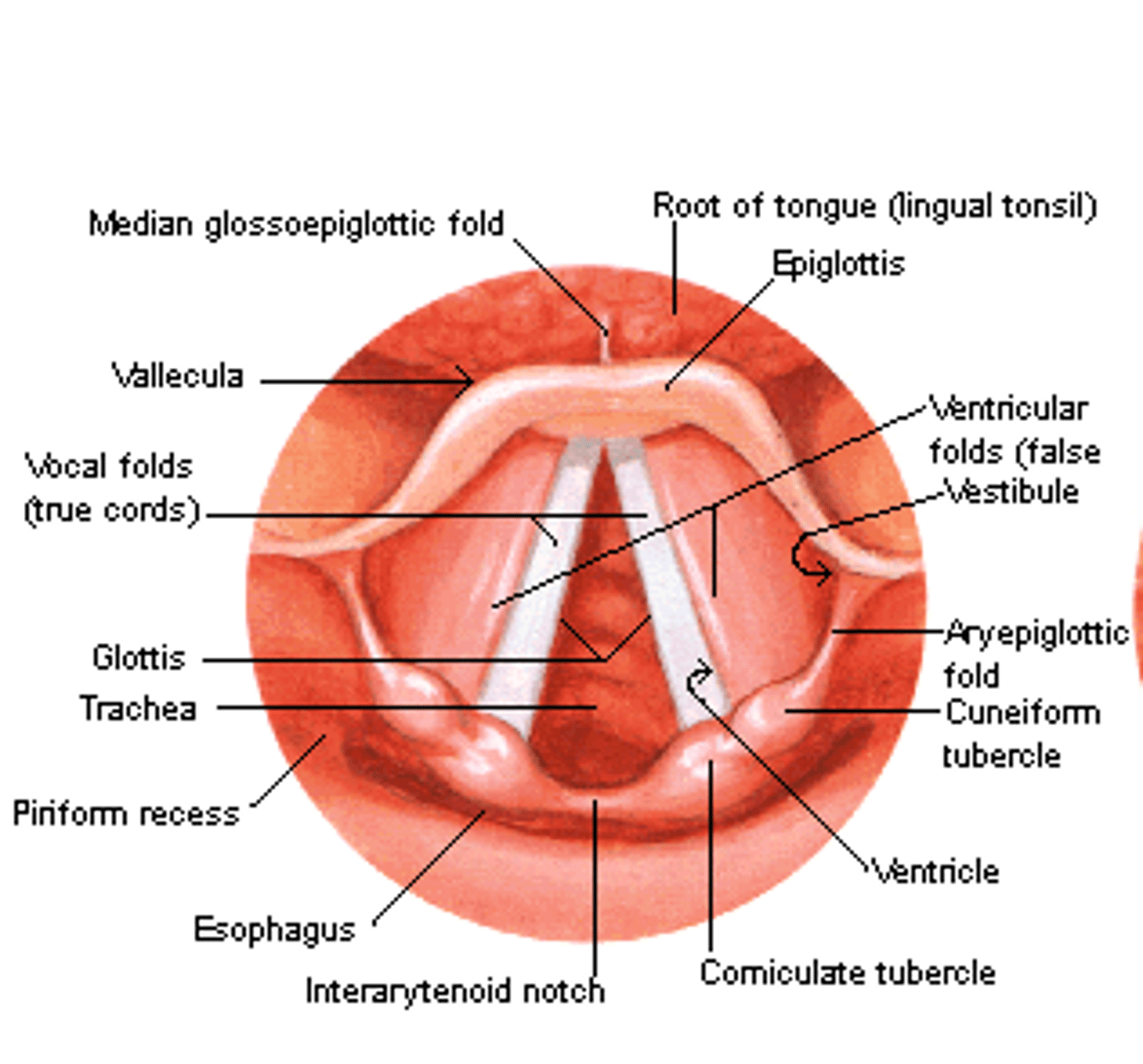
rima glottidis
aperature/space between vocal folds
opening between vocal cords
Abduction and adduction of vocal cords
Variations in Shape of Rima Glottidis
forced respiration= talking loudly
phonation=normal talking
Action of Vocal Cords - intrinsic muscles
control vocal cords
Action of Vocal Cords - innervation
recurrent laryngeal n - off of vagus n
Intrinsic muscles of vocal cords action and innervation
control vocal cords
Recurrent laryngeal nerve (branch off vagus nerve...recurrent because have to go back upwards)
Action of Vocal Cords - taut cords
high pitched sounds
Action of Vocal Cords - slack cords
lower pitched sounds
Action of Vocal Cords - adult males
- longer and thicker
- vibrate more slowly
- produce lower pitched sounds
Action of Vocal Cords - loudness
determined by how much air is forced through between the vocal cords
what forms sound? what forms words?
cords produce crude sounds that are formed into
words by the pharynx, oral cavity, tongue & lips