Introduction to Myocardial Perfusion Imaging (MPI) with Radioisotopes: Guidelines, Techniques, and Interpretation
1/48
There's no tags or description
Looks like no tags are added yet.
Name | Mastery | Learn | Test | Matching | Spaced | Call with Kai |
|---|
No analytics yet
Send a link to your students to track their progress
49 Terms
What is the primary purpose of Myocardial Perfusion Imaging (MPI)?
To show the distribution of blood flow (perfusion) in the myocardium of the left ventricle.
How is the radiopharmaceutical administered in MPI?
Intravenously.
What does tracer uptake in myocardial tissue indicate?
It is proportional to the blood supply in that region.
What are the two states assessed in MPI?
Cardiovascular rest and stress.
What condition does MPI help identify related to coronary arteries?
Areas of reduced myocardial blood flow associated with narrowed or blocked coronary arteries.
What does NICE recommend MPI for?
As a non-invasive functional test for myocardial ischemia.
What are alternative modalities to MPI mentioned by NICE?
Stress endocardiography, first-pass contrast-enhanced MR perfusion, or MR imaging for stress-induced wall motion abnormalities.
What is the significance of a narrowed coronary artery during stress?
Demand outstrips supply through the narrowed artery, leading to ischaemia.
What is the difference between ischaemia and infarction in MPI?
Ischaemia is a reversible defect with adequate flow at rest but not at stress; infarction is a fixed defect with inadequate flow at both rest and stress.
What characterizes normal myocardium in MPI?
Good perfusion at stress and rest, with normal muscle contraction.
What is hibernating myocardium?
Muscle that is alive but has very poor perfusion, insufficient to contract.
Define ionising radiation.
Waves or particles with enough energy to liberate electrons from atoms in tissue.
What is a radionuclide?
A radioactive nuclide.
What does the half-life of a radionuclide refer to?
The amount of time for the initial activity of a given radionuclide to be reduced by half.
What is a radiopharmaceutical?
A pharmaceutical labelled with a radionuclide for diagnostic or therapeutic purposes.
What is the typical duration for the radionuclide to clear from a patient?
Approximately 24 hours.
What are the most common radiopharmaceuticals used in MPI?
Tetrofosmin (Myoview©) and MIBI (Cardiolite©).
What is the main stressing technique used in nuclear medicine for MPI?
Pharmacological stress using agents like adenosine or dobutamine.
What is the effect of adenosine during pharmacological stress?
It dilates coronary arteries to increase myocardial blood flow.
What preparation is required for patients prior to MPI?
Patients must fast for at least 2 hours before all radiopharmaceutical injections.
What is the purpose of fasting before MPI?
To minimize gastrointestinal blood flow and prevent interference with the radiopharmaceutical.
What is the role of a gamma camera in MPI?
To image radioactive emissions and show the location of the radiopharmaceutical within the patient.
What is the typical duration of scans in MPI?
Approximately 15 minutes.
What does the term 'effective dose' refer to in nuclear medicine?
The energy imparted from ionising radiation in tissue, corrected for the type of radiation and type of tissue exposed.
What should patients avoid for 12 hours before an MPI test?
Caffeine, including tea, coffee, decaffeinated tea and coffee, cola, and all forms of chocolate.
What type of medications should be avoided prior to an MPI test?
Heart rate-limiting drugs, such as beta blockers, dipyridamole, calcium channel blockers, and theophyllines.
What is the purpose of a gamma camera in MPI?
To perform acquisitions while the patient is scanned, ideally with arms above the head to minimize image attenuation.
How long does each stress/rest scan take in MPI?
About 15 minutes.
What happens if there is a large collection of radiopharmaceutical near the heart during an MPI scan?
Scans are repeated to avoid blurring into the cardiac uptake in the reconstructed image.
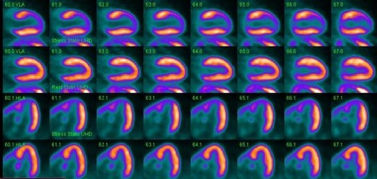
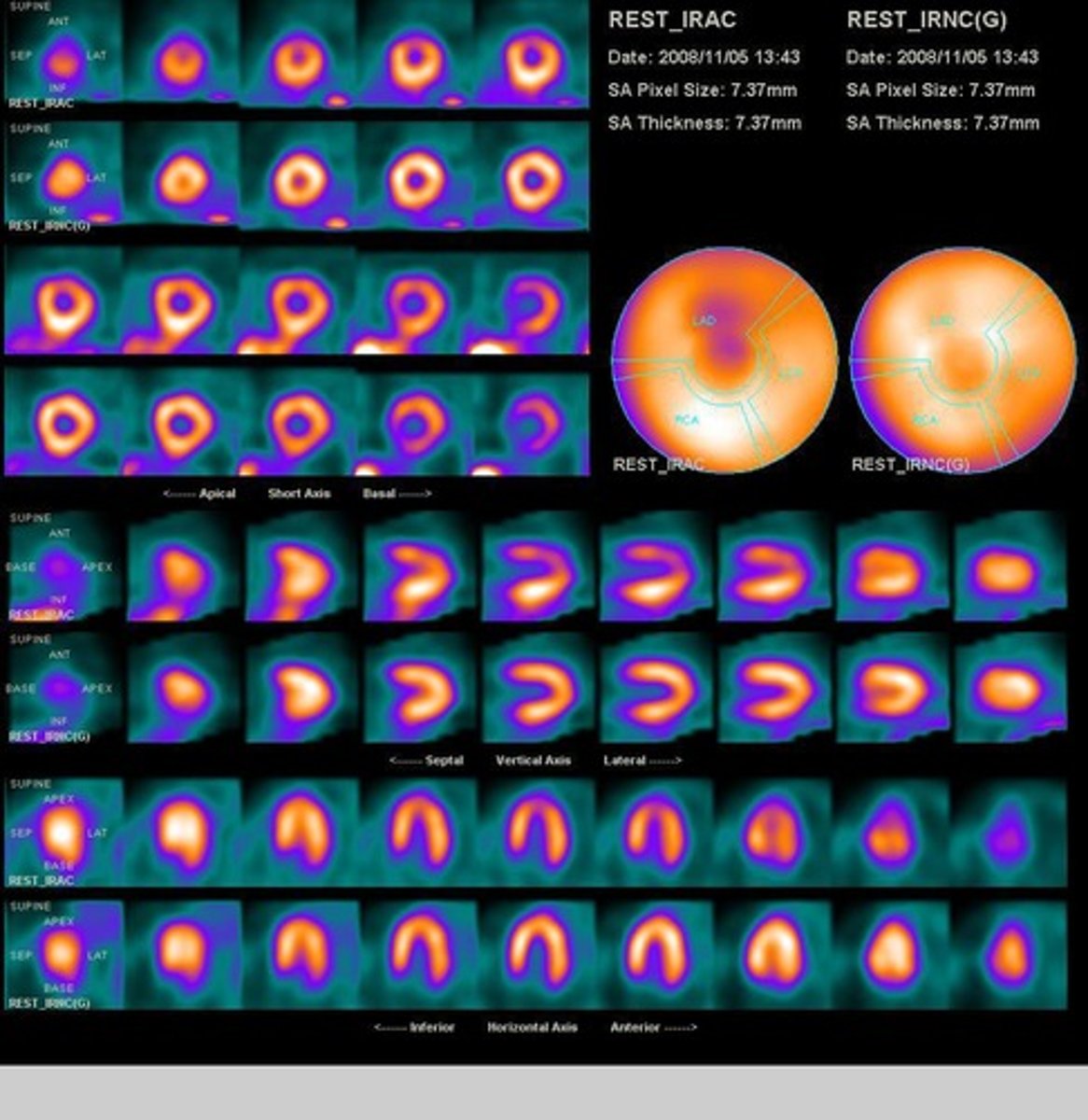
What imaging technique allows for a 3D image of left ventricular (LV) perfusion?
SPECT imaging.
What does a bulls-eye plot represent in MPI?
It displays 3D perfusion information in a 2D format.
What does MPI ECG-Gating provide?
Additional functional information by synchronizing SPECT acquisition to the patient's ECG.
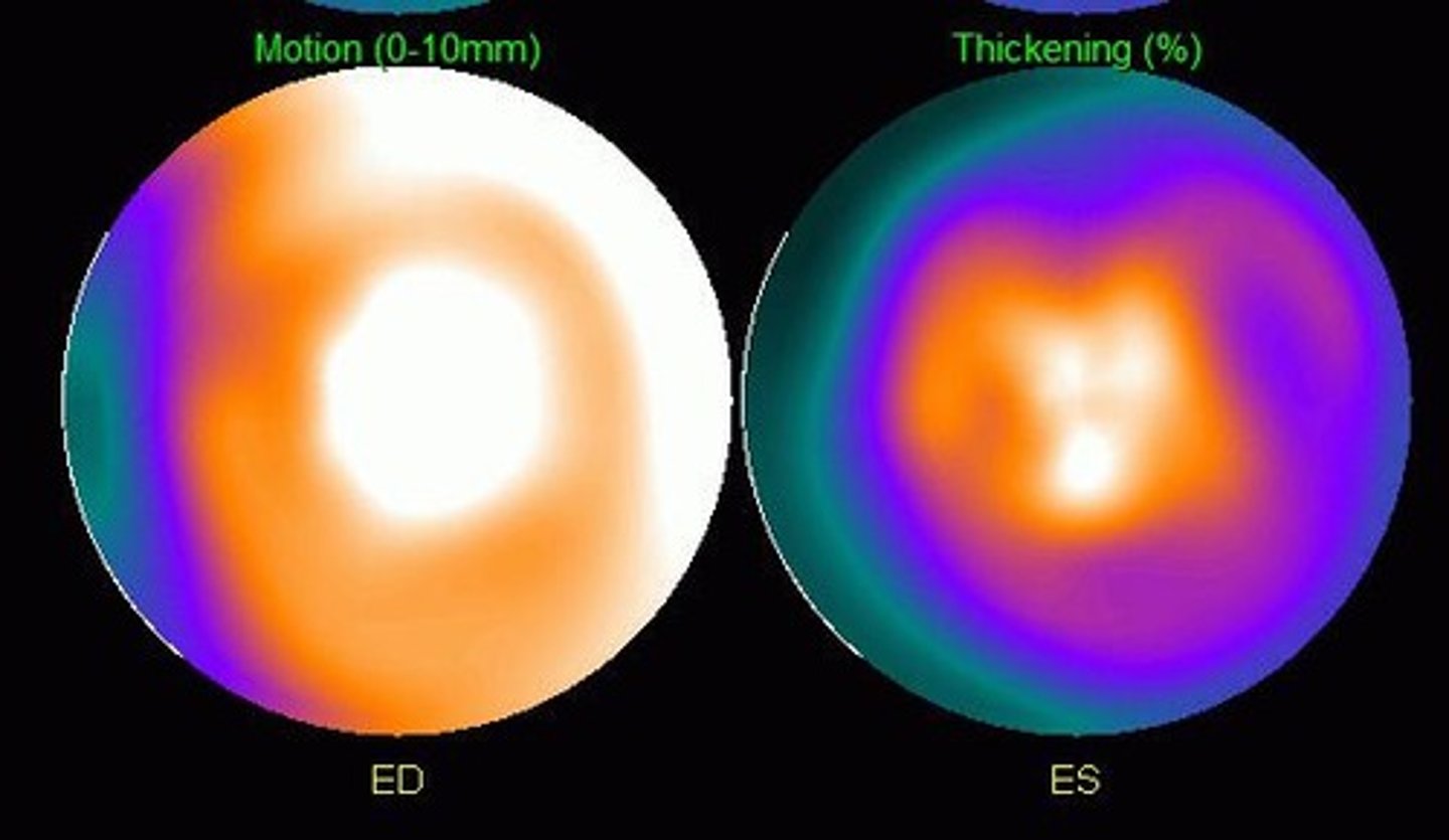
What can be determined from volume curves in MPI ECG-Gating?
End diastolic volume (EDV) and end systolic volume (ESV) to calculate ejection fraction (EF).
What is the formula for calculating ejection fraction (EF)?
EF = (EDV - ESV) / EDV × 100%.
What is a common artifact in MPI due to gamma ray attenuation?
Breast attenuation affecting the anterior and lateral wall images.
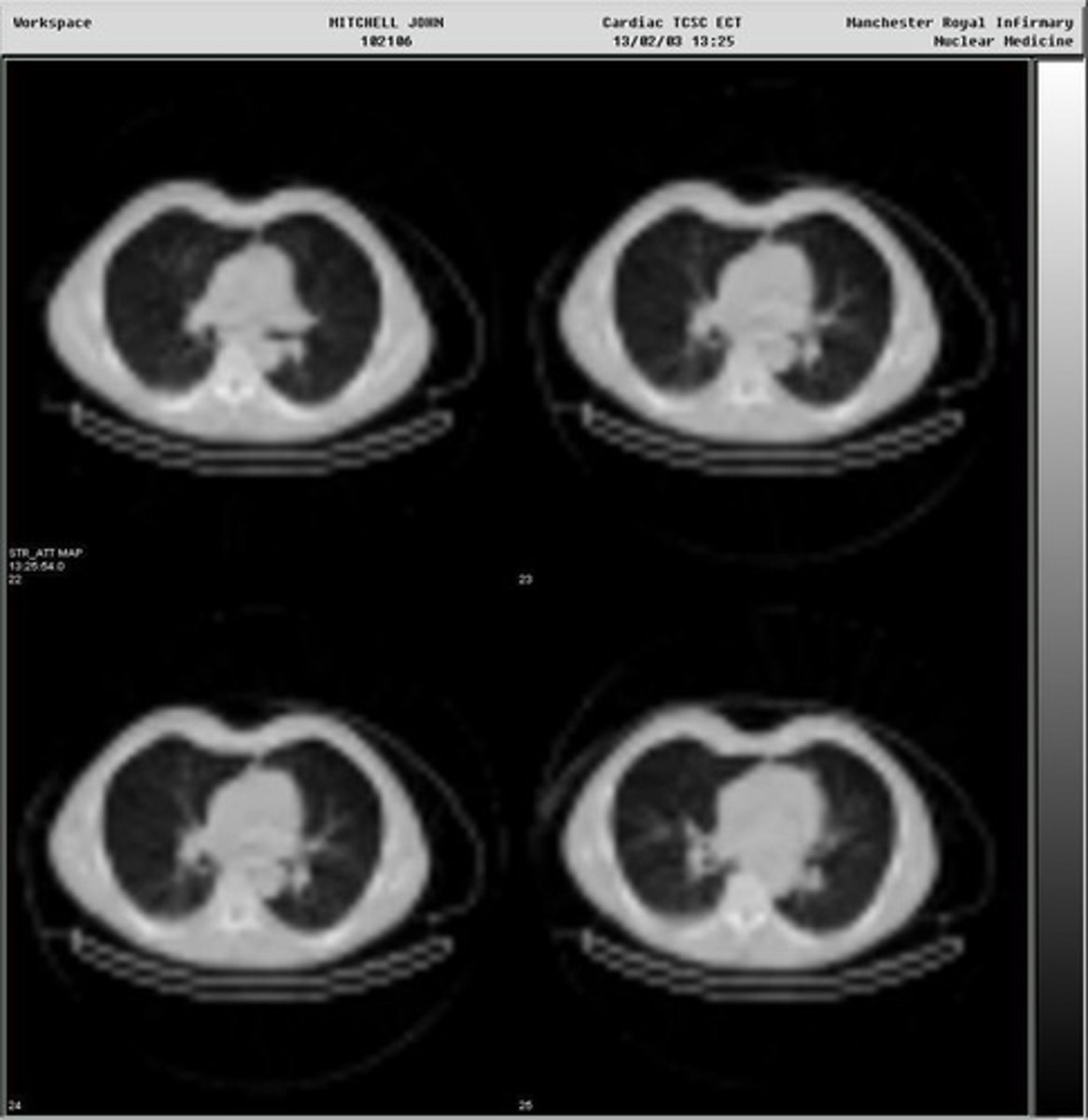
What is required for non-uniform attenuation correction in MPI?
A patient-specific attenuation map, often obtained from a low dose CT scan.
What can cause mis-registration between SPECT and CT data?
System registration errors or patient movement between scans.
What does Regulation 11 of IRMER state regarding exposure justification?
Exposures must be justified by the practitioner as showing a sufficient net benefit.
What does Regulation 12 of IRMER emphasize about radiation doses?
Doses must be kept as low as reasonably practicable consistent with the intended purpose (ALARP).
What is the significance of the annual radiation dose from UK natural background radiation?
It is approximately 2 mSv, providing context for radiation doses in medical imaging.
What is the outcome of myocardial infarction in terms of perfusion?
Absent perfusion at the apex and severely reduced perfusion in the lateral and inferior walls.
What indicates viable myocardium in MPI?
Adequate blood flow at rest but insufficient at stress, which can improve with revascularization.
What does reduced wall motion indicate in MPI?
It may indicate an infarct.
What is the typical resolution of SPECT imaging?
Approximately 15 mm.
What is the significance of wall thickness in cardiac contraction?
Wall thickness increases as the heart contracts, typically around 10 mm.
What is the effect of patient movement during an MPI scan?
It causes blurring in the image, which can be minimized with straps and arm rests.
What is the purpose of acquiring a low-resolution CT after an MPI scan?
To create a noise-free attenuation correction map for SPECT reconstruction.

What does the term 'attenuation' refer to in the context of MPI?
The reduction of gamma rays as they pass through the patient's body, which can create image artifacts.
What is the role of the gamma camera's positioning during MPI?
The closer the detectors are to the heart, the better the image quality.