StemUp: AQA A level Biology 3.3.4.1 Mass transport in Animals
1/58
There's no tags or description
Looks like no tags are added yet.
Name | Mastery | Learn | Test | Matching | Spaced | Call with Kai |
|---|
No analytics yet
Send a link to your students to track their progress
59 Terms
What are haemoglobins? (1)
- A group of chemically similar proteins
- Which are found in the red blood cell of many different organisms
What type of structure does haemoglobin have? (2)
- Quaternary structure
- Because it contains 4 polypeptide chains joined together
Describe the role of haemoglobin and red blood cells in the transport of oxygen (4)
1. Oxygen diffuses through the epithelium of the alveolus and the endothelium of the capillary during gas exchange
2. The oxygen enters the red blood cells and combines with haemoglobin to form oxyhaemoglobin
3. Oxygen is carried away by the blood, which maintains a concentration gradient
4. In the body tissues, oxyhaemoglobin releases oxygen which diffuses into the respiring cells
What happens to oxygen once it enters the red blood cells? (1)
Forms oxyhaemoglobin
How is the concentration gradient for oxygen maintained in the blood? (2)
- Oxygen is carried away from the lungs
- By the blood
What happens to oxyhaemoglobin in the body tissues? (3)
- Oxyhaemoglobin releases oxygen
- Which then diffuses into the respiring cells
- Oxygen is used up by the respiring cells
Draw and label a typical oxyhaemoglobin dissociation curve (4)
- Loading tension
- Unloading tension
- Partial pressure of oxygen found in tissues
- Partial pressure of oxygen found in lungs
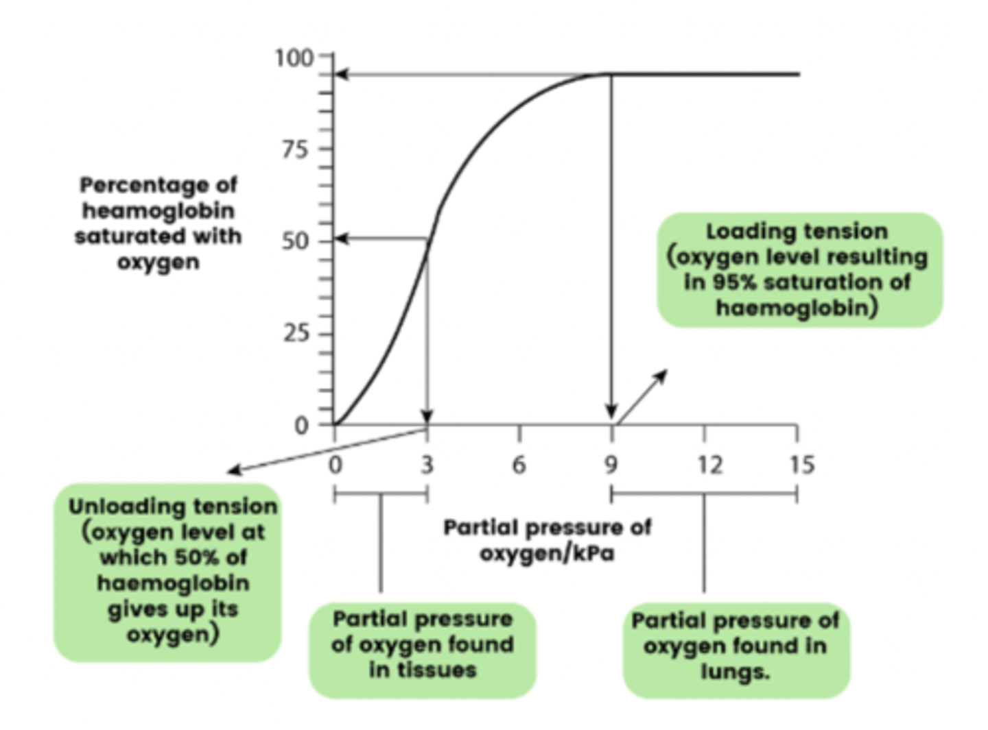
Describe, using the oxyhaemoglobin dissociation curve, what is happening in the lungs (3)
- Haemoglobin has a higher affinity for oxygen in the lungs
- It quickly binds to oxygen molecules
- Due to the high partial pressures of oxygen
Describe, using the oxyhaemoglobin dissociation curve, what is happening in the tissues (3)
- Haemoglobin has a lower affinity for oxygen in the tissues
- It quickly unloads some of its oxygen to the rapidly respiring cells
- Due to the low partial pressures of oxygen
What gives oxyhaemoglobin dissociation curve its unique shape? (2)
- Sigmoid shape
- Due to the cooperative nature of oxygen binding
Describe the the cooperative nature of oxygen binding (4)
- The binding of the first oxygen molecule
- Changes the tertiary / quaternary structure of the haemoglobin
- This uncovers the binding site of the next haem unit
- Allowing the next oxygen molecule to bind
Draw how the Bohr Effect alters the oxyhaemoglobin dissociation curve (2)
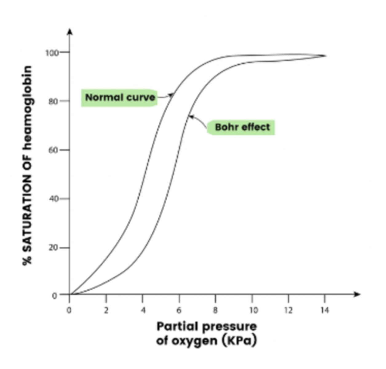
Describe the bohr effect's alteration on the oxyhaemoglobin dissociation curve (2)
- An increase in CO2 levels (e.g. during exercise) depresses the dissociation curve
- This moves the curve to the right
- Haemoglobin releases more oxygen to the respiring tissues
Explain the bohr effect's alteration on the oxyhaemoglobin dissociation curve (3)
- Occurs due to a decrease in pH
- As carbon dioxide will dissolve in the plasma
- To form carbonic acid (H₂CO₃)
What kind of advantage is the Bohr effect? (1)
Physiological advantage (e.g. for athletes)
What does it mean if the oxygen dissociation curve for an organism's haemoglobin shifts to the left? (2)
- The organism / their haemoglobin tends to store oxygen and respire less
- This is because haemoglobin will have a higher affinity for oxygen at lower partial pressures of oxygen present in its surroundings
What are two examples of a haemglobin where its oxygen dissociation curve shifts to the left? (2)
- Myoglobin
- Fetal haemglobin
NOTE: On the graph, HbF is fetal haemglobin and Mb is myoglobin
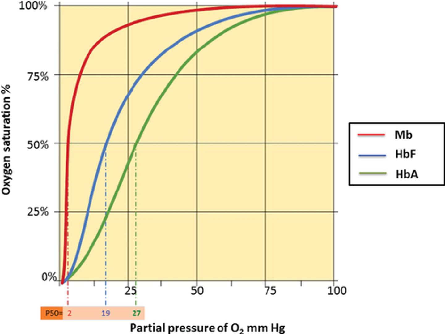
What does it mean if the oxygen dissociation curve for an organism's haemoglobin shifts to the right? (2)
- Organism tends to be more active and respire more
- This is because haemoglobin will have a lower affinity for oxygen at low partial pressures of oxygen
- Allowing more oxygen to be released
Draw and label a diagram to show the double circulation of blood in a human body (6)
NOTE: Unfortunately, you will need to learn most of the labels on this diagram
Excluding:
- Hepatic artery
- Hepatic vein
- Hepatic portal vein
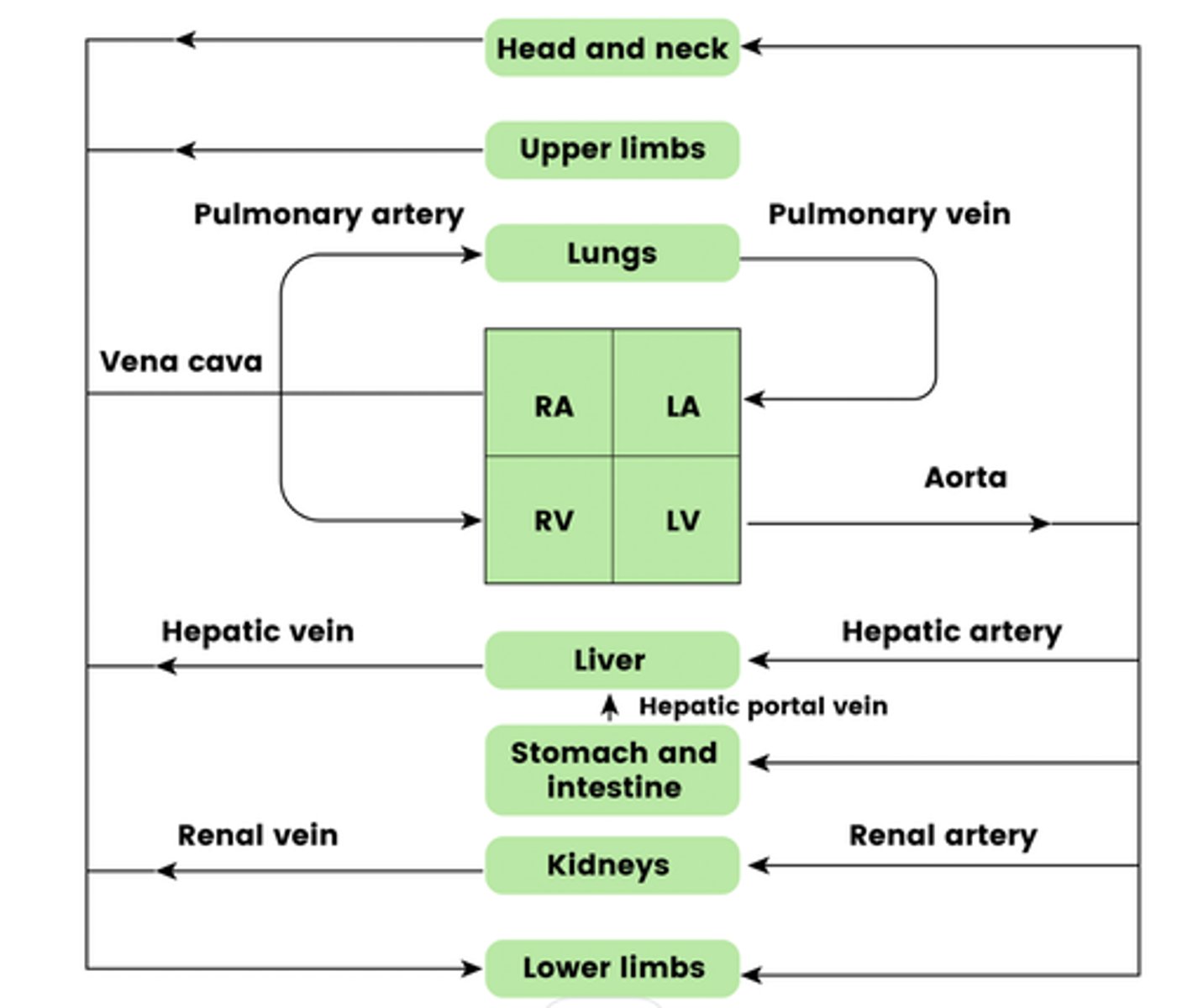
What blood vessel carries blood to the heart muscles? (1)
Coronary arteries
Draw and label a diagram of the structure of the human heart (11)
Chambers:
- Left atrium
- Left ventricle
- Right atrium
- Right ventricle
Blood vessels:
- Pulmonary arteries
- Vena cava
- Aorta
- Pulmonary vein
Valves:
- Semi lunar valve
- Atrioventricular valve
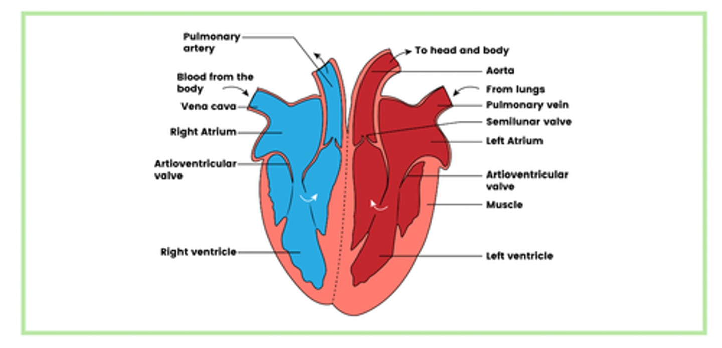
What are the 4 chambers of the heart? (4)
- Left atrium
- Left ventricle
- Right atrium
- Right ventricle
What is the function of the vena cavae? (3)
- Carries deoxygenated blood
- From the body (except the lungs)
- To the right atrium
What is the function of the pulmonary artery? (3)
- Carries deoxygenated blood
- From the right ventricle
- To the lungs
What is the function of the pulmonary veins? (3)
- Carries oxygenated blood
- From the lungs
- To the left atrium
What is the function of the aorta? (3)
- Carries oxygenated blood
- From the left ventricle
- To the rest of the body.
Why is the muscular wall of the left ventricle thicker than the right ventricle? (3)
- When the heart contracts
- It produces a greater pressure
- As it needs to pump blood to all parts of the body
What is the cardiac cycle? (1)
The sequence of events that occur during one heart beat
Draw and label a graph of the cardiac cycle (10)
All parts of the diagram are key to understand and remember

What happens at around 0 to 0.1s on our cardiac cycle diagram? (3)
- Left atrium contracts
- Blood flows into the left ventricle
- AV valve is open

What happens at around 0.1s on our cardiac cycle diagram? (4)
- Left ventricle contracts
- Volume in the left ventricle decreases
- Pressure inside the ventricle increases above that in the atrium
- This closes the AV valve

What happens at around 0.15s on our cardiac cycle diagram? (3)
- Pressure in left ventricle increases above that in the aorta
- Which opens the semi-lunar valve
- Blood flows into the aorta

What happens at around 0.4s on our cardiac cycle diagram? (3)
- Left ventricle relaxes
- Pressure in the aorta increases above that in the ventricle
- Semi-lunar valve closes
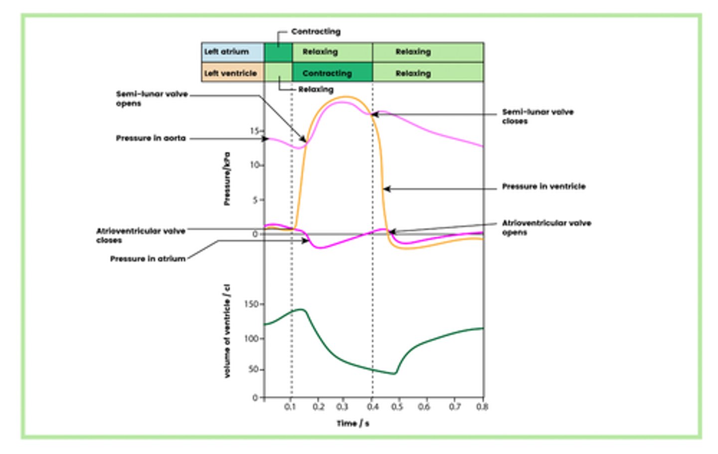
What happens at around 0.45s on our cardiac cycle diagram? (3)
- Pressure inside the ventricles falls below that in the atrium
- Atrioventricular valve opens
- Which allows blood to flow into the ventricles

How do you determine heart rate using the cardiac cycle? (1)
Take how it takes for the cycle to repeat and divide it by 60
E.g. for the diagram above, its 0.8s

Draw the cardiac output equation triangle
Where:
CO = cardiac output
SV = stroke volume
HR = heart rate
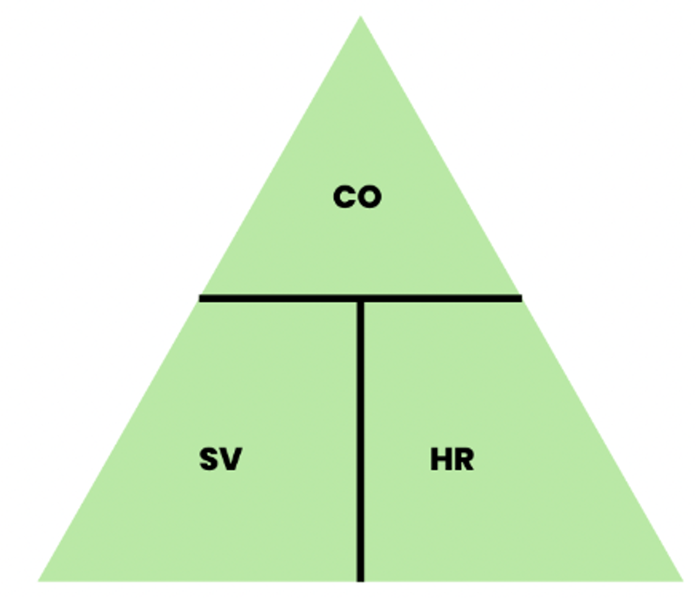
What is meant by cardiac output? (1)
The volume of blood pumped out of one ventricle per minute
What is the unit for cardiac output? (1)
cm³ min⁻¹
What is meant by stroke volume? (1)
The volume of blood expelled from the ventricle per contraction
What is the unit for stroke volume? (1)
cm³
What is meant by heart rate? (1)
The number of contractions per minute
What is the unit for heart rate? (1)
min⁻¹
What are the 4 main types of blood vessels in our body? (4)
- Arteries
- Arterioles
- Veins
- Capillaries
Draw a diagram that links the main types of blood vessels in our body
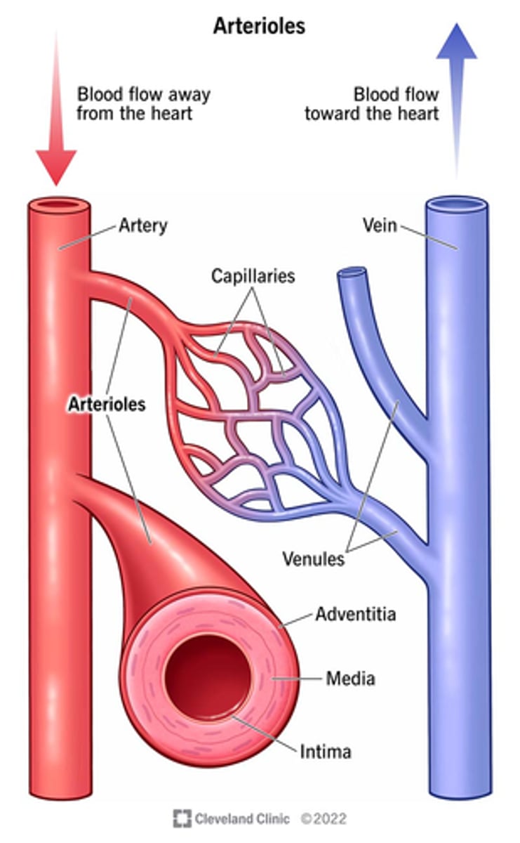
What is the function of the arteries? (1)
- To carry blood away from the heart
- At a high pressure
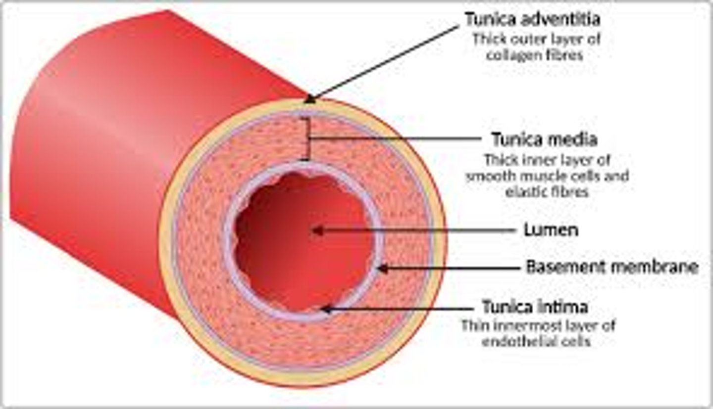
What are the features of the arteries? (3)
- Thicker wall and smaller lumen then veins
- Contain more elastic fibres and smooth muscle fibres than veins
- Transports blood at a higher pressure than the veins
NOTE: You do not need to know any of the features of the arteries, its just there as a visual guide
What is the function of the arterioles? (2)
- Controls the flow of blood to different tissues or organs
- By contraction or relaxation of the smooth muscle in their wall
What are the features of the arterioles? (5)
- Possess a higher proportion of smooth muscle than elastic fibres
- Contraction of the smooth muscle causes narrowing (vasoconstriction) of the arteriole
- Which reduces blood flow to the capillaries
- Relaxation of the smooth muscle causes widening (vasodilation) of the arteriole
- Which increases blood flow to the capillaries
What is the function of the veins? (2)
- Carries blood towards the heart
- Under low pressure
What are the features of the veins? (5)
- The lumen is larger than in arteries
- So that even at low pressure, blood flows back to the heart at the same rate that it leaves along the arteries
- Contracting muscles in the legs and body press on the veins and squeeze the blood along
- Veins have semi-lunar valves at intervals
What is the function of the semi-lunar valves at intervals in the veins? (2)
- Prevents back-flow of blood
- Ensuring it travels in one direction towards the heart
What is the function of the capillaries? (2)
Where the exchange of substances with blood and tissue occurs
What are the features of the capillaries? (6)
- The walls of the capillaries are one endothelial cell thick
- Which gives a very short diffusion pathway for the exchange of substances with the tissues
- There are gaps between the endothelial cells
- Which increase the permeability of the capillary
- There are very large numbers of capillaries and they are highly branched
- Giving a large surface area for exchange with the tissues
How thick are the walls of the capillaries? (1)
One endothelial cell thick
Why is the thickness of the capillary walls important? (1)
Gives a very short diffusion pathway for the exchange of substances with the tissues
What feature of the capillaries increases their permeability? (1)
Gaps between the endothelial cells
How do the capillaries provide a large surface area for exchange of substances? (2)
- Large numbers of them
- Highly branched
Describe how tissue fluid is formed (4)
1. Contraction of the ventricles produces a high hydrostatic pressure
2. This forces water and some dissolved substances out of the blood capillaries to the surrounding tissues
3. Filtered plasma forms tissue fluid which surrounds the body cells
4. Oxygen, glucose, minerals etc diffuse into the body cells
5. Carbon dioxide, urea and other metabolic waste diffuse out
Describe how filtered plasma is reabsorbed into the blood (4)
1. Plasma proteins will remain in the blood (won't enter the tissues)
2. This reduces the water potential of the blood
3. Water moves back into the blood by osmosis
4. The rest of the filtered plasma is returned to blood by the lymphatic system