B3.2 - transport systems
1/27
There's no tags or description
Looks like no tags are added yet.
Name | Mastery | Learn | Test | Matching | Spaced | Call with Kai |
|---|
No analytics yet
Send a link to your students to track their progress
28 Terms
Adaptions of capillary for exchange of materials
Pores (fenestrations) to increase permeability for rapid exchange
Large surface area due to branching and narrow diameters
Tissues w/ high oxygen/nutrient requirements (muscles) have high density capillary networks
Single layer of flattened endothelial cells to reduce diffusion distance
Tissue fluid (fluid coming out of capillaries into tissues → water, oxygen, glucose, ions)
Capillaries near arterioles release tissue fluid more readily, capillaries near veins uptake more readily (differences in external vessel pressures)
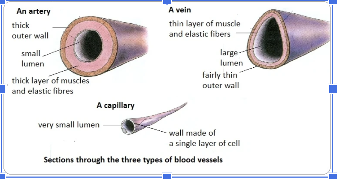
Structure of artery
Thick, muscular wall
Narrow lumen (hollow inside the artery)
Circular in shape → maintain shape well
‘Ridges’ on inside → ‘inner surface corrugation’
Visible wall fibers
structure of veins
Thin wall (rely on skeletal muscle surrounding veins to contract blood back to the heart)
Wide lumen
Somewhat flattened
No inner surface corrugation → little muscle
No wall fibers
easy way to remember arteries, veins, capillary comparisons
transport systems and their directions
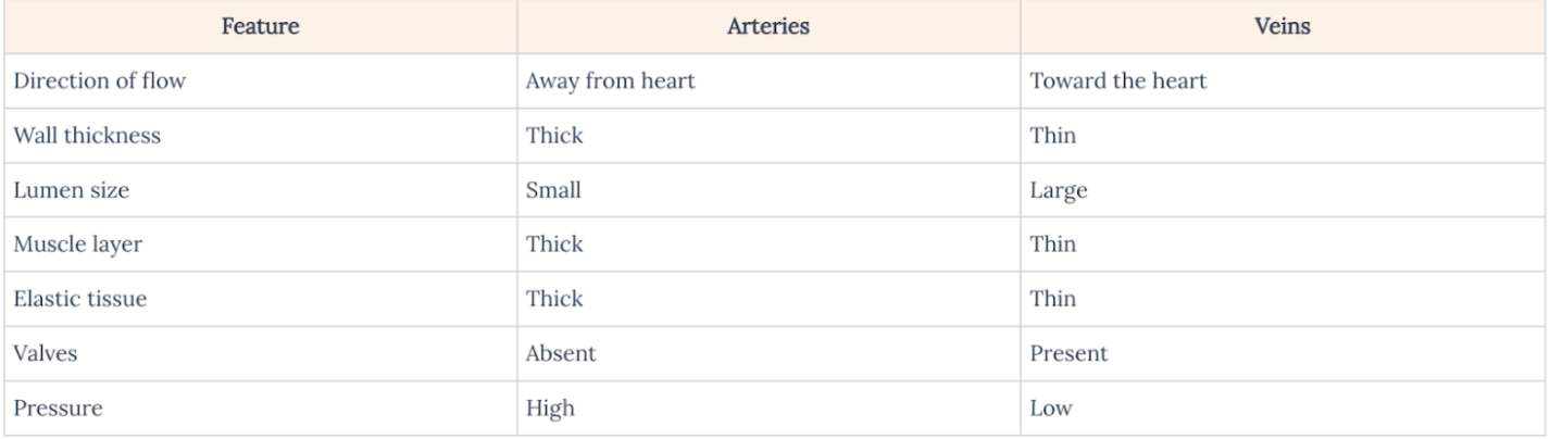
Adaptations of Arteries for Transport of Blood From Heart
Narrow lumen → small area maintains high pressure to pump blood
Thick muscular wall (with collagen/elastic fibres)
Contracts to narrow the lumen (vasoconstriction)
Relaxes to widen the lumen (vasodilation)
Collagen → maintain elasticity, make sure arteries can withstand high pressure
Elastic fibers → Acts like a pressure reservoir, helping to even out blood flow
Stretches during systole (when heart pumps)
Recoils during diastole (heart relaxation) → less energy for a full contraction
Pulses
Can be taken on wrist or neck/with digital pulse reader
Not the same as heartbeat (heart muscle contractions) but the arteries contracting → same timing, can be used interchangeably
Adaptations of Veins for Return of Blood to Heart
Thin flexible walls → allows external skeletal muscle to contract/squeeze vein so blood can be pumped back to the heart
Valves → one-way flaps, close to prevent backflow
Wide lumen → creates low pressure → easier for skeletal muscles to squeeze
Coronary Heart Diseases (occlusion of coronary arteries)
Coronary Arteries: arteries branching out from the aorta that supply oxygenated blood to the heart tissue
Coronary Heart Disease (CHD): coronary arteries become narrowed/blocked with plaque (cholesterol/other lipids)
Myocardial infarction (heart attack): coronary artery is totally blocked
Risk factors: Smoking, obesity, lack of exercise/genetics, Hypertension, poor diet, age
Transporting Water from Roots to Leaves during Transpiration (entire process)
Xylem: carries water from roots to shoots (1 upward direction)
relies on cohesion and adhesion
Transpiration: water evaporates through stomata → causes tension pulling water up from the roots → water drawn out pores of xylem vessels and through cell walls
Capillary action: water moving through/along a material against the force of gravity
COHESION: creates a transpirational pull upwards as water evaporates through stomata (no energy used) → ensures a continuous column of water
Adaptations of Xylem Vessels for Water Transport
Made of dead, hollow cells → maintains continuous water column + water contact (i.e. allows cohesion to exist)
Lack of cell contents
Lignin (polysaccharide) → structural support i.e. prevents xylem collapse and withstands tensions
Pits in the xylem → water can pass between xylem and phloem
Absence of end walls → for unimpeded flow
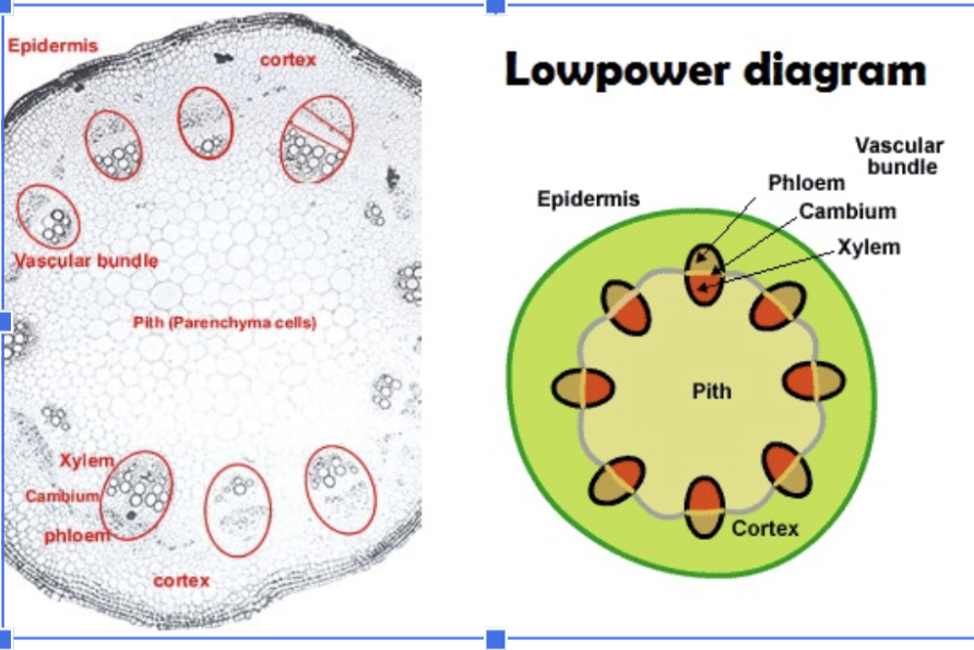
Features of Dicot Stems and their locations
Vascular bundles: bundles of xylem + phloem → region closer to the inside is the xylem, phloem is closer to the outside
Pith: core of the stem → ‘bulks out’ the stem
Cortex: layer under the epidermis → storage/structural support/photosynthesis
Epidermis: outer layer of stem → waterproofing/protection
Features of dicot roots and their functions
Phloem in between the xylem star
Endodermis → inner skin of cells that water passes through to access the xylem
Cortex → unspecialized cells ‘bulk out’ root → strength/higher surface area
Epidermis → absorbs water/minerals from soil using root hairs (long, narrow outgrowths)
Pith: core of the stem → ‘bulks out’ the root
Release and reuptake of tissue fluid in capillaries —> their structure that allows them
Plasma: fluid consisting of substances (e.g., glucose, amino acids, mineral ions, vitamins, hormones, and plasma proteins) dissolved in water
Capillary walls are adapted to allow plasma to leak out into spaces between tissue cells
Plasma proteins (too large) → remain in the blood / small molecules pass out of blood and form tissue fluid
Tissue fluid is formed by pressure filtration of plasma in capillaries close to arterioles
promoted by the higher pressure of blood from arterioles → allows release and reuptake of tissue fluid
Lower pressure in venules allows tissue fluid to drain back into capillaries
Exchanging Substances Between Tissue Fluid and Tissue Cells
Oxygen passively diffuses into cells (concentration gradient)
Glucose moves into cells using sodium-glucose cotransporters (against the concentration gradient) → secondary active transport
Energy from sodium / glucose (PASSIVE)
CO2 + waste moves into capillaries passively (concentration gradient)
Drainage system of excess tissue into lymph ducts
Not all (~85%) of tissue fluid returns to capillaries
Remaining 15% returns into the lymphatic system → prevents oedema (swelling)
Drains back into heart/blood/circulatory system
All tissues have narrow, blind-ended lymphatic vessels with permeable walls → merge repeatedly into left and right lymph ducts
presence of valves and thin walls with gaps in lymph ducts
Tissue fluid enters lymphatic vessels → lymph
Lymph drains into subclavian veins → vena cava → right side of the heart
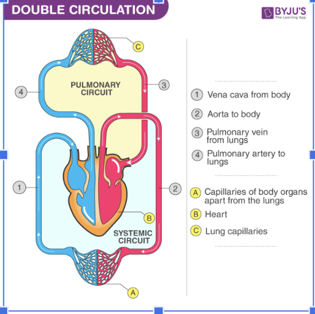
Double Circulation of Mammals
Double circulation (pulmonary and systemic circulation)
High-pressure body blood/systemic loop (to reach all organs)
Require separate low-pressure pulmonary blood loop to the lungs → allows oxygen to move from alveoli into capillaries & prevents capillaries from bursting
Single circulation of bony fish
Single circulation
Blood is pumped from the heart → gills → body → heart (enough remaining pressure to pump oxygenated blood to body and heart)
water pumped over gill filaments
oxygen diffuses from water → gills → blood, CO2 diffuses from blood → gills → water
Water flow reduces risk of capillaries bursting → high-pressure blood can exist in gills
What do the Atria, ventricules, atrioventricular, and semilunar valves do?
Atria → contract simultaneously to squeeze blood into ventricles
Ventricles → contract simultaneously to pump blood into arteries
Thicker ventricular walls compared to atria (left is thicker than right → pumps blood at higher pressure for systemic circulation)
Atrioventricular (AV valves) → prevent backflow into the atria
Semilunar (pulmonary + aortic) valves → prevent backflow into the ventricles
other mamallian heart adaptions (besides atria, ventricular, atrioventricular, and semilunar valves)
Cardiac muscle → muscles forming walls of atria and ventricles → coordinated myogenic contractions (electrical signals propagated throughout wall of heart)
Septum → wall separating right/left ventricles AND right/left veins → prevents oxygenated (LEFT) and deoxygenated (RIGHT) blood from mixing
Coronary Vessels (coronary arteries and veins in wall of heart)→ bring oxygenated blood to heart tissue and drain deoxygenated blood away
SA (sinoatrial ‘pacemaker’) node (wall of right atrium)→ initiates heartbeat
Interval between SA node signals determine heartbeat rate
Generates electrical impulse → atrial contraction
AV node (lower back section of right atrium) → gets electric signal to ventricles (coordinates heartbeats)
Receives signal from SA node → delays impulse → atria finish contracting → ventricular contraction
Know structure of heart
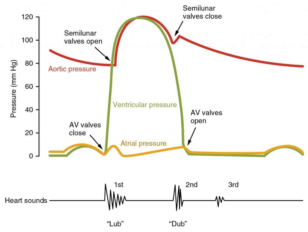
Stages in the Cardiac Cycle (Atrial Systole)
Atrium contracts → rapid but small increase in pressure
Ventricles relax → AV valves open, semilunar valves close
Ventricles filled with blood → small increase in pressure
Blood pressure in artery decreases to its minimum
Stages in the Cardiac Cycle (Ventricular systole)
AV valves close (first heart sound “lub”), semilunar valves open
Blood pushed into arteries
pressure in ventricles > arteries
Atria slowly relaxes, filled with blood → pressure increases
Ventricular diastole
Ventricle relaxes → pressure < arteries (rapidly decreases)
Semilunar valves close (second heart sound “dub”), AV valves remain closed
AV valves open
Ventricle pressure < atrium
blood enters ventricle → slow increase in pressure
Know graph for cardiac cycle
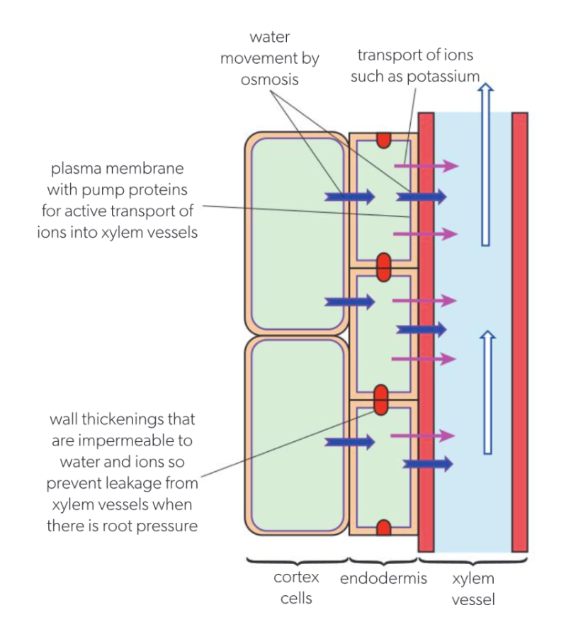
Root pressure in xylem by active transport of mineral ions
Water only moves upwards when transpiration (pulling force initiated by stomata evaporation) occurs
Sometimes transpiration is not possible e.g., high humidity or spring bfr deciduous plant leaves open (closed stomata at night, loss of leaves)
If pulling force is unable, plants rely on root pushing force → positive pressure potential in xylem
Plants establish pressure in rootsbyactively transporting mineral ions into root → triggers osmosis (high solute conc. in xylem) → pressure increase
Phloem Sieve Tubes + Companion Cells for Translocation of Sap (definitions)
Sap: fluid containing water and nutrients, transported within vascular tissues
Phloem: bidirectional transport of carbon compounds (sucrose) from source to sink
Source: where carbon compounds are made (leaf)
Sink: where carbon compounds are stored/used (root, shoot, flower, fruit)
Sieve plates: channels through which transport occurs → have pores for phloem sap to flow through
Reduced cytoplasm and organelles, no nucleus for sieve tube elements → cant generate energy → relies on companion cells for active transport
Sieve tube elements: long, elongated cells forming continuous tubes for nutrient transport (leaves to rest of plant)
Companion Cells: help transport carbon compounds from sources to phloem cells, from phloem cells into sinks
Many mitochondria + cytoplasmic plasmodesmata which allows ATP/carbon compounds to pass through
Process of translocation of sap
Phloem Loading: carbon compounds actively transported into the phloem sieve tubes
Water moves via osmosis from the xylem to the phloem → creates area of high pressure
Pressurized Movement: Carbon compounds, water (phloem sap) are forced from areas of high pressure to low pressure
Sink Pumping: compounds pumped from phloem into sink
Without high solute concentration, water moves back into xylem through osmosis
*these adaptations ease the flow of sap