Module 6 Urinary System Lab Practice
1/106
Earn XP
Description and Tags
NASDFNiuaweugawehgpqwieoiqwoepoqweqwopeiqwpoeiqwpoeipwoqeiqwpeioqweiq
Name | Mastery | Learn | Test | Matching | Spaced | Call with Kai |
|---|
No analytics yet
Send a link to your students to track their progress
107 Terms
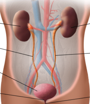
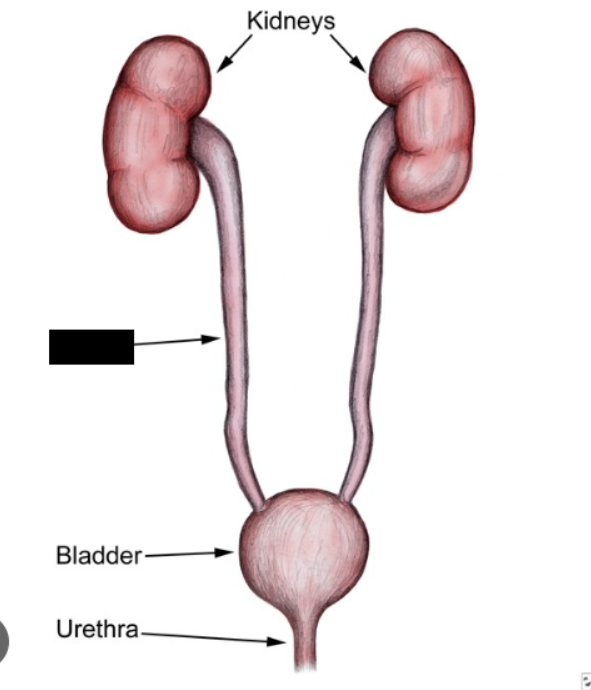
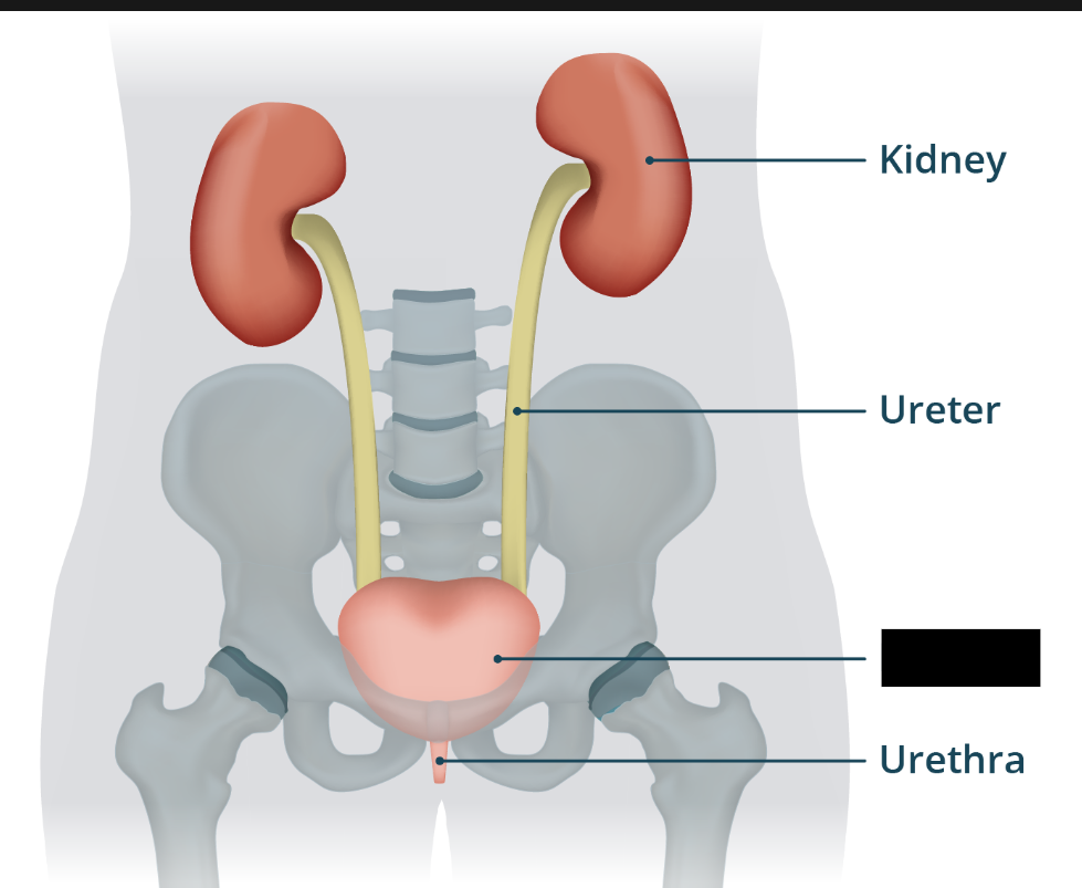
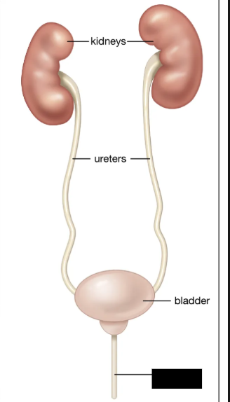
kidneys ureters urinary bladder urethra
The urinary structures include the ________, ________, ____ ____, and ________.
Urinary System
System that functions to maintain fluid, electrolyte, and pH balance, as well as remove toxins from the blood.
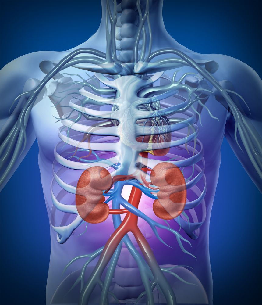
Kidneys
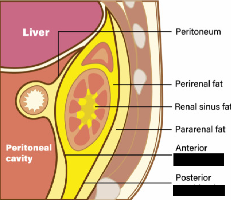
Paired organs located behind the peritoneal membrane (retroperitoneal). Bean-shaped, and about the size of an adult fist. Located laterally in the flank area (side of the torso), about the level of the twelfth thoracic vertebra to the third lumbar vertebra.
perineal fat
Adipose tissue called ______ ___ surrounds each kidney.
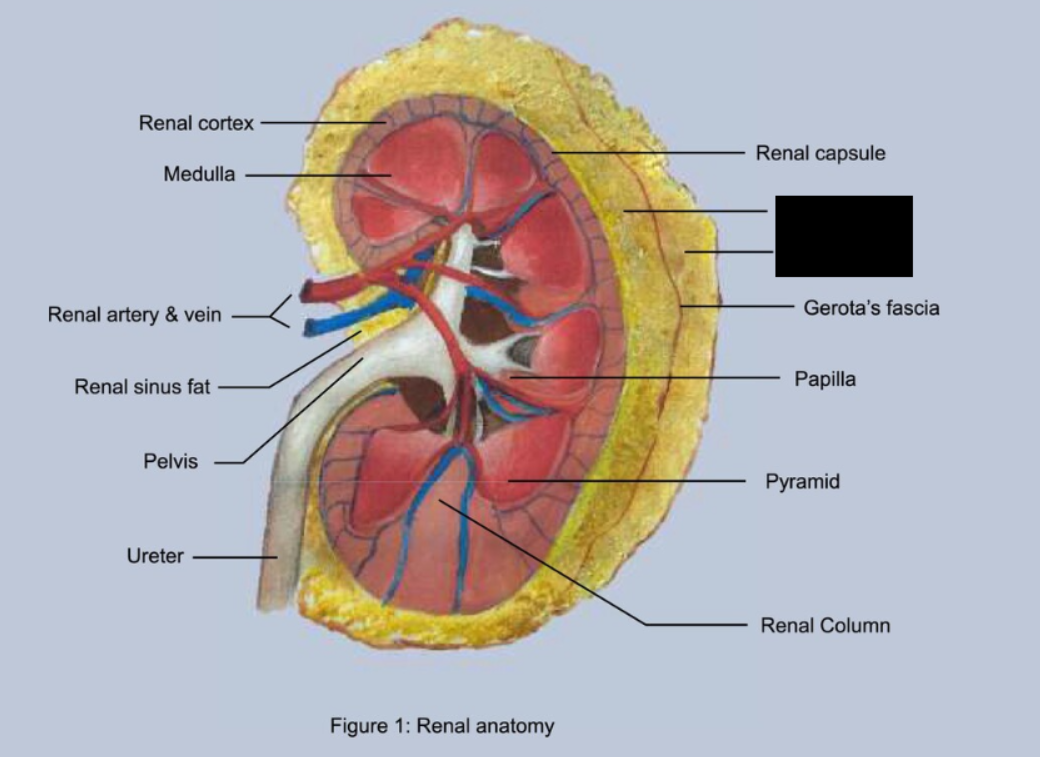
renal capsule
The outer layer of the kidneys consists of a layer of fibrous connective tissue called the ____ ________
renal fascia peritoneum
The kidneys are partially held in place by connective tissue connections called _____ _____ that connect to the outer portion of the __________.
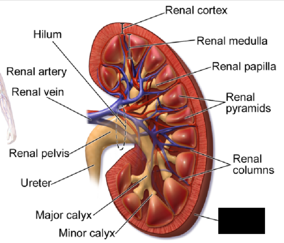
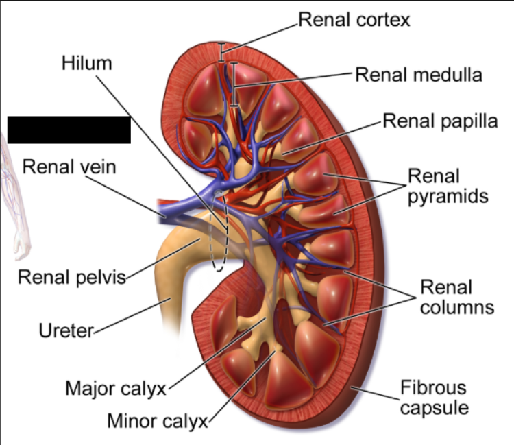
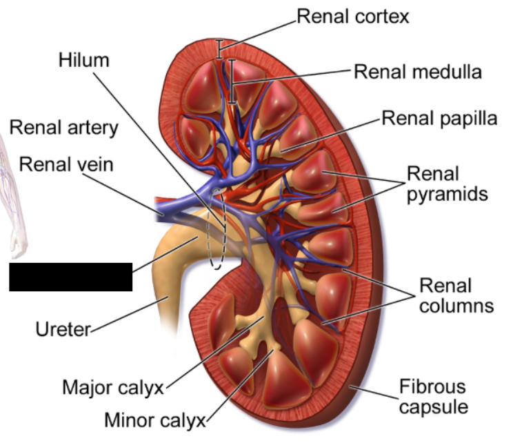
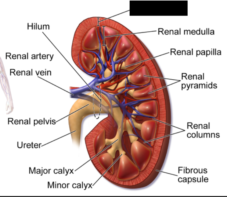
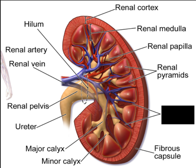
hilum
Each kidney has an indentation called a _____ that opens to a renal sinus where the renal artery, vein, and ureters enter and exit the kidney.
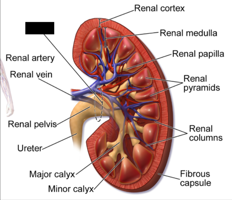
Renal Artery
An artery that supplies each kidney with blood. Branches off the abdominal aorta and extends into the hilum of the kidney. Forms smaller branches called segmental arteries that form smaller interlobar arteries.
Renal Vein
Major blood vessels that carry filtered, deoxygenated blood away from the kidneys and return it to the heart via the inferior vena cava.
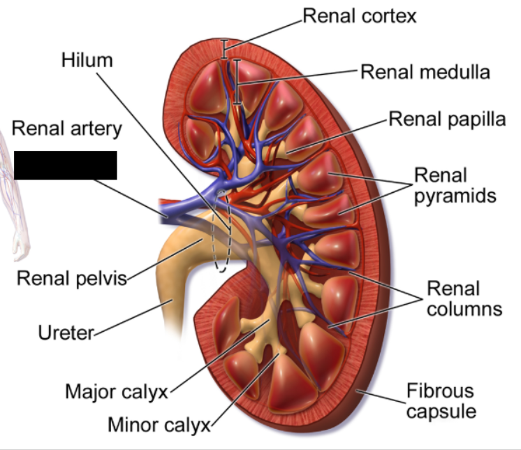
Renal Pelvis
A funnel-shaped structure located at the center of each kidney. Its primary function is to collect urine produced by the kidney and funnel it down into the ureter, which then transports it to the bladder.
Ureters
Tubes that carry urine from the kidneys to the bladder. They exit the kidneys at the hilum and extend inferiorly and medially to the bladder. They have a smooth muscle layer capable of producing peristaltic contractions that occur every 2 to 3 minutes. The parasympathetic nervous system increases these contractions, and the sympathetic nervous system inhibits them.
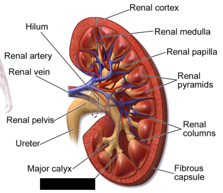
Major Calyx
a funnel-shaped tube in the kidney that collects urine from smaller chambers called minor ______ and drains it into the renal pelvis. Each human kidney typically has two to three.
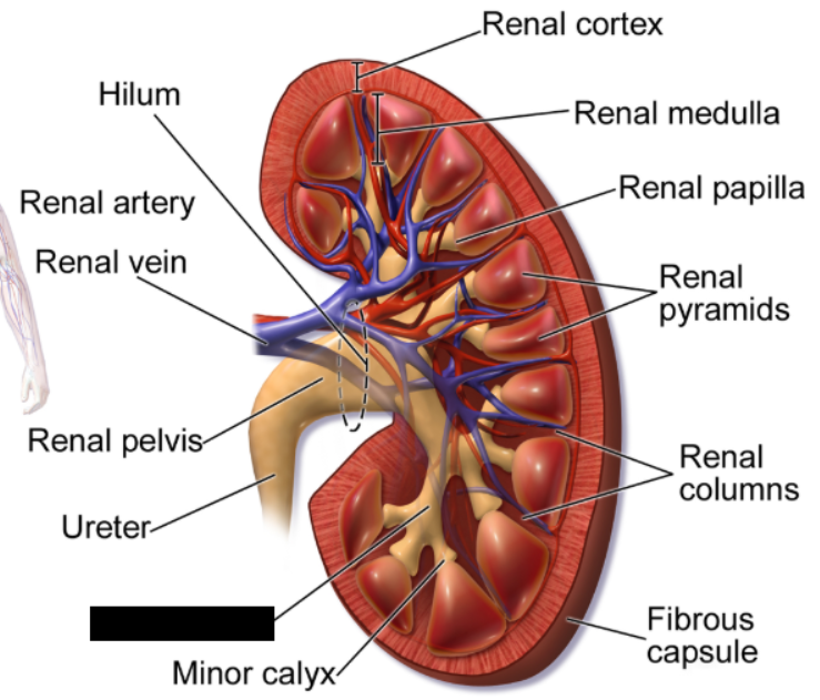
Minor Calyx
a small, cup-shaped structure in the kidney that collects urine directly from the renal pyramids. Several minor ______ merge to form major _______, which then drain into the renal pelvis and ureter.
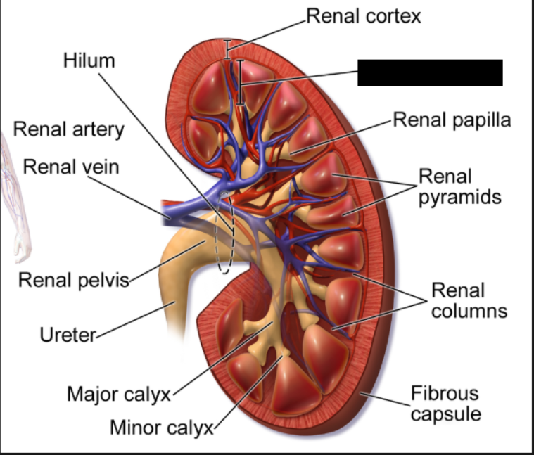
Renal Cortex
the outer, granular layer of the kidney located between the renal capsule and the deeper renal medulla. It plays a crucial role in ultrafiltration, blood regulation, and hormone production (aldosterone)
Renal Medulla
The innermost region of the kidney, located just beneath the renal cortex. It plays an indispensable role in maintaining the body's fluid, electrolyte, and blood pressure balance by regulating urine concentration
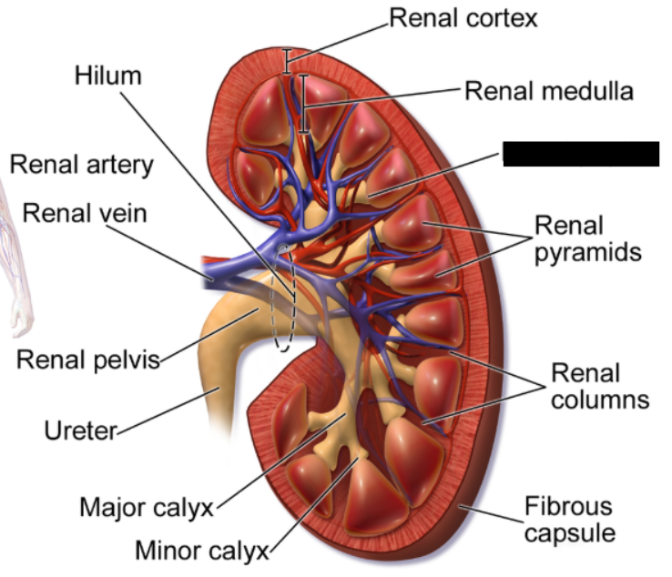
Renal Papilla
the apex (pointed tip) of a renal pyramid inside the kidney. It functions as the final exit point for urine, filtering fluid from the pyramid's collecting ducts into the minor calyx
Renal Pyramids
cone-shaped tissues located in the inner region of the kidney, called the renal medulla. Humans typically have 7 to 18 of these structures per kidney. Their primary function is to transport urine from the outer, blood-filtering region of the kidney to the ureters
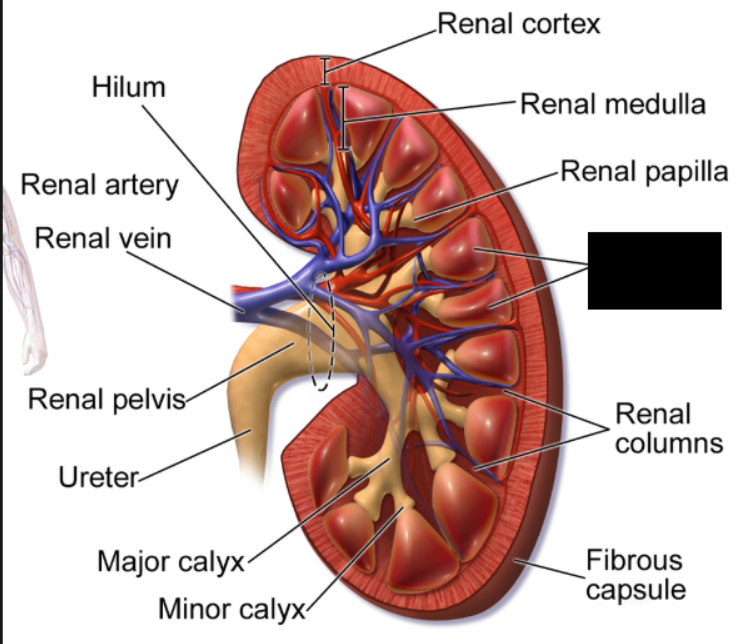
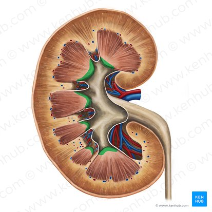
Renal Columns
inward extensions of the renal cortex that project down between the renal pyramids. They provide structural support, anchor the cortex, and act as pathways for the renal blood vessels, nerves, and urinary tubes.
superior
The left kidney is actually positioned a bit more _______ compared to the right kidney
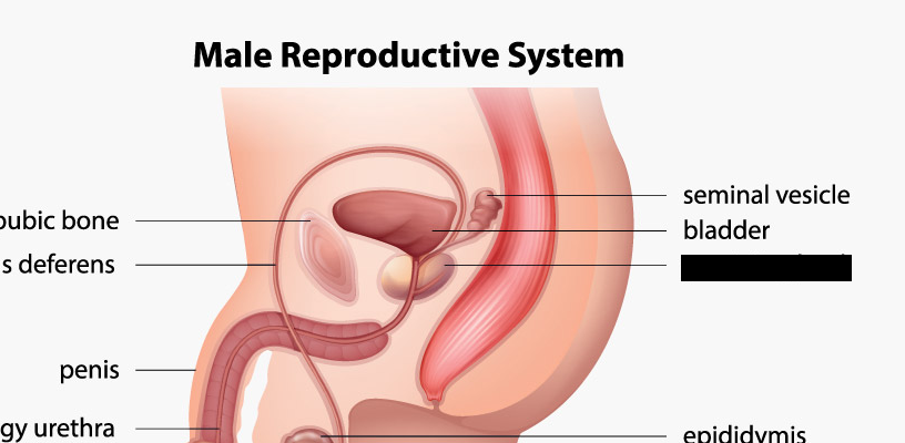
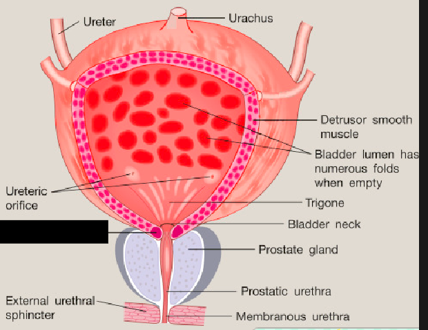
prostate gland
The ______ _____ is exclusive to males, and is just inferior to the urinary bladder. It is a walnut-sized gland in the male reproductive system located just below the bladder and surrounding the urethra. Its primary function is to produce seminal fluid, which nourishes and transports sperm during ejaculation
Renal Papillae
The apex (pointed tips) of renal pyramids inside the kidney. It functions as the final exit point for urine, filtering fluid from the pyramid's collecting ducts into the minor calyx.
Segmental Arteries
The first branches of arteries after the renal artery. They branch off to form interlobar arteries.
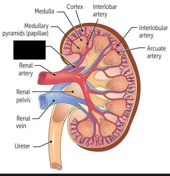
Interlobar Arteries
Arteries that branch off from segmental arteries. They flow through the renal columns and branch to form arcuate arteries.
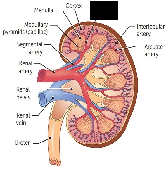
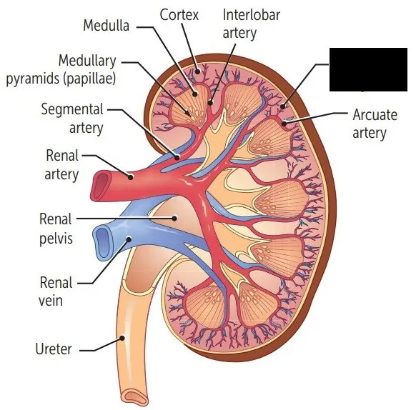
Arcuate Arteries
Arteries that branch off from interlobar arteries.They are located between the cortex and medulla. They branch to form interlobular arteries that travel in the cortex.
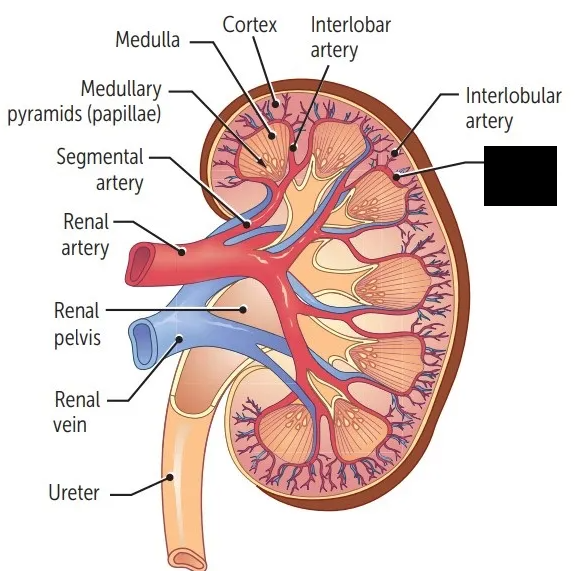
Interlobular Arteries
Arteries that branch off arcuate arteries. They travel in the cortex, and branch off to form afferent arterioles that supply the functional unity of the kidney, known as the nephron.
renal segmental interlobar arcuate interlobular afferent arterioles
What is the order of division of the renal artery?
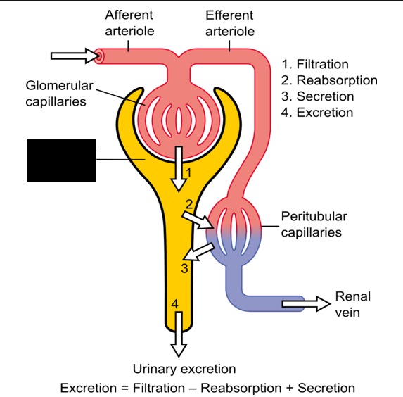
Afferent Arterioles
Branches from interlobar arteries that supply the functional unity of the kidney, known as the nephron.
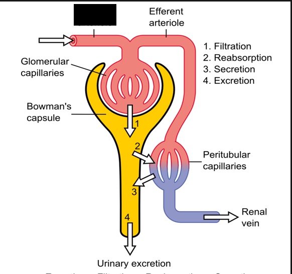
Efferent Arteriole
Exits the nephron and feeds interlobular veins that empty into arcuate veins. The arcuate veins then empty into the interlobular veins, which empty into the renal vein
Renal Nerves
The nerve supply of the kidneys. Sympathetic postganglionic neurons from the celiac plexus and inferior splanchnic nerves.
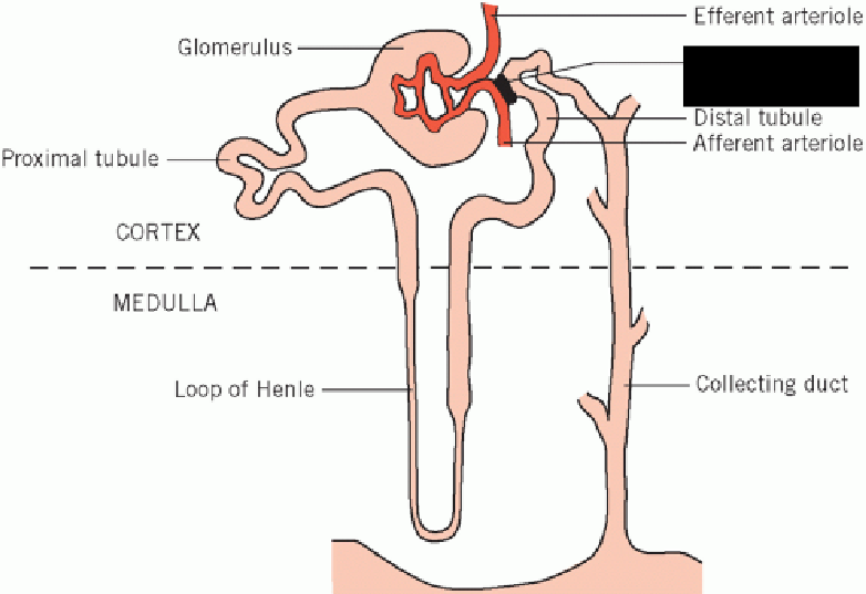
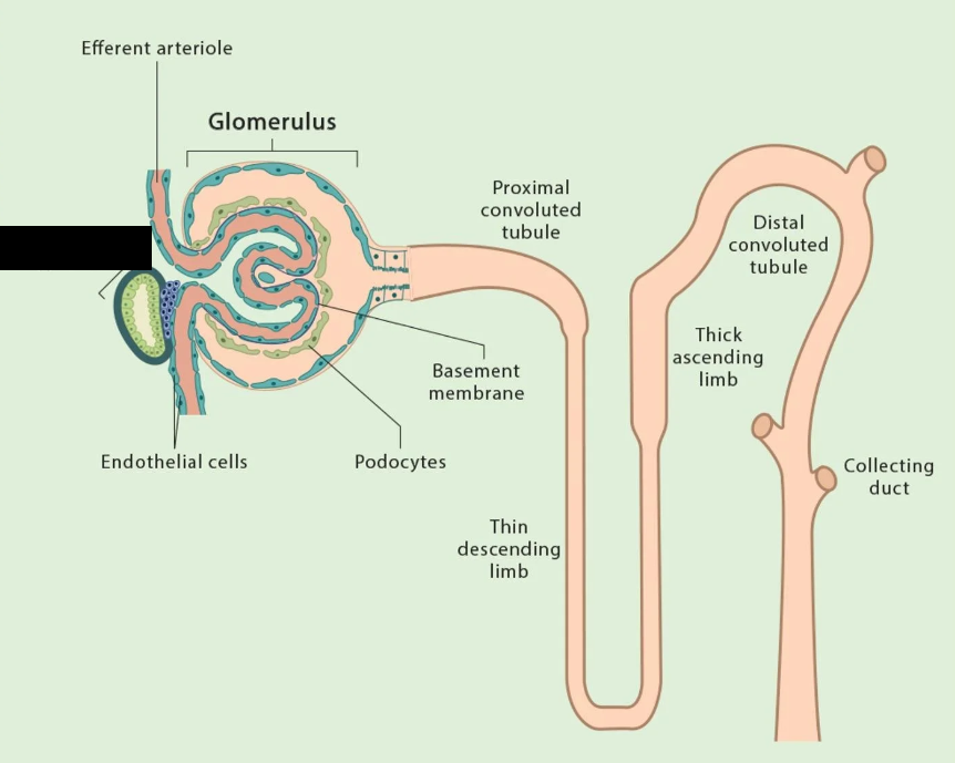
Nephron
The functional unit of the kidney; a microscopic structure. There are over one million of them in one kidney. Some lie near the medulla and are called juxtamedullary ________, which extend deep in the medulla. They consist of a renal tubule and renal corpuscle.
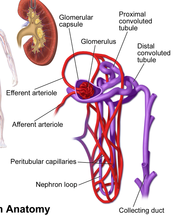
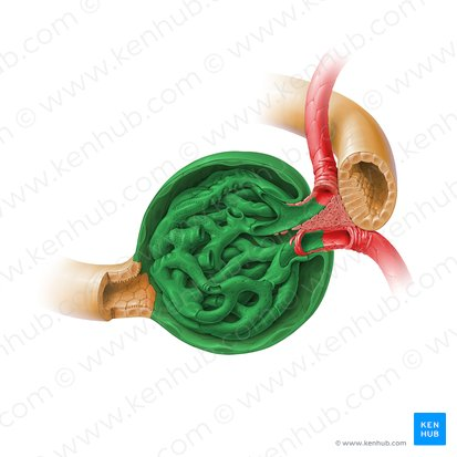
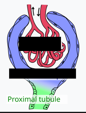
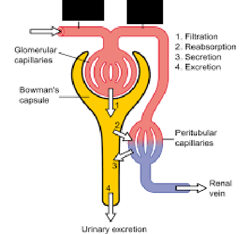
Renal Corpuscle
A spherical structure that consists of a capillary network called the glomerulus surrounded by a fibrous capsule called the glomerular capsule (Bowman’s capsule)
afferent efferent peritubular capillaries
The capillary network is fed by an _______ arteriole, and blood exiting the nephron flows through the _______ arteriole and ________ ________ that surround the nephron.
glomerulus glomerular capsule
The _______ and _______ _______ are where filtration occurs in the nephron
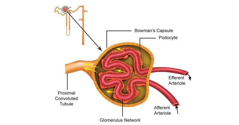
Glomerular Capsule
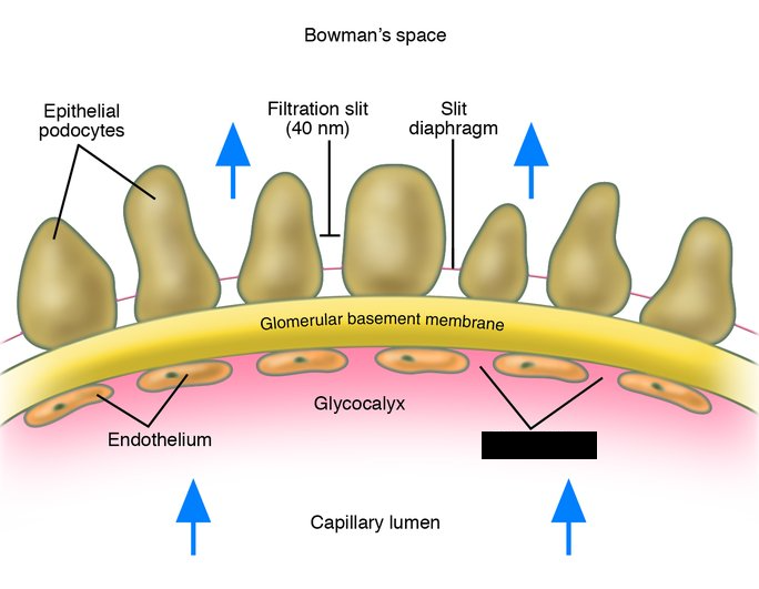
Also called the Bowman’s capsule, a cup-shaped sac at the beginning of the nephron in the kidneys. Consists of two layers: an outer or parietal layer, which consists of simple squamous epithelium, and an inner or visceral layer, containing special cells called podocytes
Podocytes
Cells in the inner layer of the Glomerular Capsule that surround the glomerular capillaries. Small openings between them, called filtration slits, act as holes in a filter.
Fenestrae
Small openings in glomerular capillaries that act as a filter, combined with the filtration slits between podocytes.
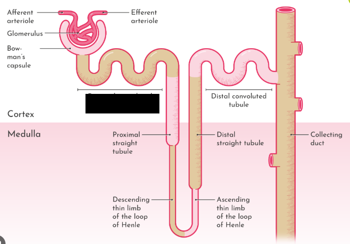
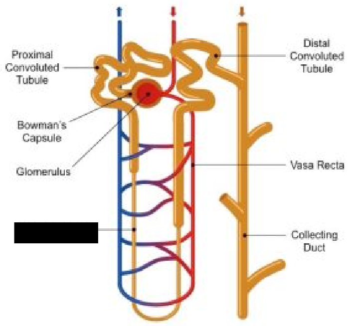
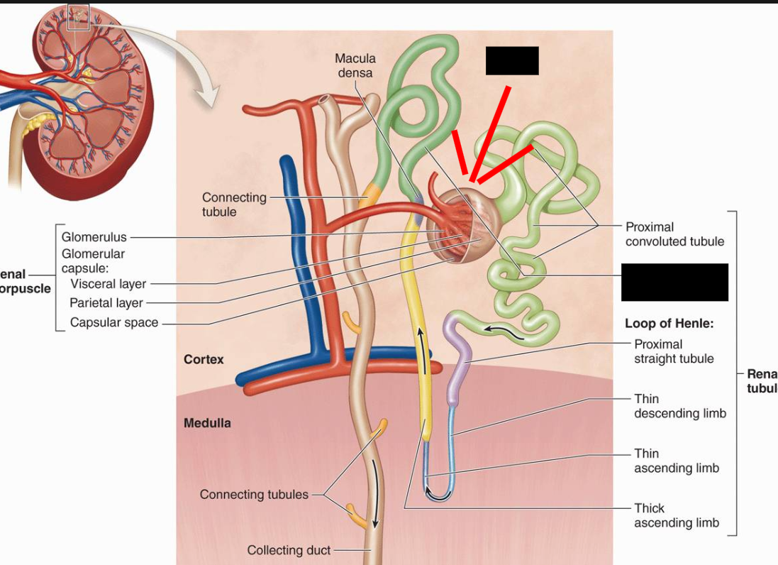
Proximal Convoluted Tubule
The first part of the renal tubule where the filtrate from the glomerular capsule initially flows through
Renal Tubules
Tubes in the kidneys that process fluid filtered from the blood. Lined with cuboidal epithelium and a basement membrane, with the excepting of the ascending limb of the nephron loop. Performs reabsorption and secretion of substances. Substances are selectively moved from them to the peritubular capillaries, or from the peritubular capillaries to the tubule.
nephron loop
After fluid moves out of the renal tubule, it moves through the ______ ____ (loop of Henle).
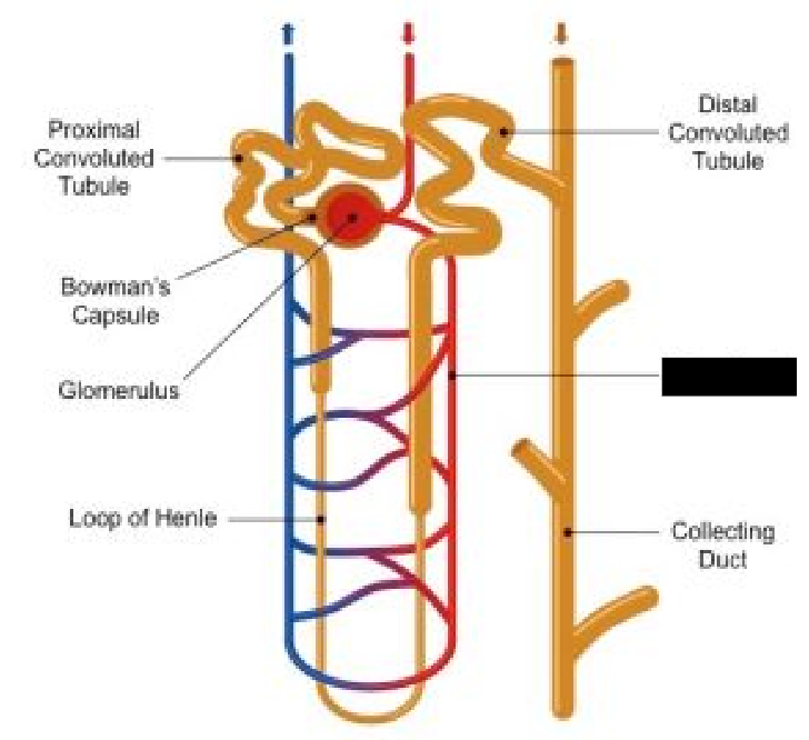
Nephron Loop
A U-shaped tube in the nephron. Receives liquid from the proximal convoluted tubule. It has a descending and ascending limb, each with different tissue characteristics. The ascending limb is lined with simple squamous epithelium in the lower section, which becomes simple cuboidal epithelium in the thick section.

Vasa Recta
Capillaries that surround the nephron loop.
distal convoluted tubule juxtaglomular apparatus
After flowing through the nephron loop, the fluid flows through the _____ _____ _____ , and past the _____ _____
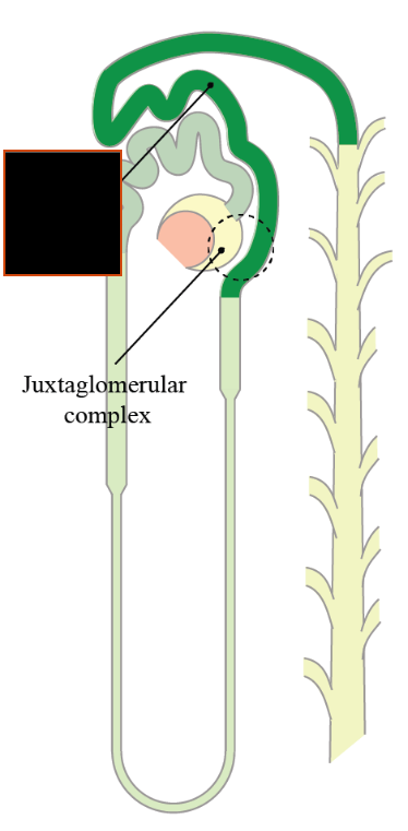
Juxtaglomular Apparatus
Monitors blood solute concentration as well as concentration of urine. Aids in regulating the fluid volume and blood pressure. Consists of a group of cells that reside between the distal convoluted tubule and the afferent arteriole.
distal convoluted tubule
Fluid (Urine) drains from the ____ ________ ____ to the collecting duct of the nephron.
collecting duct
Urine from many nephrons drains into one ________ ____. The collecting duct merges with other ________ ____ at the renal papilla.
renal papilla minor calyces major calyces renal pelvis
Urine flows from the _____ _______ to the _____ _______, which combine to create _____ _______, which then combine to form the _____ _______.
ureter urinary bladder urethra out the body
Urine flows into the _____ and to the _____ _____ , _____ , and _____ _____ _____ .
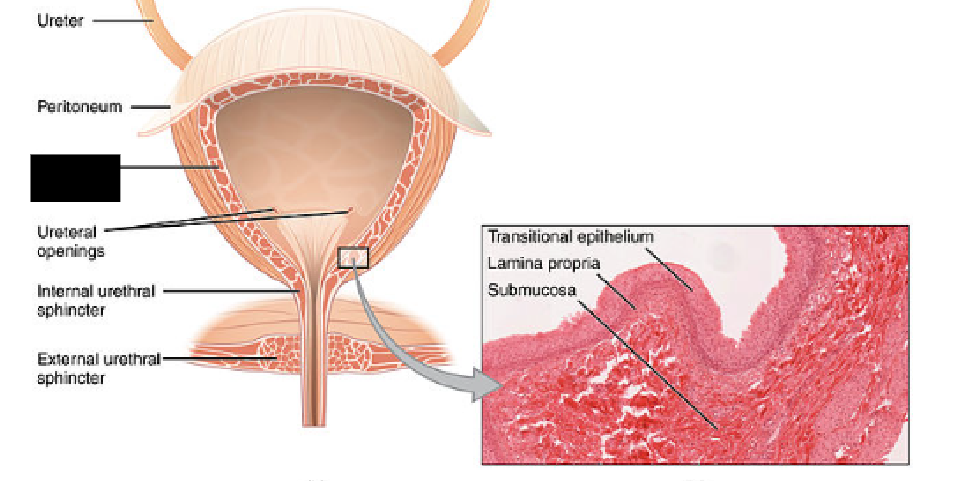
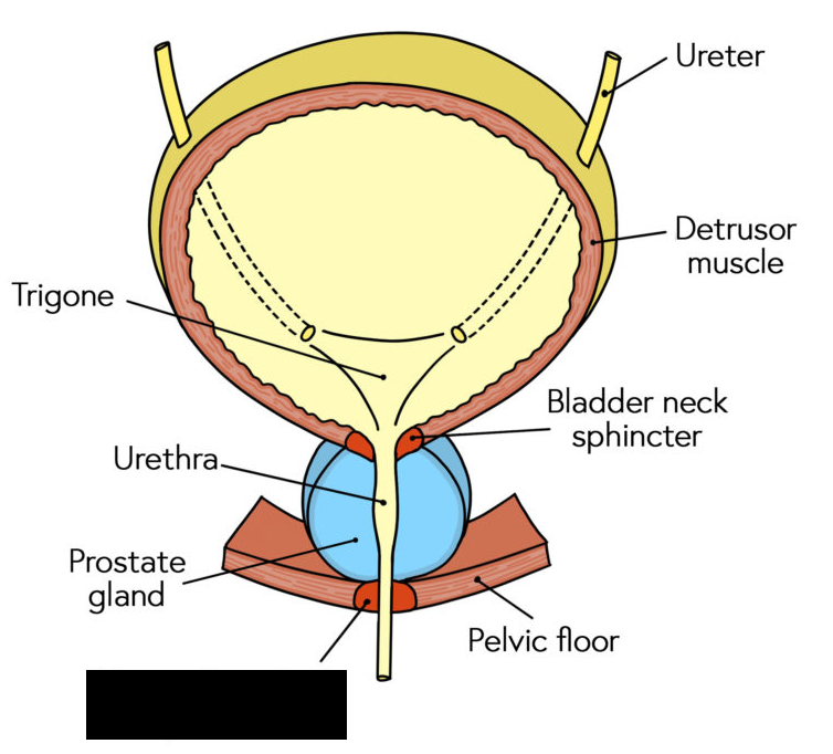
Urinary Bladder
A hollow organ that resides in the pelvic cavity. The ureters connect at the posterolateral surface. It can store up to one liter. At around 300ml, the urge to urinate becomes evident.
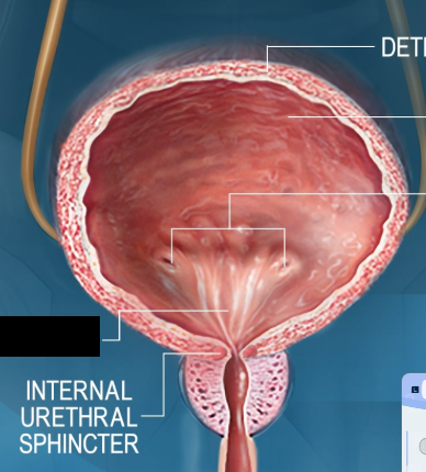
Detrusor Muscle
A thick smooth muscle layer of the bladder. Contraction of it increases the internal pressure of the bladder and causes urine to be expelled.
Urethra
Carries urine from the bladder out the body.
Trigone
The area on the inside of the bladder between the two ureter connections and the urethra.
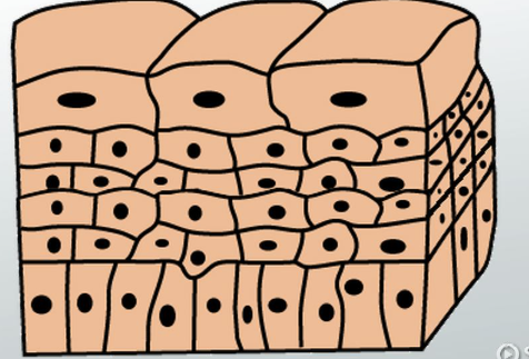
transitional epithelium
Both the urinary bladder and ureters are internally lined with __________ __________ , a special kind of epithelium that allows for the cells to slide past each other during the distension of the bladder.
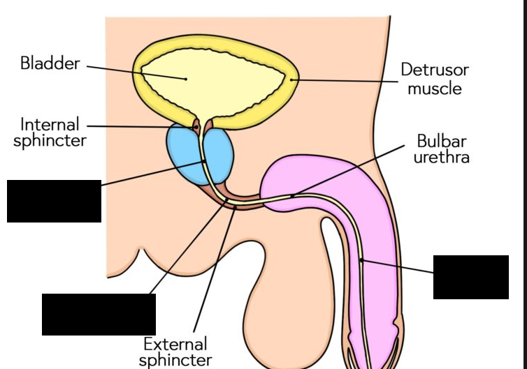
internal urinary sphincter
Male bladders contain an area of smooth muscle and elastic tissue called the ______ _______ _______, which isn’t present in females. It keeps semen from entering the urinary bladder during intercourse
external urinary sphincter
Both males and females have an ______ _______ _______ located in the urethra that controls the flow of urine.
prostatic urethra membranous urethra penile urethra
The male urethra consists of 3 parts.
The ______ _______ exits the bladder and extends to the inferior prostate gland, where it becomes the ______ _______. It then enters the penis where it becomes the ______ _______.
Benign Prostatic Hyperplasia
A condition of the prostate where the prostate enlarges and sometimes blocks the urethra, resulting in difficulty urinating. Possible in males only, since females dont have a urethra.
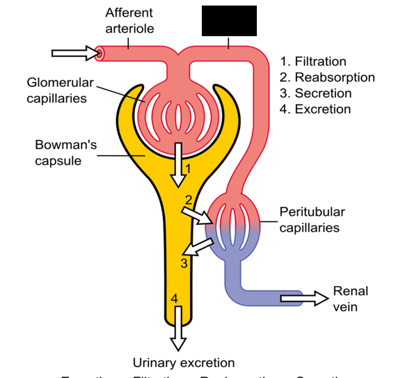
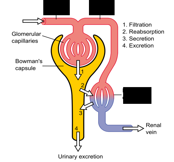
Filtration Tubular reabsorption Tubular Secretion
There are 3 primary processes of urine formation: ________, _________ ________(moving substances from the kidney to the blood), and ________ ________(moving substances from the blood to the kidney)
blood
Tubular reabsorption employs a number of mechanisms to move substances back into the _____.
blood urine
Tubular secretion also uses mechanisms to move substances from the _____ to the _____.
erythropoietin
The kidneys secrete ___________ in response to low oxygen levels, which travels to the bone marrow to stimulate the production of red blood cells
123
The kidneys make a lot of filtrate each day; about ___ml per minute, which adds to about 180 liters per day.
1 2
Most of the filtrate is reabsorbed into the body, leaving around _-_ liters of urine per day.
Net Filtration Pressure
The pressure gradient present that helps substance pass through filters in the kidneys. The combination of a series of pressure that exist in the renal corpuscle. Directly proportional to the glomerular filtration rate. If ___ ____ ____ increases or decreases, so does the glomerular filtration rate, as well as the filtrate produced.
Glomerular Capillary Hydrostatic Pressure
The blood pressure inside the capillaries. Usually about 50 mm Hg and must be greater than the pressure inside the glomerular capsule, or glomerular capsular hydrostatic pressure., which is the main pressure. Controlled in part by the diameter of afferent and efferent arterioles.
smaller
Efferent arterioles have a ______ diameter from afferent arterioles
The diameter works to decrease blood flow through efferent arterioles, increasing pressure inside the glomerular capillaries.
glomerular capillary hydrostatic pressure
Changing diameter of afferent and efferent arterioles changes ______ ______ ______ ______ . If the diameter of afferent arterioles increase, or if the diameter of efferent arterioles decrease, capillary pressure will increase.
Afferent efferent
_______ arterioles carry blood inside the glomerulus for filtration, while _______ arterioles carry blood away from the glomerulus.
Glomerular Capsular Hydrostatic Pressure
Pressure inside the glomerular capsule, created by fluid inside the capsule as well as downstream in the tubules. Usually about 10 mm Hg. Works against filtration.
Colloid Osmotic Pressure
Pressure produced by the presence of plasma proteins in the blood called colloids. They produce a pulling force, causing water to move back into the glomerular capillaries. About 30 mm Hg.
capillary capsular Colloid osmotic pressure
Net filtration pressure is calculated with this formula:
Net Filtration Pressure = Glomerular ________ hydrostatic pressure – Glomerular ________ hydrostatic pressure – ________ ________ ________
Combining the normal values gives us:
NFP = 50 mm Hg - 10 mm Hg - 30 mm Hg = 10 mm Hg
So the net filtration pressure is about 10 mm Hg.
vasoconstrict
When systemic blood pressure (The force of circulating blood against the walls of the body’s arteries) increases, afferent arterioles ________ , keeping glomerular capillary hydrostatic pressure constant. They do the opposite when systemic blood pressure is low.
Sympathetic Nervous System
The _________ ______ ______ affects the afferent arterioles by causing it to vasoconstrict under intense sympathetic activity, such as exercising strenuously or being under shock.
Symporters
Special transport proteins that are present in some cells that line the kidney tubules. Sodium and glucose move through these, with sodium providing movement via a gradient, and glucose goes through the transport protein as well. Amino acids and vitamins also are transported back into tubular cells via _________.
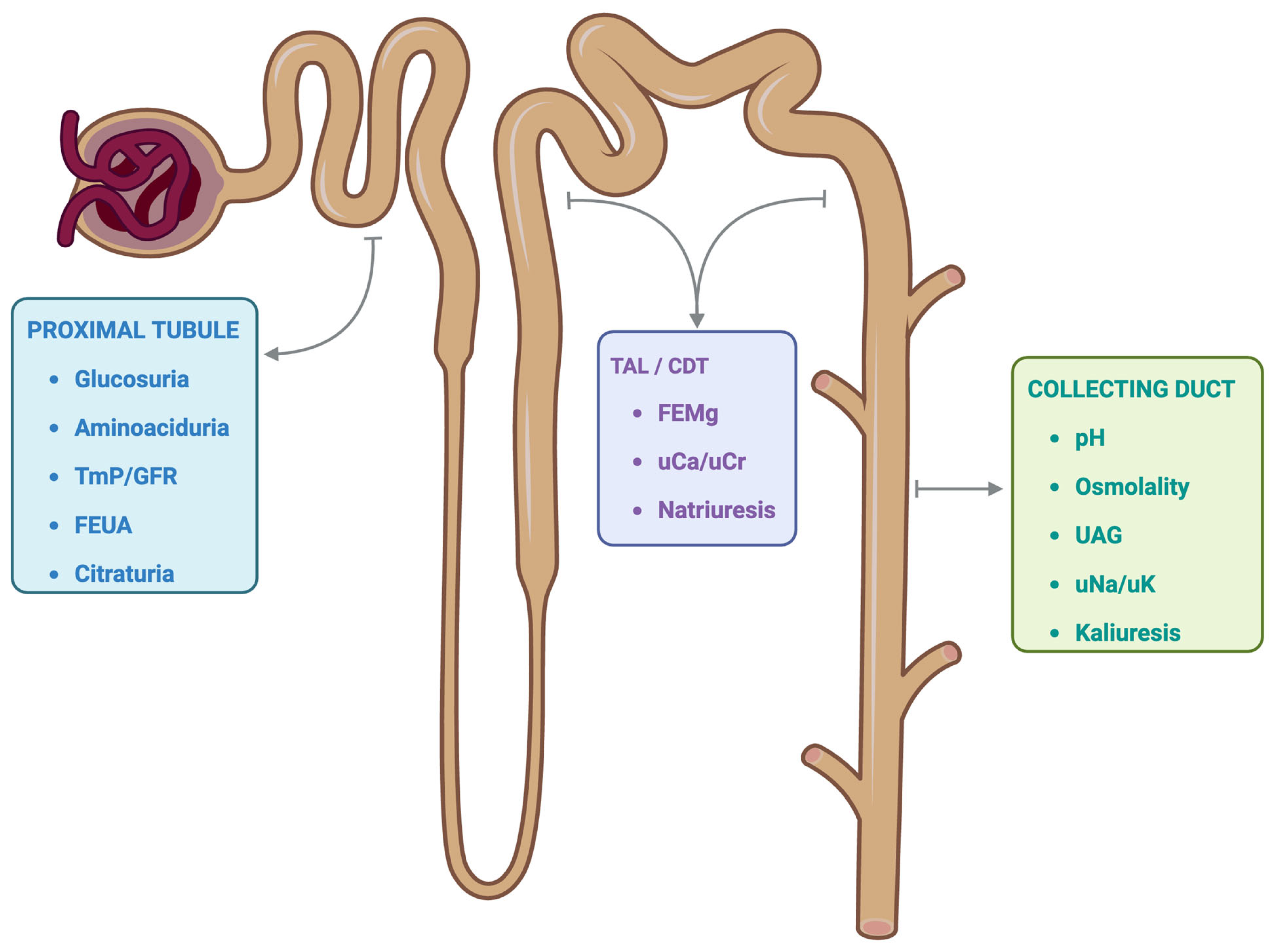
glucosuria
If an excess of glucose was produced by the filter, it would exceed the number of symporters. The glucose would then flow into the urine and out the body, in a condition called _________ , which occurs in people with diabetes
passively
Sodium, calcium, magnesium, and potassium all move ________ from the tubules to the blood.
water
Since so much sodium moves into the interstitial space surrounding the tubules, _____follows sodium via osmosis. This is how _____ is reabsorbed from the filtrate.
Sodium-Hydrogen Antiporter
An instance of passive transport in tubular secretion. A protein that uses the sodium gradient to move sodium from the tubule to the inside of the cell, while moving excess hydrogen ions out of the cell and into the tubule. Sodium provides the movement so that hydrogen can be transferred into the tubule
tubular fluid
Ammonia, potassium, penicillin, and para-amino hydrochloric acid are all substances secreted into the _______ ____. They are all not normally produced in the body.
leaky
Certain tubule cells are “_____” to sodium and potassium. When aldosterone attaches to receptors on these cells, it increases their permeability to sodium and potassium.
Atrial Natriuretic Hormone
A hormone secreted by the wall of the right atrium in the heart in response to atrial stretch. It has the opposite action of aldosterone, and inhibits sodium and water reabsorption in the kidney tubules.
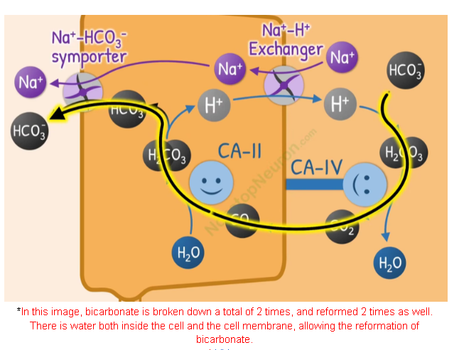
Bicarbonate Reabsorption
There are a couple of steps associated with bicarbonate ions moving from the kidney tubules to the blood.
Hydrogen is secreted in the tubule by the way of the sodium-hydrogen antiporter
Hydrogen runs into bicarbonate ions to form carbonic acid, which dissociates into carbon dioxide and water.
Carbon dioxide then diffuses into cells lining the lumen of the tubule, and combines with water in the presence of carbonic anhydrase to form carbonic acid yet again.
The carbonic acid dissociates into hydrogen and bicarbonate ions again, and the hydrogen gets antiported back out to keep the process looping, while the bicarbonate moves out of the cell and into the blood.
Juxtaglomerular Apparatus
Also called the JG apparatus, is a group of cells residing at the junction of the afferent arteriole and distal ascending limb of the nephron loop. It consists of 2 different types of cells. They are located on the afferent arteriole side.
Angiotensin II
A substance formed by the renin-angiotensin system that promote vasoconstriction and secretion of the adrenal cortex hormone Aldosterone.
Macula Densa Cells
Cells in the juxtaglomerular apparatus that monitors the filtrate in the kidney tubule. Usually, they keep open the afferent arteriole by secreting nitric oxide synthetase secretion, which promotes nitric oxide secretion (widens blood vessels). This secretion is inhibited if too much filtrate is produced (too much urine). Additionally, they also secrete adenosine, which promotes vasoconstriction of the afferent arteriole.
hypotonic
Fluid exiting the nephron loop is ______. It enters the distal convoluted tubule, which is surrounded by the interstitial space and peritubular capillaries, which are isotonic.
If the tubule is impermeable, dilute urine is produced
If the tubule is more permeable, then water moves to the more concentrated interstitial space and blood. ADH controls this balance of water.
polyuria
Pathologies such as diabetes or some medications can produce a larger urine output, known as ______.
Oliguria
A urine output of less than 500 ml/day
Anuria
A urine output of less than 100 ml/day
Green
_____-tinted urine may be evident of bile pigments or bacterial infection
Dark
_____ urine might indicate metal or phenol poisoning, or even severe bleeding in the kidneys
urea
Urine can develop an ammonia order after standing due to the breakdown of ____.
acetone
An ______ odor in the urine may indicate diabetes.
4.6 8.0
The pH of urine varies between _._ and _._
1.001 1.035
Urine’s specific gravity (a expression of it’s density compared to water; with water being represented by the number 1) is around _.___ to _.___
Micturition Reflex
Reflex signaled by the bladder walls being stretched. Stretching of the bladder sends impulses to sensory neurons in the pelvic nerves to the sacral segments of the spinal cord. Parasympathetic impulses then cause the bladder to contract.
pons cerebral cortex
Motor impulses for micturition originate in a micturition center in the ____. The center also receives input from the _______ _____, so one can have control over micturition.
Renal Clearance
Represents how much blood has to pass through the kidney to completely remove a substance. Specifically, it is the volume of blood plasma from which a substance is completely removed in one minute. It reflects the 3 processes of urine formation.
C U V P
Renal clearance can be calculated with the formula (_) = __/_, where
_ is renal clearance
_ is the concentration of the substance in the urine
_ is the rate of urine output
_ is the concentration of the substance in plasma
The result is the amount of blood plasma that is cleared of the substance every minute.
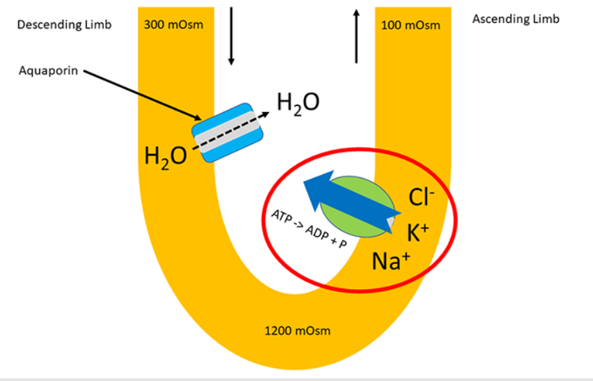
countercurrent multiplier
The nephron loop creates a large concentration gradient using a mechanism called a ____________ ______.
First, an isotonic (about 300 milliosmole) solution moves in through the descending limb
The descending limb is permeable to water. There are aquaporins present in the channel, so water flows out of the descending limb into the interstitial space, since it is very concentrated at the location.
As water moves out of the loop, concentration of the solution becomes about 1200 milliosmole.
The solution then moves to the ascending limb, which contains active transport protein channels that move sodium, potassium, and chloride out the ascending limb.
The concentration after the transport of molecules in the ascending limb is now about 100 milliosmole.
The ____________ part of the ____________ ______ is demonstrated by water moving in one direction, while a solute current moves in an opposing direction.
Benedict’s Test
A test used to find the presence of glucose in a solution. The more amber/dark brown the solution is after the test, the more glucose present. Benedict’s solution is added with a bit of heat to the test.