Parasitiology Exam 1
1/34
There's no tags or description
Looks like no tags are added yet.
Name | Mastery | Learn | Test | Matching | Spaced | Call with Kai |
|---|
No analytics yet
Send a link to your students to track their progress
35 Terms
Protozoa
single cell
Helminths
multi-celled
Ectoparasites体外寄生虫
insects, arachnids, etc.
arthropods that attach or burrow into the skin and remain there for relatively long periods of time
Pseudopodia (Protozoan Locomotion)
Protruding of pseudopodia
formation of new adhesions
development of traction
release of old adhesions
Definitive host
organism where reproductive cycle occurs (may serve as a reservoir)
Intermediate host
harbors parasite for transition period, often when asexual cycle occurs
Accidental host & Dead end host
atypical host in which the organism may not develop, or just may not be able to complete lifecycle
who is at risk of getting parasite infection
minorities, immigrants and poor/disadvantaged people
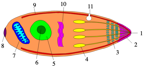
Apicomplexan structure
polar ring: critical for parasite morphology and host cell invasion
microenemes: crucial for host cell recognition, gliding motility, and active invasion
Rhoptries: specialized secretory organelle that release their contents during host cell invasion.
Recrudescence
recurrence of asexual parasite which (for any reason) originates from the same parasites responsible for the initial illness.
Relapse
recurrence of asexual parasite from hypnozoite(s) after earlier elimination of iRBC stages.
Recurrence
renewed asexual parasite, which is easily recognized if blood stages appear following drug treatment (can be due to new infection, relapse or recrudescence).
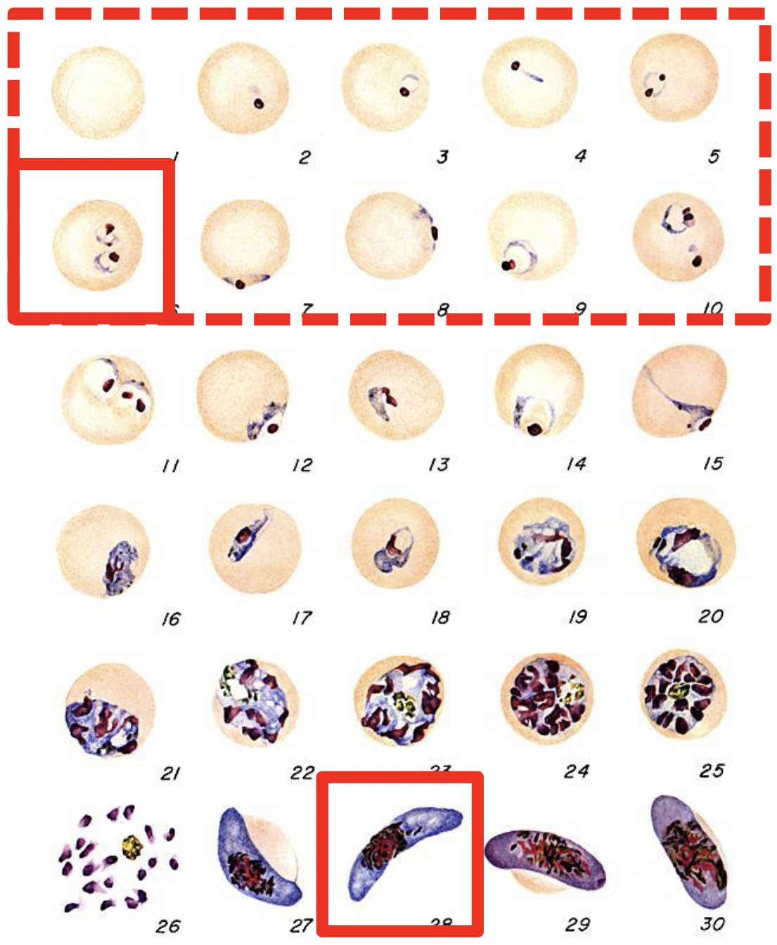
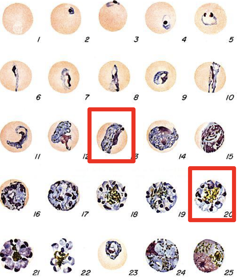
P. falciparum
Only rings observed
Banana-shaped gametocyte
16-32 merozoites
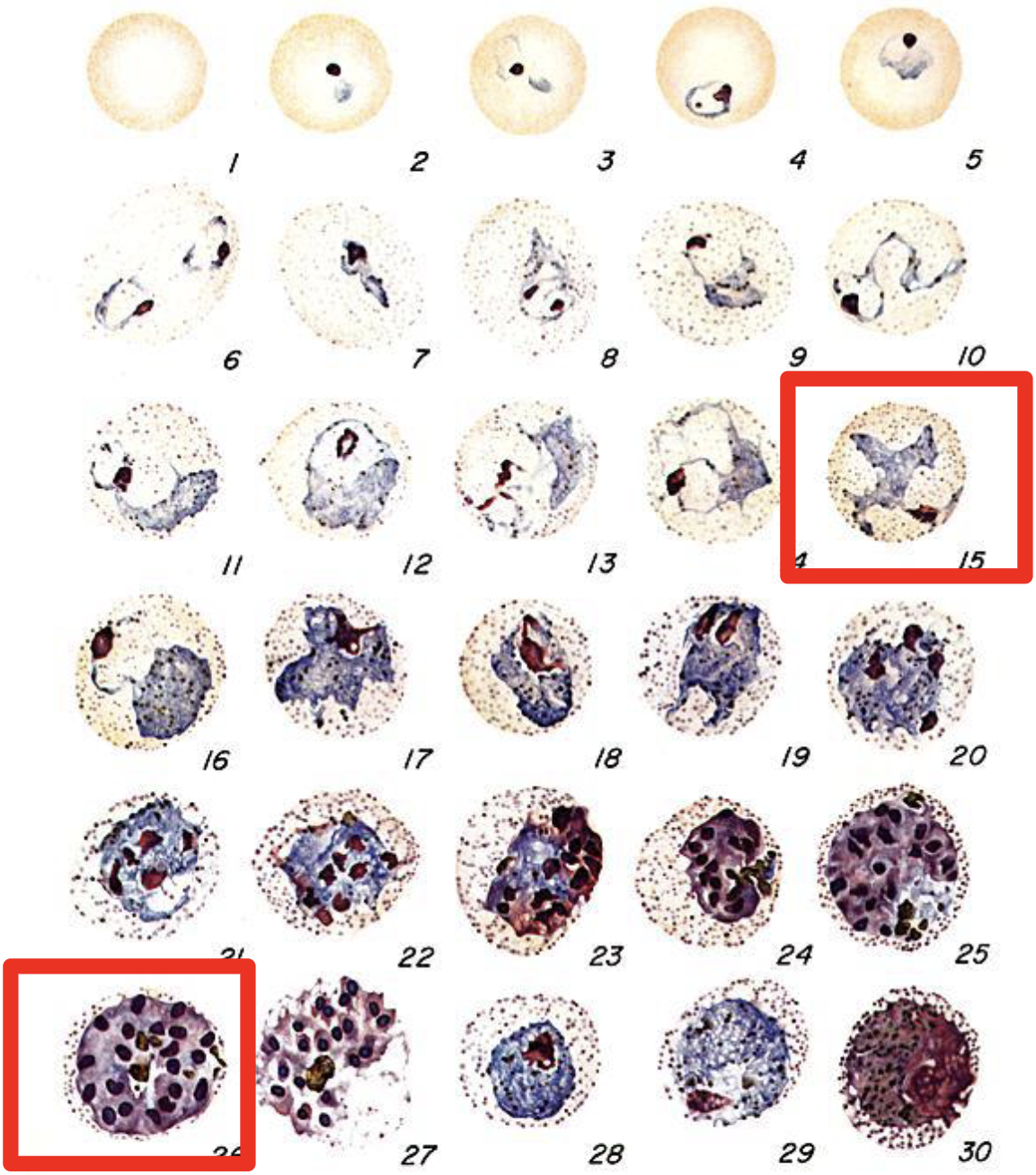
P. vivax
>12 merozoites
Schuffner’s dots /stippling
Ameboid troph
Require Duffy receptor (=“entry point” on RBCs)
P. ovale
Up to 12 merozoites per schizont
Ovale-shaped RBCs
Schuffner’s dots/stippling
Don’t require Duffy receptor (=“entry point” on RBCs)
P. malariae
Band-form trophozoites
Only ~8 merozoites per schizont (often in rosette)
Recrudescing (in blood)
Looks same as P. knowlesi
Why do ‘buffy coats’ work for diagnosing blood parasites?
Density of most blood parasites leads them to accumulate just below the WBCs.
Sporozoite
The infective stage transmitted to a new host.
Trophozoite
The feeding/growing stage of the parasite before reproduction.
inside RBCs.
Merozoite
The daughter cell produced by replication.
Spreads infection from cell to cell.
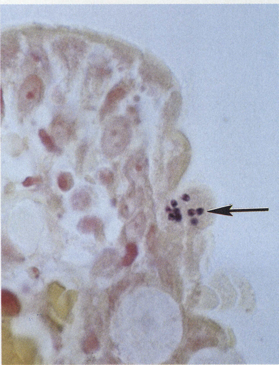
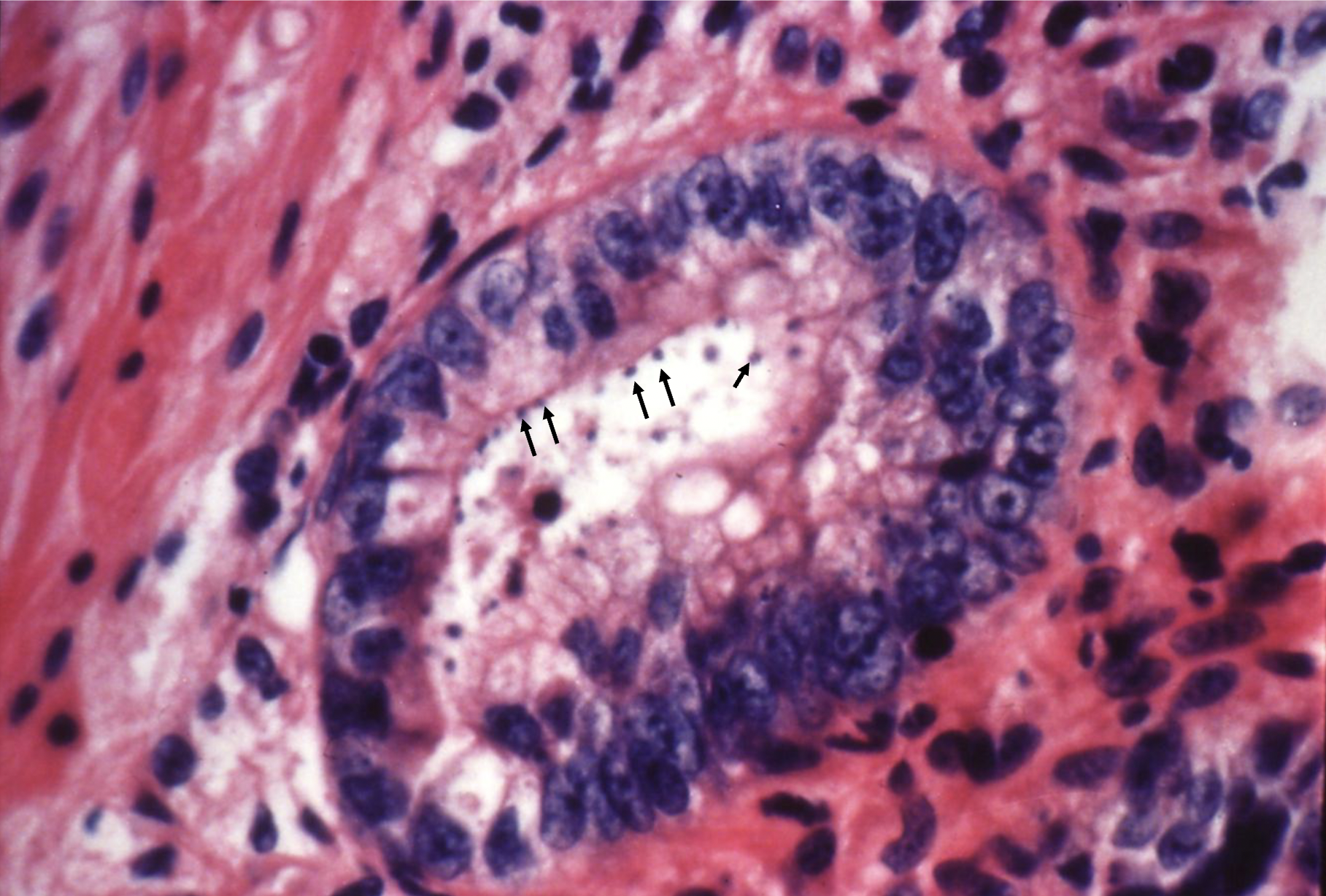
Cryptosporidium parvum/hominis
Intracellular, extracytoplasmic blue ‘beads’ in the brush border
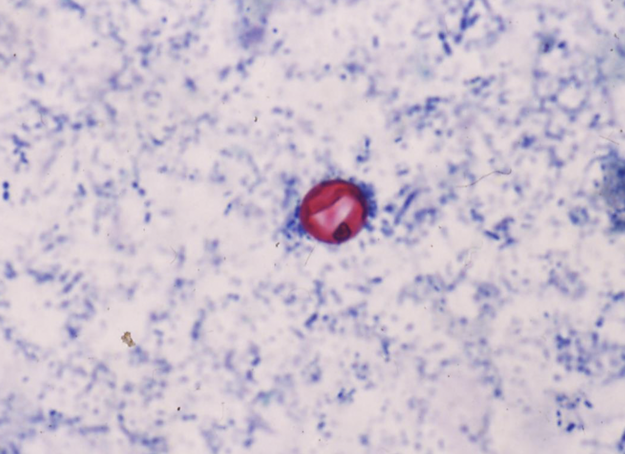
Cyclospora cayetanesis on modified acid-fast
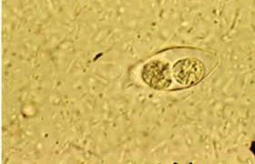
Cystoisospora belli’s oocyst under Iodine Stain
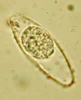
Cystoisospora belli’s sporoblast/sporocyst under Iodine Stain
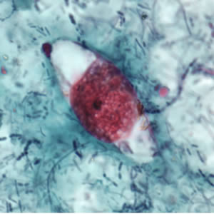
Cystoisospora modified acid fast staining
Pink/red/purple oocysts
blue-green background
Typical ellipsoidal shape
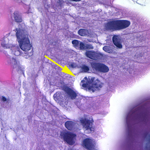
Cystoisospora on Hematoxylin and Eosin (H&E) stain
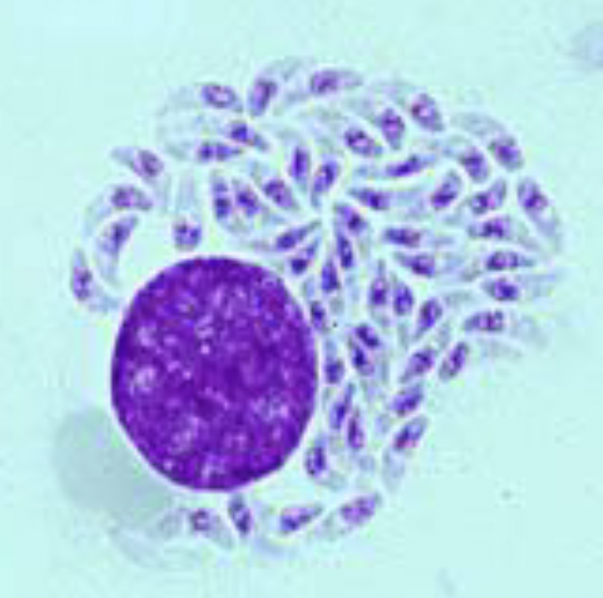
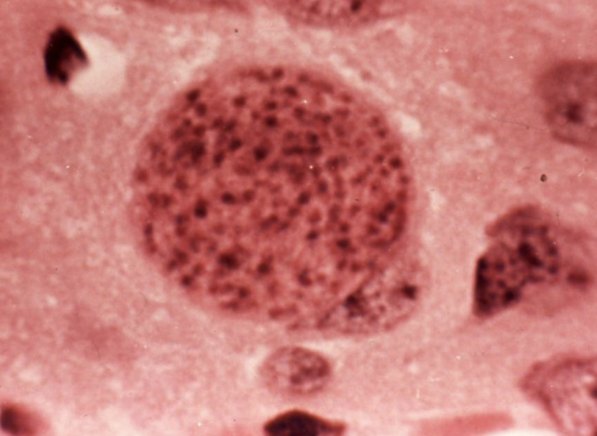
Toxoplasma gondii: tachyzoites
Toxoplasma gondii: tissue cysts containing bradyzoites
Toxoplasmosis: if a patient is IgM is positive, but IgG is negative, what to do next?
suspect Acute infection or false-positive
perform another serological test in 1–2 weeks
- if the result remain the same → false-positive IgM
because if there is an infection, the body is going to start producing IgG in 1-2 wks of infection
- if the result of IgG change to positive → then it is acute infection
becuase Negative IgG = no infection seen at all
Toxoplasmosis: if a patient is both IgM and IgG positive, what to do next?
suspect recent infection within 12 months or False-positive
send the specimen to a reference lab for further testing like IgG/IgM avidity test.
Toxoplasmosis: if a patient is IgG is positive, but IgM is negative, what does that mean?
Infected with Toxoplasma more than 1 year.
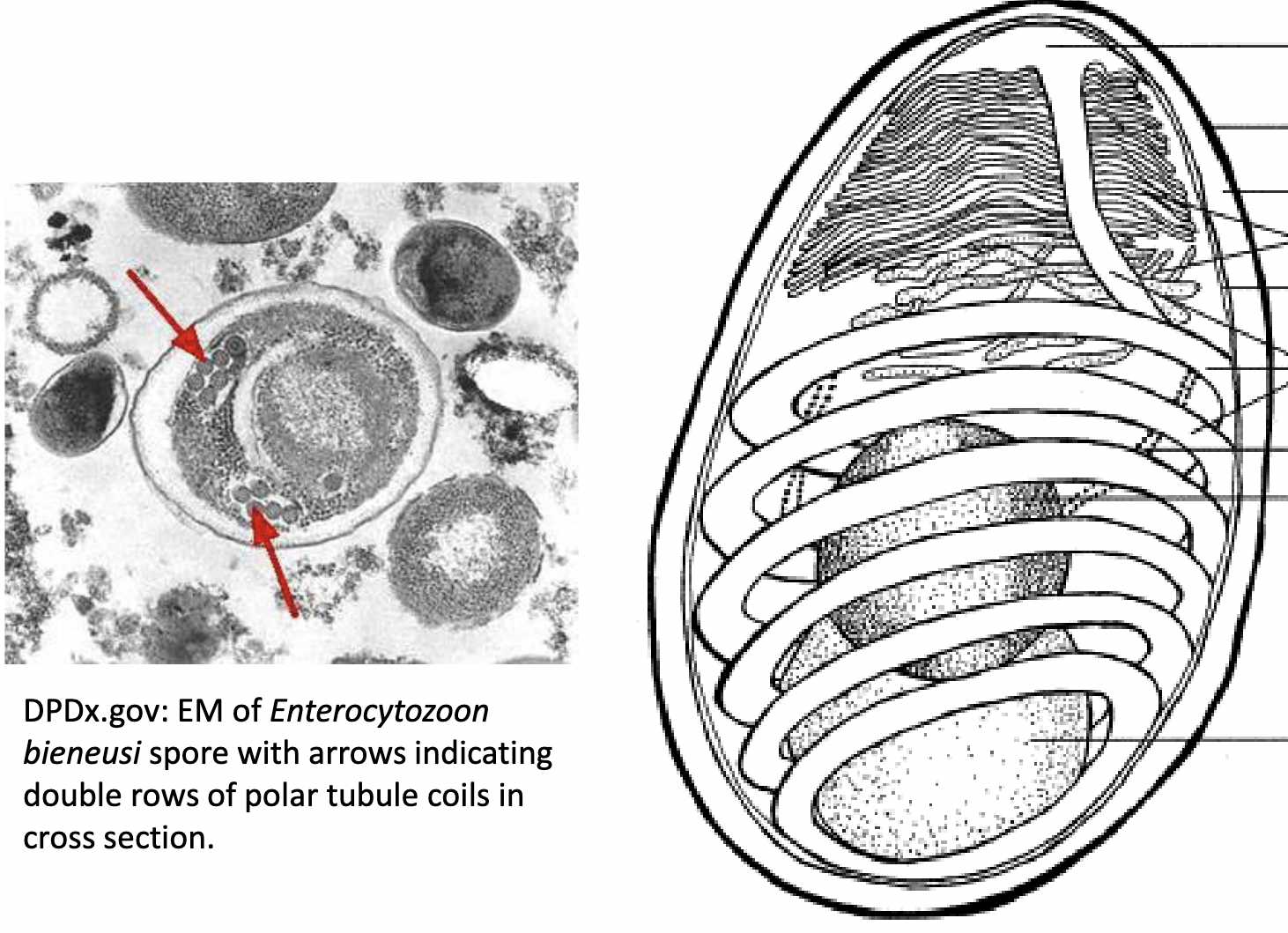
Microsporidium
characteristic coiled polar filament
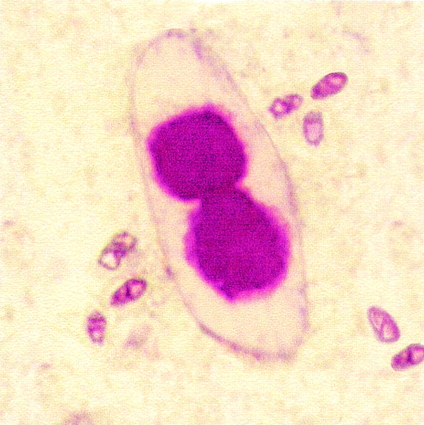
Acid Fast Trichrome Stain of Microsporidium
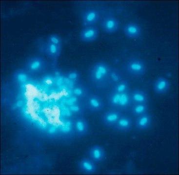
Chitin stain of Microsporidium
Microsporidia – Jejunal Biopsy