Neuro Midterm Practical
1/40
There's no tags or description
Looks like no tags are added yet.
Name | Mastery | Learn | Test | Matching | Spaced | Call with Kai |
|---|
No analytics yet
Send a link to your students to track their progress
41 Terms
what is gray matter composed of? what does it form?
brain (outer) → cell bodies + dendrites (pink-gray due to blood supply)
forms nuclei (specialized w/ functionally related cell bodies) + cortex (layered surface covering CNS)
what is white matter composed of? tract?
brain (center) → axons (w/ myelin sheath), fatty + white
fasciculus, funiculus, lemniscus, peduncle
tract → specialized axon bundle w/ common origin + destination
what are the sulci that divide 6 lobes in each cerebral hemisphere?
central → frontal vs parietal
lateral → fronto-parietal vs temporal
parietooccipital → parietal vs occipital
cingulate → parietal vs limbic
what are the cortical regions of the frontal lobe?
primary motor (precentral gyrus) → contains cell bodies of desc. path + initiates voluntary movement
premotor + supp. motor (precentral + post frontal gyri) → planning + initiate voluntary movement
motor speech (broca) → opercular + triangular inf. frontal gyrus (L. hemisphere)
prefrontal → executive fxn, personality, insight/foresight
what are the components of the parietal lobe?
postcentral gyrus (primary somatosensory cortex) → tactile + proprioception (sensory localization)
inf. parietal lobe (L. hemisphere) → language comp. (w/ temporal)
remaining → spatial orientation + attention direction
what are the components of the temporal lobe?
primary auditory cortex (sup. + small area of sup. gyrus) → process auditory info
sensory speech (wernicke) → post. portion of sup. gyrus (L. hemisphere)
visual info. process (inf. surface) → higher-order visual processing
learn + memory (med) → complex learning + memory
occipital lobe, limbic lobe, insula function and location?
occipital → visual fxn, primary visual cortex (calcarine sulcus + surrounding cortex)
limbic → emotional response, drive-related behavior + memory formation (w/ hippocampus)
insula → sensory + emotional processing, covered by opercula
diencephalon function? divisions?
sensory processing, homeostasis, regulate various physiological fxn w/in brain
thalamus, hypothalamus, subthalamus
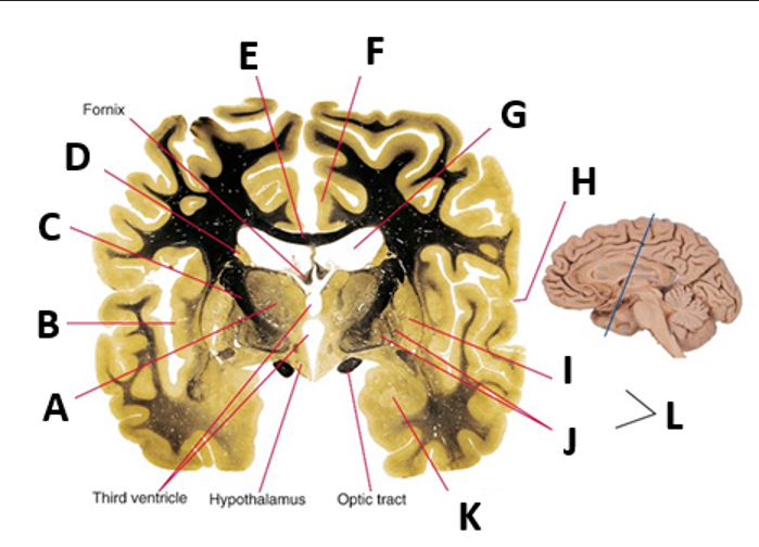
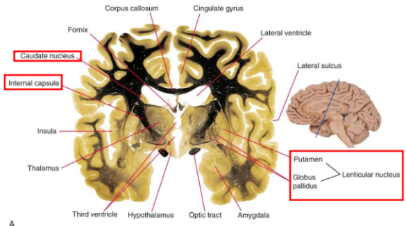
thalamus function? hypothalamus function?
thalamus → every path conveying subcortical info to cerebral cortex stops here (except olfactory)
hypothalamus → control autonomic fxn (visceral response, temp. reg., limbic system)
which nerves emerge from midbrain? cerebral peduncle function?
CN III (oculomotor) + CN IV (trochlear)
cerebral peduncles carry desc. projection from cerebral cortex
which cranial nerves emerge from basal pons? middle cerebellar peduncle function?
CN V (trigeminal) lat., CN VI (abducens) midline, CN VII (facial) + CN VIII (vestibulocochlear) cerebellopontine angle
middle cerebellar peduncle carries input to brain
which structures does the medulla contain?
pyramids → carry fibers destined for spinal cord
olive → protuberance post. to pyramids
CN XII (hypoglossal) emerge from pyramid-olive sulcus
cerebellum function? lobes?
coordinate movement + maintain balance
ant. (ant. to primary fissure) → coordinate trunk + limb movement
flocculonodular → receive input from vestibular system, control eye movement + posture
post. → largest, receives most cortical input, coordinate voluntary movement, balance/control, timing/precision
location of basal nuclei/ganglia? function?
neuronal cell body masses beneath cerebral cortex in each hemisphere
motor control + other
caudate nucleus → C shape, frontal to temporal
lenticular nucleus → putamen + globus pallidus, lat. + partial ant. to thalamus
what is the internal capsule of the basal nuclei/ganglia?
white matter, thick fiber sheet separating lenticular nucleus from thalamus + caudate nucleus
contains fibers interconnecting cerebral cortex, thalamus, caudate nucleus
function of amygdala and hippocampus?
emotional processing, memory formation, spatial navigation w/in limbic system
amygdala → emotional processing + memory
hippocampus → memory formation + spatial navigation
when does the notochord form? what does it initiate? what does it turn into?
first
initiates neural tube formation (via SHH)
induction of ectoderm → neural plate
becomes nucleus pulposus (IV disc)
what does the neural tube give rise to? what is produced after the neural tube fuses? what are the 3 dilations?
CNS (brain + spinal cord)
neural tube fuses → roof plate makes BMP + floor plate makes SHH
primary brain vesicle
prosencephalon
mesencephalon
rhombencephalon
what does the prosencephalon (forebrain) form? what forms the ventricles?
telencephalon → cerebral hemispheres (hippocampus, basal ganglia, olfactory cortex
vesicles → lateral ventricles (btwn lat. + 3rd = interventricular foramen of Monro)
diencephalon → hypothalamus, thalamus, pineal gland, neurohypophysis, retina
cavity = 3rd ventricle
what does the mesencephalon (midbrain) form?
ant. (visual) + post. (auditory) colliculi
cavity = cerebral aqueduct (of sylvius) → connects 3rd + 4th ventricles
what does the rhombencephalon (hindbrain) form? what occurs if there is failure of the rhombic lips to form properly?
metencephalon → pons + cerebellum
failure to dev. → greatest effect on motor fxn
myelencephalon → medulla oblongata
cavity = 4th ventricle
failure of rhombic lips to form = coordination problems
what does the cavity of the spinal cord become? what occurs after the neural tube closes?
central canal
neural tube close → neuroepithelial cells divide (3 layers)
ventricular, intermediate, marginal
what does ventricular zone (neuroepithelial layer) give rise to?
layer of ependymal cells lining central canal
primitive nerve cells (neuroblasts) → all neurons + glial cells of spinal cord
what does the intermediate zone (mantle layer) consist of? what originates from here?
neurons + glial cells of gray matter (spinal cord)
contains developing alar plate + basal plate
basal plate → hypoglossal nerve + nucleus ambiguus
what does the marginal zone contain? what does it form?
nerve fibers (axons) of neuroblasts of mantle layer + glial cells
forms white matter of spinal cord thru myelination of axons
what forms after neuroblasts are added to the mantle layer (intermediate zone)?
ventral thickenings (basal plates) → ventral motor horn cells, motor areas
dorsal thickenings (alar plates) → form sensory areas
where does the conus medularis extend to? where do nerves exit?
@ birth → L3, @ adult → term. L1-L2
lumbar, sacral, coccygeal nerves “pulled up” relative to IV foramina they exit from (disc herniation effect)
what do they centricles contain?
initially → amniotic fluid
later → choroid plexus develops w/in = CSF
what do neural crest cells give rise to? if migration fails? what does each spinal nerve and ganglion associate with?
PNS (peripheral nerves + enteric ganglia)
failure of migration → greatest impact on myelination in PNS
w. somite of developing embryo
non neural derivatives of neural crest cells? what can disruption in neural crest cells cause?
aortic arch, pharyngeal arch, adrenal medulla, chromaffin cells, melanocytes, schwann cells
disruption in neural crest cells → syndromes affecting face, heart, metabolism, nervous system
e.g: cleft lip/palate, treacher collins, tetralogy of Fallot albinism, pyloric stenosis, Hirschsprung disease
when does the neural plate fold? what does it give rise to? what do some cells differentiate into?
wk 4 → lat. edges elevate to form folds (fuse = neural tube)
cranial = brain, caudal = spinal cord, lumen = ventricular system of brain + central canal of spinal cord
some cells diff. into neural crest cells
what do the basal plate and alar plate in the brainstem give rise to?
basal → motor cranial nerve nuclei
alar → sensory cranial nerve nuclei
what are neural tube defects (NTD)? what can detect early NTD? what can prevent it? what can it present as?
failure of ant. or post. neuropore to close → congenital defect
elevated a-feto protein in amniotic fluid
folic acid
open defect in lumbar area
what is holoprosencephaly (HPE)? characteristics? mild characteristic?
loss of midline structure = malformation of brain + face
sulci + gyri absent
some mild cases → presence of single central incisor
what is anencephaly? characteristics?
upper NTD via failure of cranial/ant. neuropore of neural tube fusing during wk 4
failure of brain to fully develop (rudimentary brain present) + failure of bony cranial skull to form
incompatible with extrauterine life (survive only few hours)
what is spina bifida occulta? how is it “hidden”? when does it occur? characteristics? how can it be prevented?
vertebral arches fail to form properly → create vertebral defect in lumbosacral region
most common + mild form (involves 1+ vertebrae)
wk 3-8
tuft of hair + dimple over lower lumbar region
folic acid
if the anterior and posterior neuropore doesn’t close during wk 4 what NTD conditions could occur?
meningocele → meninges protrude thru vertebral defect + form CSF filled sac
meningomyelocele → meninges/spinal cord protrude + form CSF filled sac
rachischisis (myeloschisis) → post. neuropore of neural tube fails to fuse
what is hydrocephalus? what obstruction causes this? what does it impact?
abnormal accumulation of CSF in 3rd ventricle
obstruction of aqueduct of Sylvius (aqueductal stenosis) → prevents CSF of lat. + 3rd ventricle from passing into 4th ventricle + subarachnoid space (usually resorbed)
impacts midbrain (cerebral aqueduct)
what is arnold-chiari malformation? symptoms?
caudal herniation of cerebellar vermis/tonsils + medulla oblongata thru foramen magnum
symptoms → movement abnormalities, abnormal eye movements, motor + sensory deficit
compress medulla + CNIX, CNX, CNXII → difficulty swallowing, vibration during respiration (laryngeal stridor), diminished gag reflex, hoarseness