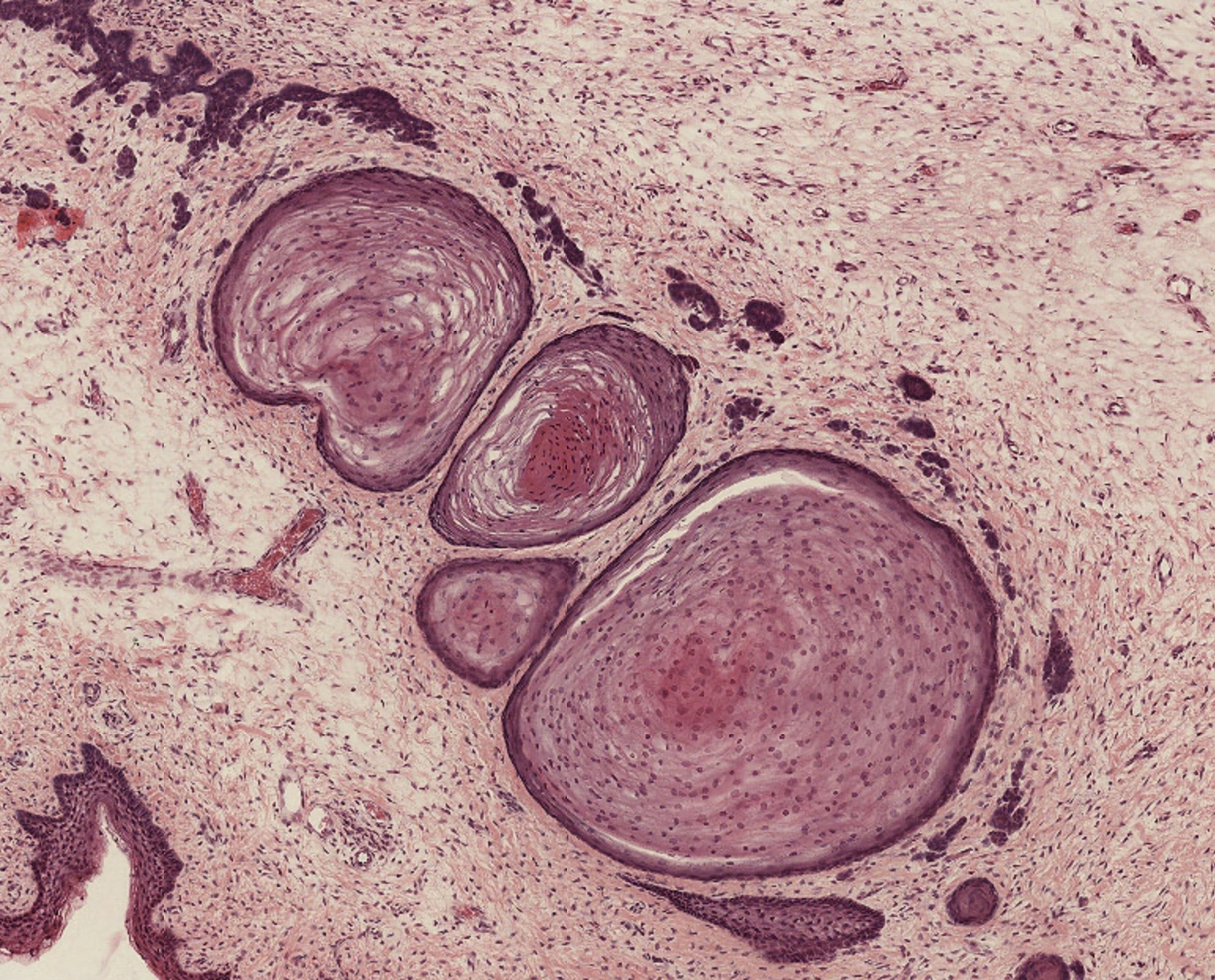
Tooth development histology
1/16
There's no tags or description
Looks like no tags are added yet.
Name | Mastery | Learn | Test | Matching | Spaced | Call with Kai |
|---|
No analytics yet
Send a link to your students to track their progress
17 Terms
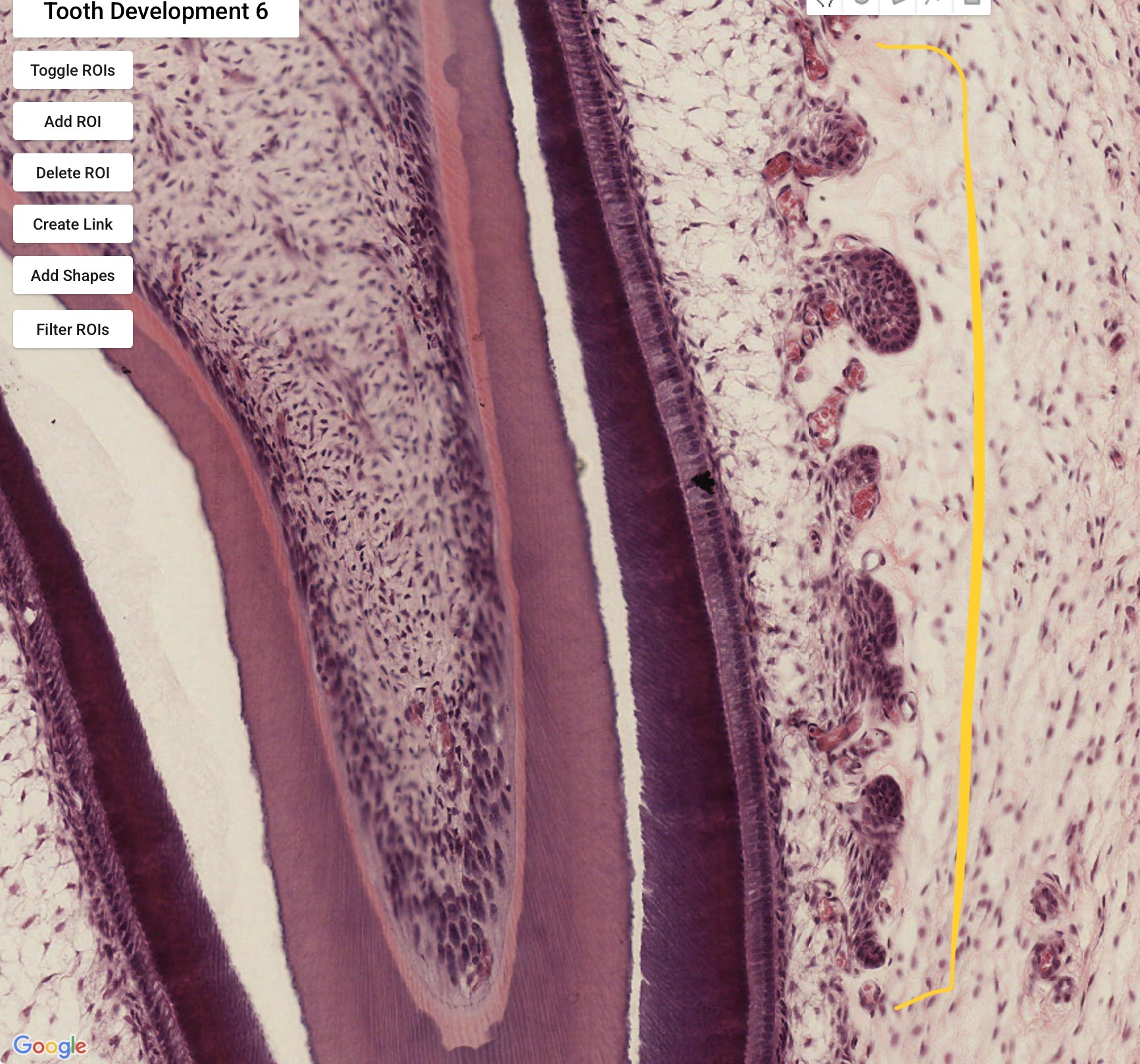
Folding of outer enamel epithelium around capillaries to allow for blood supply - as the layer of enamel formed from the IEE means it cannot access blood supply from the dental papilla

What is circled?
What could the white space be?
Enamel prisms
Enamel which has completely dissolved, so no staining as there is no matrix

Odontoblasts (which form dentine inwards)
IEE (which forms enamel outwards)
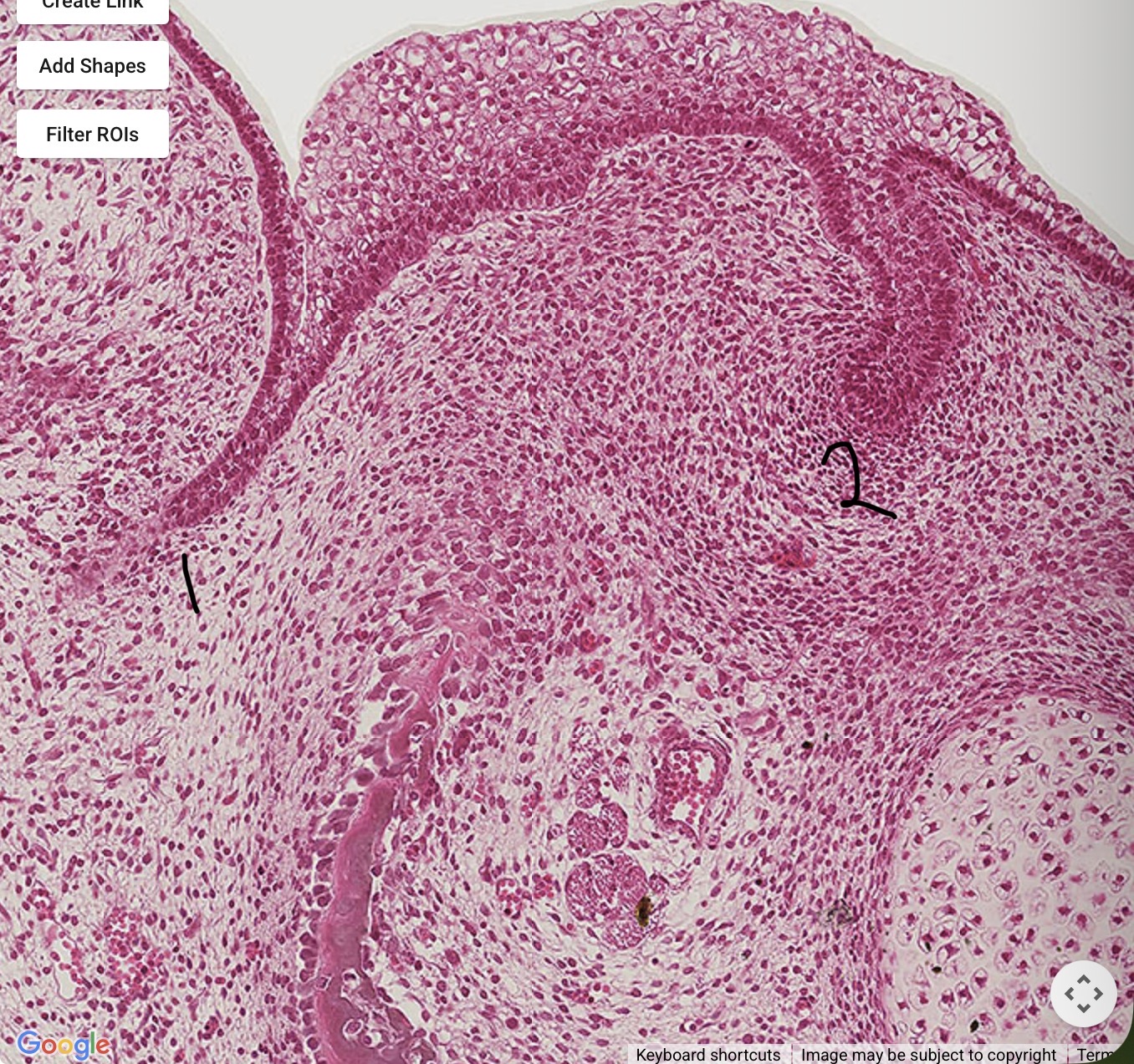
Vestibular lamina
Dental lamina
Enamel organ surrounding dental papilla
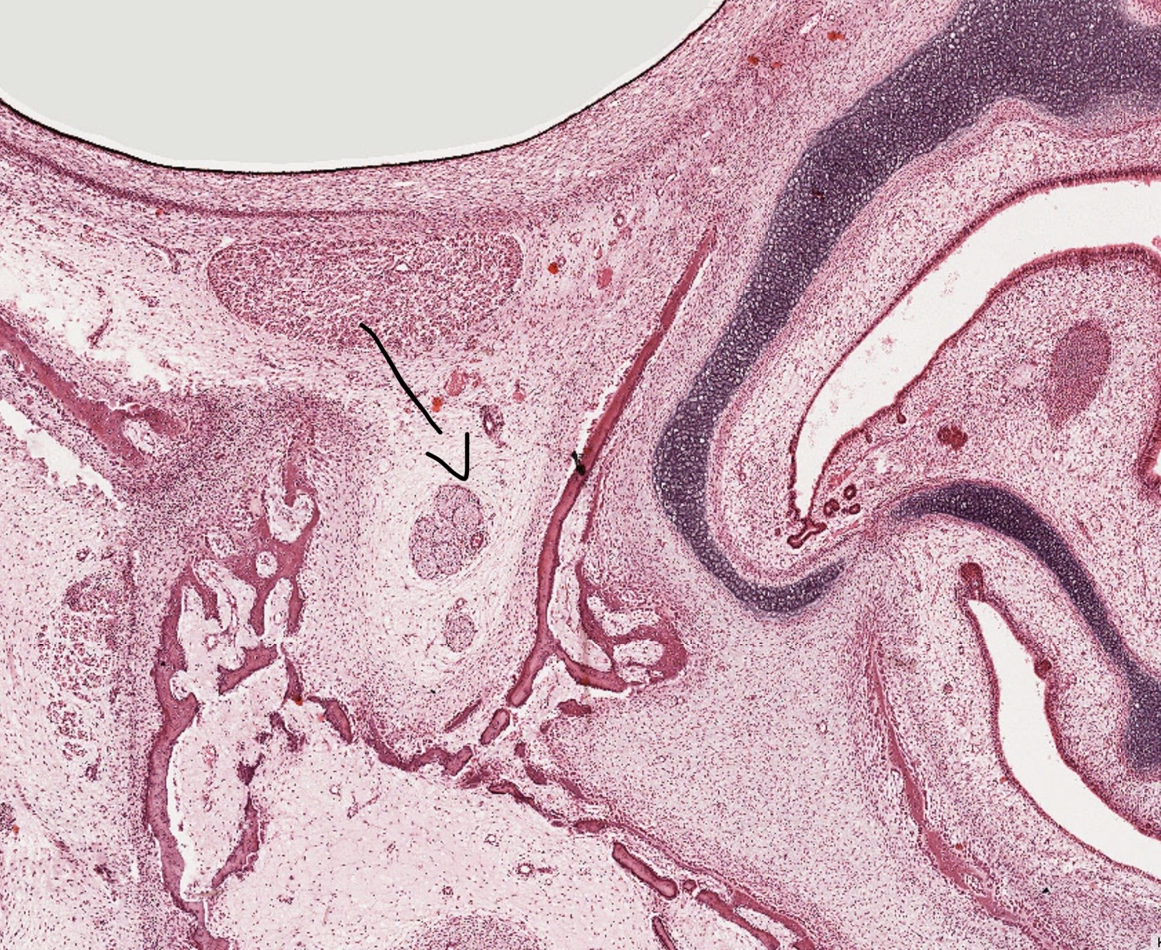
Infraorbital nerve
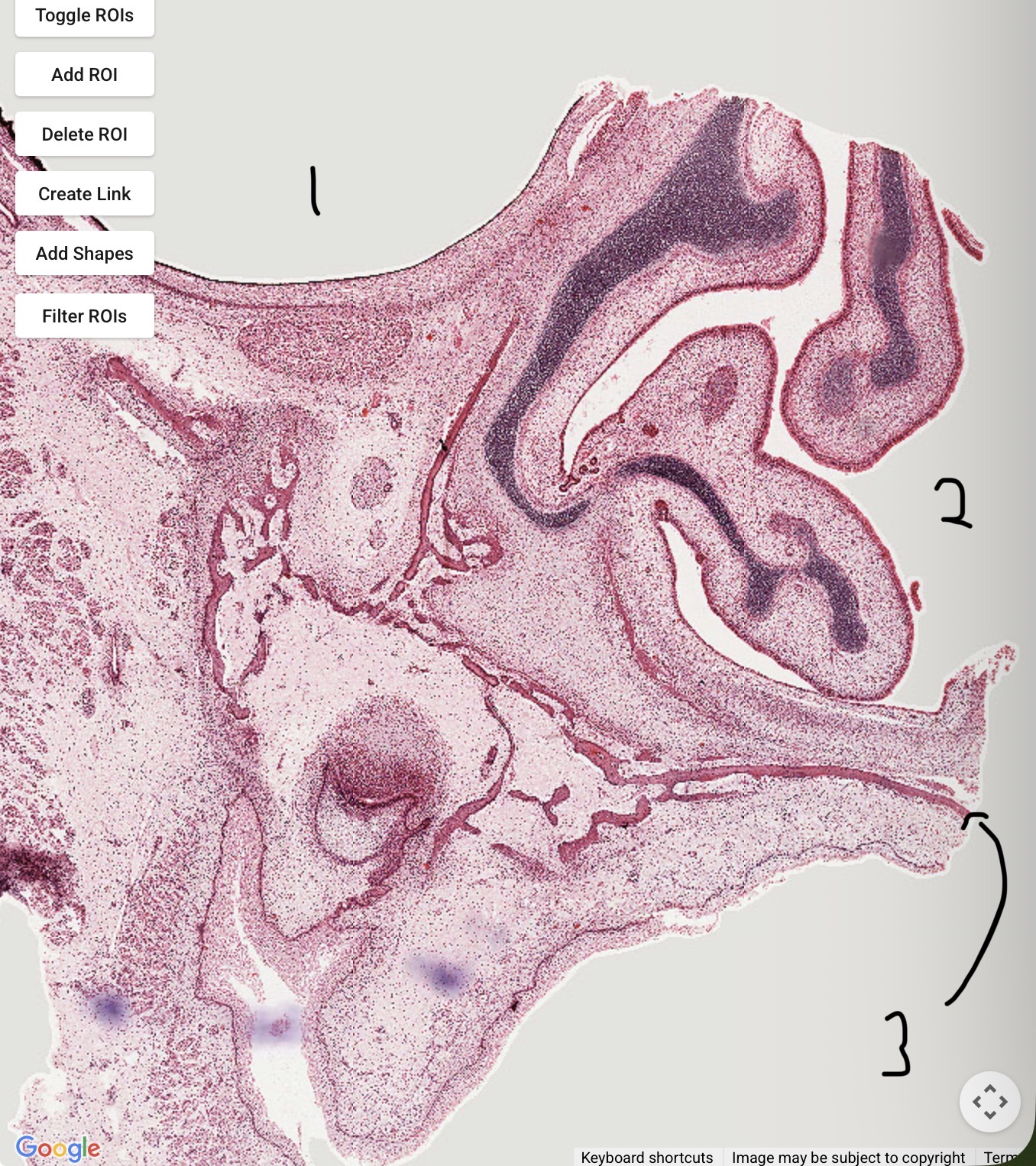
Orbit
Nasal concha
Hard palate
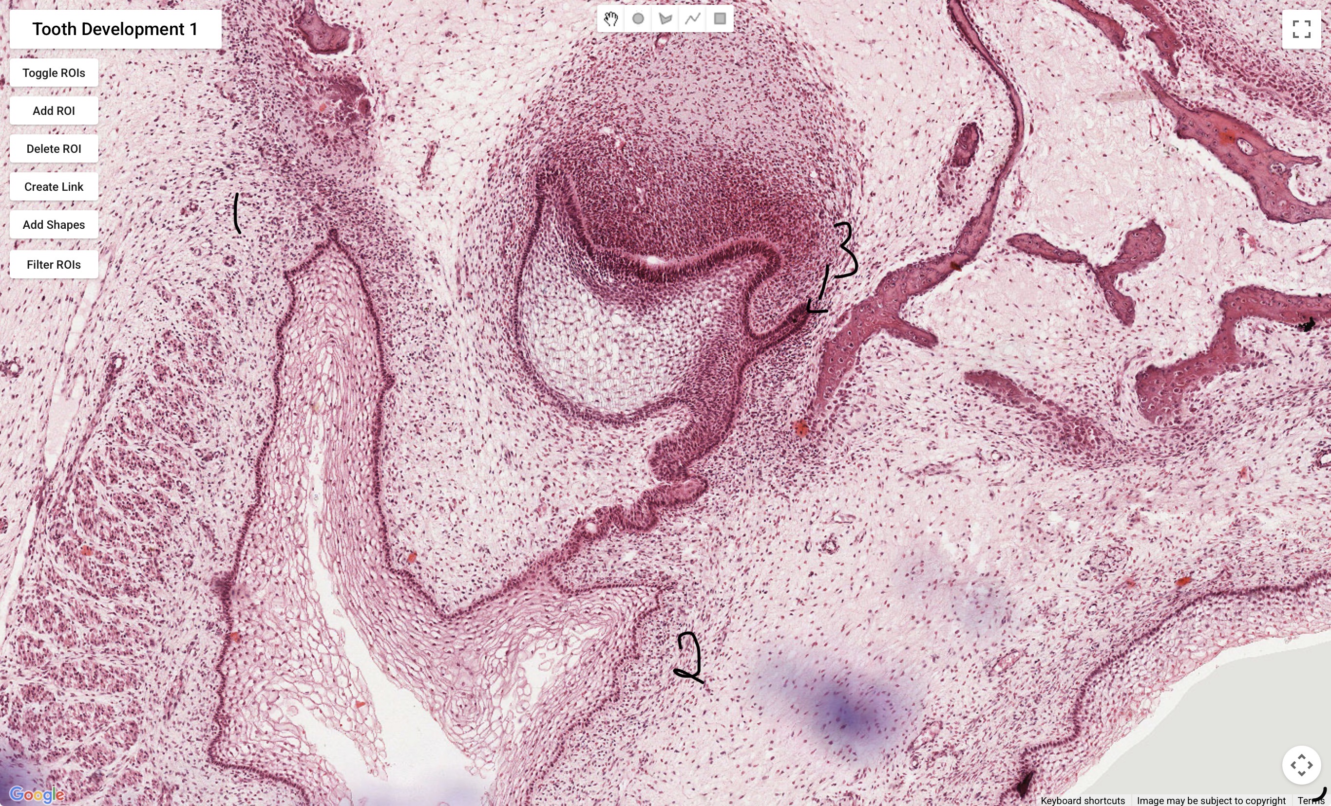
Vestibular lamina
Dental lamina
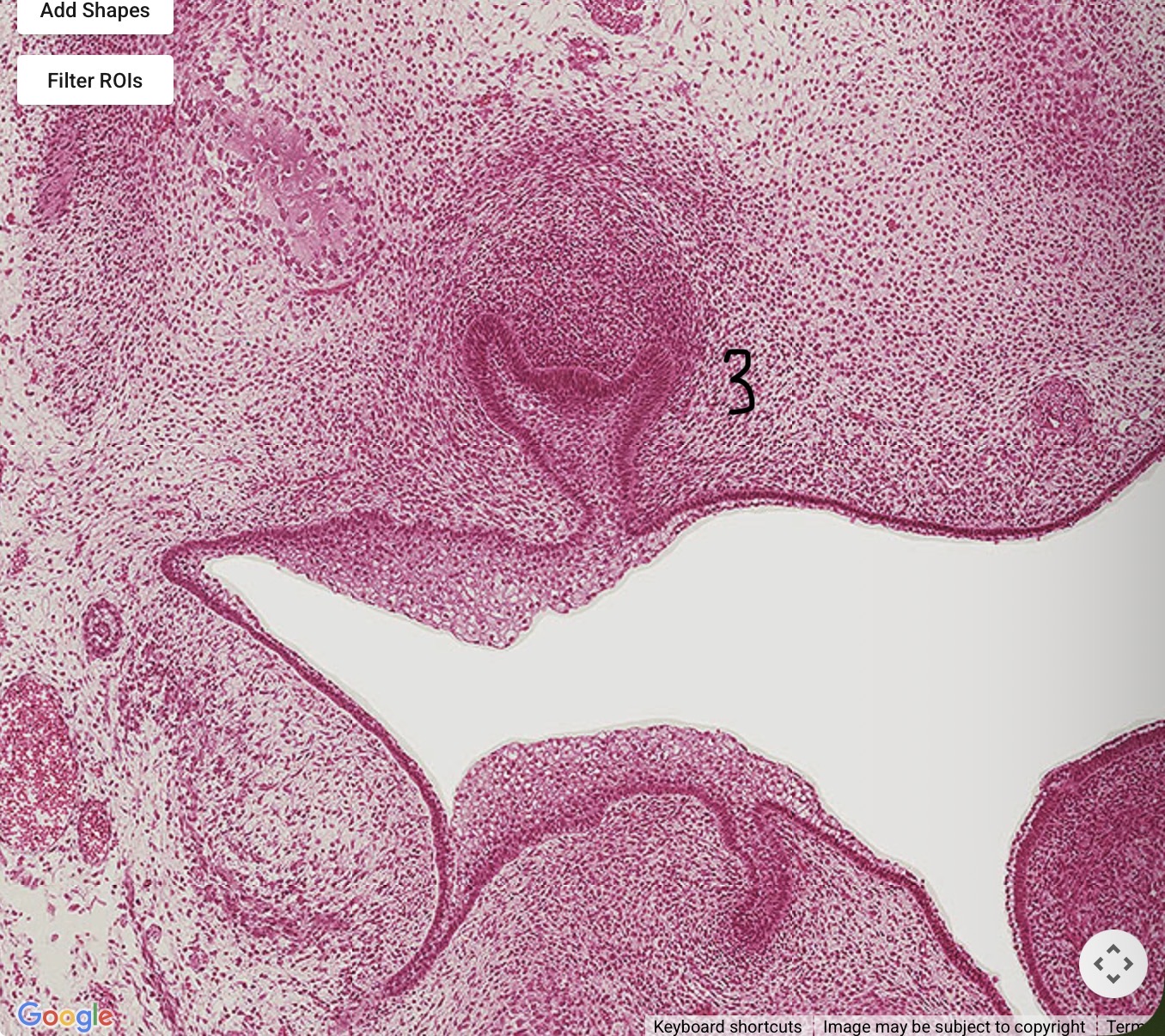
Successional lamina - derived from and lingual to the dental lamina

Dental follicle
OEE
IEE
Cervical loop
Dental papilla
Stratum intermedium
Stellate reticulum
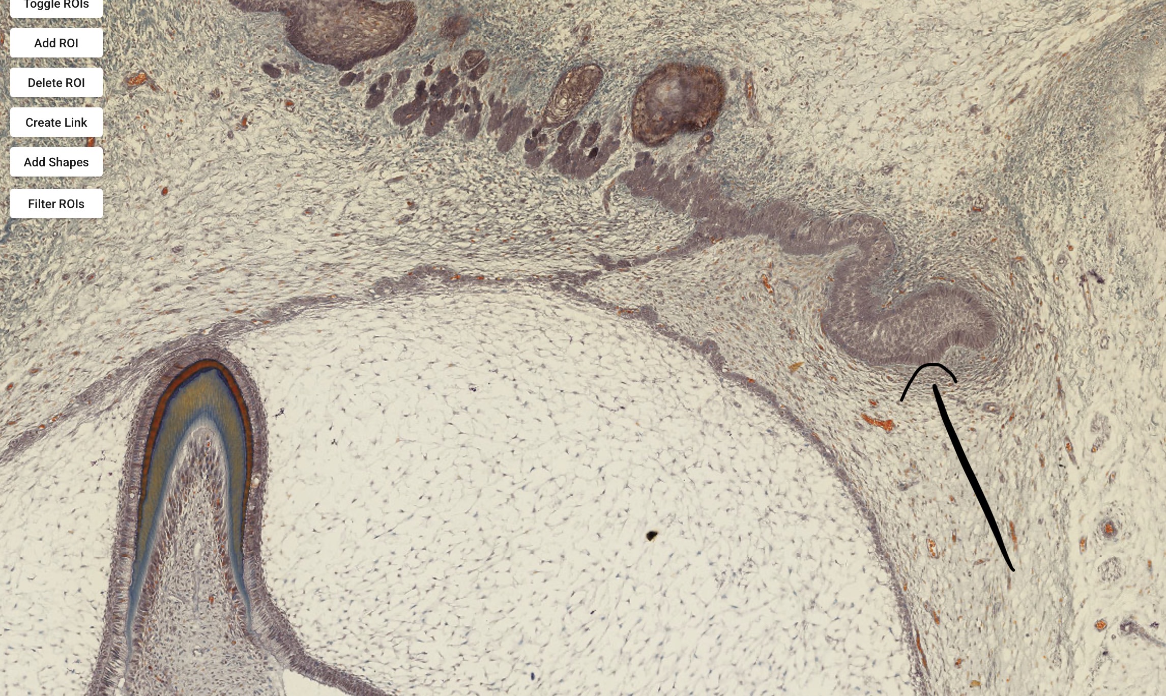
Successional lamina
What is the tooth germ composed of?
The enamel organ, dental papilla, and dental follicle
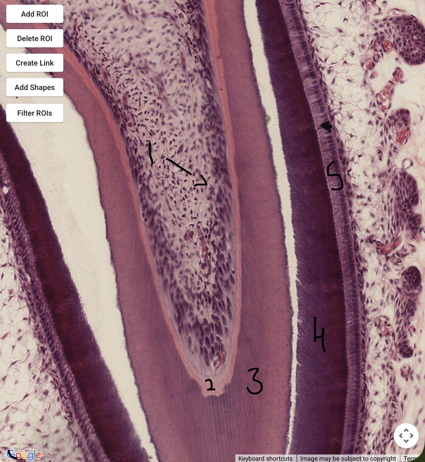
Odontoblasts
Predentine
Dentine
Enamel
Ameloblasts

Bone
Epithelium
Cell nests

Dental papilla
Dentine - purple dots are the odontoblast processes of dentinal tubules
Enamel
Ameloblasts
IEE
OEE
Successional lamina
What is the IEE continuous with?
Why is this?
The ameloblasts at the enamel surface
The IEE forms the IJE, by fusing with the OEE to form the REE, then the IJE as the tooth erupts
Give the timeline of tooth development
6 weeks - thickening of epithelium (primary epithelial band)
7 weeks - primary epithelial band splits into dental and vestibular lamina
8 weeks - tooth germ starts to form (bud stage)
9 weeks - cap stage
12 weeks - vestibular lamina formed
14 weeks - bell stage, dentinogenesis starts following breakdown of the dental lamina - cell nests and cell rests from this = cystic potential
HERS forms once crown of the tooth has been formed and root dentinogenesis has started.