BIOL 2160 EXAM IV - CHAPTER 12
1/132
There's no tags or description
Looks like no tags are added yet.
Name | Mastery | Learn | Test | Matching | Spaced | Call with Kai |
|---|
No analytics yet
Send a link to your students to track their progress
133 Terms
what are the basic components of the cardiovascular system?
Blood (fluid that carries materials to and from cells), Blood Vessels (series of tubes), and Heart (Pump)
what are the components of blood?
blood cells (erythrocytes, leukocytes, and platelets) and plasma
what does plasma mostly contain?
mostly water, and also salts and proteins (mostly antibodies).
what are erythrocytes?
red blood cells; carry oxygen bound to hemoglobin (4 iron molecules attached to it)
how does blood get their red color?
red blood cells contain hemoglobin that whenever oxygen binds to its iron molecules, it’ll turn deep red
what are leukocytes?
white blood cells; mediate immune system
what are platelets?
cell “fragments” and part of megakaryocytes; aid in blood clotting
what blood vessels transport blood away from the heart?
arteries (big) and then to arterioles (small)
what blood vessel allows exchange?
capillaries (smallest); tiny diameter forces blood to move through as slowly as possible to allow enough time for exchange to take place and this makes it the only “leaky” one out of the rest of the blood vessels
what blood vessel transport blood to the heart?
Blood leaving capillaries, on the way back to the heart, first flows into venules (small), and then into veins (big)
what are the chambers of the heart?
left and right atria, left and right ventricles
what’s a septum?
a muscular wall that separates two sides of the heart to keep the oxygenated blood on the left side of the heart from mixing with the deoxygenated blood on the right side of the heart
what is the role of deoxyenated blood?
blood RETURNING from your tissues. This blood has dropped off oxygen to your tissue cells and picked up CO2; means blood that is low in oxygen and high in CO2
what is the role of the right side of heart (right atrium and right ventricle)?
supplies deoxygenated blood to pulmonary circuit (lungs)
what is the role of the left side of heart (left atrium and left ventricle)?
supplies oxygenated blood to systemic circuit (body)
why do our veins look blue?
because there’s that’s a layer of fat around the veins diffracting the light and making them look blue
realistically, what’s the color of deoxygenated and oxygenated blood?
deoxygenated blood is a deep maroon or burgundy an oxygenated blood is bright red/cherry colored
Which chamber of the heart is the most muscular?
left ventricle
why is the left ventricle the most muscular (and also why the aorta is the largest vessel in the body)?
Because it has to pump blood the longest distance (all the way to your toes) while both atria only have to pump blood to the ventricles and the right ventricle only has to pump blood to the lungs, which are right next to the heart
what does the pulmonary artery carry?
carrying deoxygenated blood to the lungs so that it can be oxygenated
what is the aorta carrying?
carrying oxygenated blood to all of the body tissues
how does an individual get PDA (Patent Ductus Arteriosus)?
when the ductus arteriosus never closes, resulting in mixing of oxygenated and deoxygenated blood. Fairly common in premature babies
what are the results of PDA (Patent Ductus Arteriosus)?
Leads to shortness of breath and stunted growth and can lead to congestive heart failure later in life
what is Ductus Arteriosus?
a duct that connects the aorta and the pulmonary artery in embryonic development. Normally closes shortly after birth
what is the first step for the path of blood flow?
pulmonary circuit; blood vessels in lungs and blood vessels in the rest of the body (respiratory system provide oxygen to the blood passing through the lungs in the pumlonary circuit)
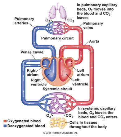
what is the second step for the path of blood flow?
systemic circuit; blood vessels in the rest of the body (oxygen gets transport to the cells in the body that are using oxygen for cellular respiration where these same cells are producing CO2 which will be diffused from the blood (pulmonary capillaries) to the lungs (alveoli) where it is exhaled)

what do heart valves do?
allow unidirectional blood flow to prevent back fllow of blood by opening; opening and closing controlled by pressure differences between chambers
what are the two sets heart valves?
atrioventricular valves and semilunar valves
what are the atrioventricular valves?
Mitral valve (left AV) and tricuspid valve (right AV)
what are the semilunar valves?
Aortic semilunar valve (left SL) and pulmonary semilunar valve (right SL)
what do the atrioventricular valves do?
prevent blood from leaving the ventricles and going back into the atria
what does the tricuspid valve do?
separates the right atrium from the right ventricle
what does the mitral (bicuspid) valve do?
separates the left atrium from the left ventricle
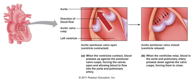
what do the semilunar valves do?
prevent blood from leaving the arteries and going back into the ventricles. ”Semilunar” means “half moon”, because that’s how they’re shaped
what does the pulmonary semilunar valve do?
separates the right ventricle from the pulmonary artery
what does the aortic semilunar valve do?
separates the left ventricle from the aorta
what are the ways to increase pressure in the heart?
add more blood, more blood = more pressure. & Contract. Once a chamber in the heart is filled with blood, the only way to build pressure is for that chamber to contract.
what area of pressure is blood always from and going to?
wherever blood moves in the heart, or in the vessels, it’s always moving to an area of lower pressure. And it’s coming FROM an area of higher pressure
how are AV valves open?
When pressure is higher in the atria than it is in the ventricles, these AV valves open and allow blood to flow to the ventricles and the ventricles are relaxed

how are AV valves closed?
When pressure is higher in the ventricles, these valves close to prevent blood from moving back into the atria and the ventricles are contracting
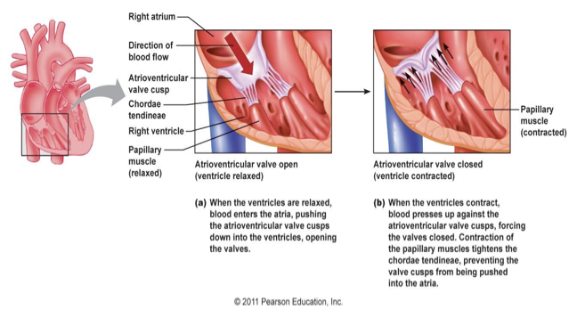
what keeps the AV valves from everting?
The papillary muscle contracts and pulls on chordinae tendineae to keep the valves from turning inside out when pressure gets really high in the ventricles. They maintain tension to keep the valves from everting.

how do SL valves open?
when pressure is higher in the ventricle than it is in the aorta; ventricles are contracting

how do SL valves close?
when pressure gets higher in the aorta than in the ventricle to keep the blood from moving back into the ventricles; ventricle is relaxed
Where is pressure highest, in veins, or in arteries?
arteries
how does the series flow through the pulmonary and systemic circuits?
Left ventricle pumps oxygenated blood through the aortic semilunar valve into aorta which then goes to all of the body cells
why is cutting an artery more dangerous than cutting a vein?
blood is coming directly from the heart here with nothing to slow it down while blood in veins is coming from capillaries so it’s slow
When you get blood taken, do they take it from a vein or an artery? Why?
vein because it it’s going into the tube, then it doesn’t matter how high the pressure is and its is the only blood vessel we can see through the skin
what is the second occurrence in the series flow through pulmonary and systemic circuits?
Aorta branches and carries blood to all organs and tissues in systemic circuit
what happens once blood is in the systemic circuit?
oxygen diffuses from the blood into the tissue cells that are using this oxygen for cellular respiration. That means pressure for oxygen is higher in systemic capillaries than it is in tissue cells. CO2 being produced by these cells diffuses into the blood and makes its way back to the right side of the heart, so that it can be pumped to the lungs. This means that CO2 pressure is higher in the tissue cells than it is in the systemic capillaries.
what is the third occurrence in the series flow through pulmonary and systemic circuits?
Blood is deoxygenated in organ capillary beds and travels back to heart through inferior and superior vena cava into right atrium; no valves are present so pressure is really high in right atrium
what is the fourth occurrence in the series flow through pulmonary and systemic circuits?
From right atrium, blood goes through tricuspid valve to right ventricle. When pressure in the right atrium is higher than pressure in the right ventricle, the tricuspid valve opens and allows deoxygenated blood to flow into the right ventricle.
what is the fifth occurrence in the series flow through pulmonary and systemic circuits?
The right ventricle pumps blood through the pulmonary semilunar valve into pulmonary artery. When pressure in the right ventricle is higher than pressure in the pulmonary artery, the pulmonary semilunar valve opens and allows deoxygenated blood to flow into the pulmonary artery. This blood will be pumped to the lungs, where oxygen will diffuse from lungs to the blood, and carbon dioxide will flow from the blood to the lungs.
what is the sixth occurrence in the series flow through pulmonary and systemic circuits?
Blood is oxygenated in the lungs and travels to left atrium through the pulmonary vein (the only oxygenated vein in the body). no valves present because of how high the pressure is
what is the seventh occurrence in the series flow through pulmonary and systemic circuits?
From left atrium, blood goes through bicuspid valve (aka mitral valve) into left ventricle. When pressure in the left atrium is higher than pressure in the left ventricle, the left AV valve, also called the bicuspid valve, or the mitral valve opens, allowing oxygenated blood to flow into the left ventricle.
what is the eighth occurrence in the series flow through pulmonary and systemic circuits?
the whole process starts over
what is the cardiac cycle (contraction of myocardium)?
the order the chambers of the heart contracts which is first both atria contract and the both ventricles contract
How does the heart achieve sequential contraction of atria and ventricles?
The heart contracts without neural input—it is myogenic
Rhythm of heartbeat is due to activity of pacemaker cells and conduction fibers
Force of heartbeat is due to activity of contractile cells
what does it mean rhythm of heartbeat?
Rhythm is how quickly it beats. This is due to electrical activity in the heart.
what does it mean by force of heartbeat?
Force is how hard the heart beats. This is due to contractile (muscle) cells.
what do cardiac muscle contain?
Contains sarcomeres—same contraction machinery as skeletal muscle, Contains gap junctions—multiple cells can contract together, and Action potentials are longer than in skeletal muscle cells
what do gap junctions do?
These direct connections between cells allow for synchronized electrical activity between cells. All of the cells in the heart are connected by gap junctions. So if you send an electrical signal through one, it will travel through all of the other cells eventually.
what are the types of cardiac muscle cells?
cardiac pacemaker cells, cardiac conduction fibers, and cardiac contractile cells
what do cardiac pacemaker cells do?
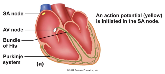
behave like neurons and produce action potentials and set rhythm of heartbeat through two nodes (cluster of cells): SA node and AV node
what is the Sinoatrial Node (SA) node?
in the top of the right atrium. It produces an action potential that makes both atria contract
what is the Atrioventricular Node (AV) node?
at the top of the ventricles. It produces a second action potential that makes both ventricles contract
what does cardiac conduction fibers do?
are like the electrical wiring in the heart. They simply allow the action potentials that were produced by the SA node and AV node to propagate throughout the heart. They don’t PRODUCE the action potential, and they don’t RESPOND to it. They simply pass it along (Transmit rhythm of heartbeat)
what do cardiac conduction fibers include?
internodal pathway, interatrial pathway, Purkinje fibers
what do cardiac contractile cells do?
behave like muscle cells. They contract when they receive the action potentials produced by the nodes and transmitted by the conduction fibers. (generate the contractile force)
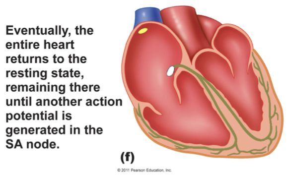
what first happens in the conduction system of the heart?
an action potential (yellow) is initiated in the SA node
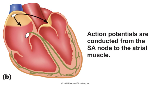
what’s the second thing that happens in the conduction system of the heart?
action potentials are conducted from the SA node to the atrial muscle
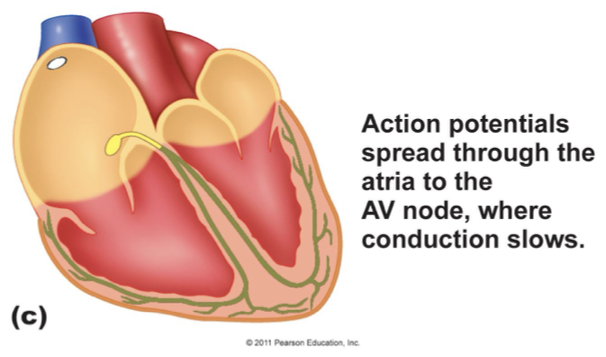
what’s the third thing that happens in the conduction system of the heart?
action potentials spread through the atria to the AV node, where conduction slows (AV nodal delay after atria contract)
what’s the purpose of AV nodal delay?
allows enough time for the ventricles to fill up COMPLETELY before they start contracting. If the delay were shortened, you would have less blood leaving the heart per heartbeat. And the heart would have to work harder.
what’s the goal of a heartbeat?
heart wants to have happen is for as MUCH blood to leave the ventricles per heartbeat, as possible. This keeps the heart from having to work as hard. In order to accomplish this, the ventricles need to be FULL of blood before they contract.
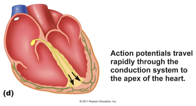
what’s the fourth thing that happens in the conduction system of the heart?
action potentials travel rapidly through the conduction system to the apex of the heart
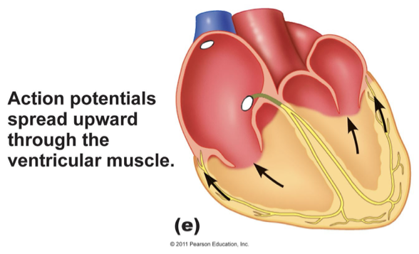
what’s the fifth thing that happens in the conduction system of the heart?
action potentials spread upward through the ventricular muscle
what’s the sixth thing that happens in the conduction system of the heart?
eventually, the entire heart returns to the resting state, remaining there until another action potential is generated in the SA node
Why is left arm pain a possible indication of a heart attack?
The nerve that signals from your heart to your brain enters the spinal cord right next to the nerve that signals from your left arm to your brain. When the nerve from the heart sends the pain signal to the brain, it releases neurotransmitter onto the appropriate synapse in the spinal cord. During this process, some neurotransmitter diffuses to the neighboring synapse and binds receptors on the nerve fiber that carries sensory information to the brain from the left arm.Because of this, the brain receives 2 different pain indication signals: one from the heart, and one from the left arm.
what is the ionic basis of electrical activity of the heart?
pacemaker cell action potentials (SA and AV node) and cardiac contractile action potentials
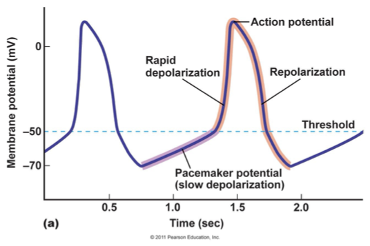
why do pacemaker cells don’t have a steady resting membrane potential?
These cells produce one action potential followed by another. There is NEVER a time where the membrane potential levels out, because there is never a time when you DON’T need your heart to beat. (Spontaneously depolarize and fire action potentials)
what does it mean if there’s a flat line?
that you’re dead because the pacemaker cell is always in the process of depolarizing, repolarizing, or hyperpolarizing
why do cardiac pacemaker cells spontaneously depolarize?
because of the activity of “funny” channels (HCN channels (HCN = hyperpolarization-activated and cyclic nucleotide-gated channels))
how are “funny” channels activated?
can be activated by cyclic-nucleotides like Cyclic AMP, but are activated in the pacemaker cells through hyperpolarization of the membrane. In other words, the undershoot / hyperpolarization at the end of one action potential opens these channels, causing a slow depolarization that results in the next action potential.
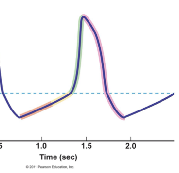
what is happening in orange?
Voltage-gated K channels close
“Funny” channels (let Na in, K out) open: these channels open at -70 mv, very negative membrane potential, the inward Na current is much bigger than the outward K current because of the driving force. depolarization (to -55 mV)
PNa↑ PK↓: there are a LOT more voltage-gated K channels in the membrane than there are funny channels. And while the membrane is STILL permeable to K, it’s just not AS permeable as it was when all of those voltage-gated K channels were open.
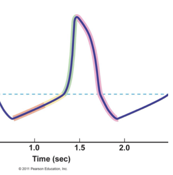
what is happening in yellow?
“Funny” channels close
T-type voltage-gated Ca channels open: t for temporary because channel is only open for a short period of time and Ca comes in quickly and depolarizes because chemical force (conc. gradient) is enormous so electrical force doesn’t matter making it able to bring membrane to threshold (-55 mV to -50 mV)
PNa↓ PCa↑

what is happening in green?
Voltage-gated L-type Ca channels open: L stand for longer duration for when the channel is open and Ca enters and depolarizes the cell further
Voltage-gated Na channels open: further depolarization causes steep rising phase (-50 mV to +60 mV)
PCa↑↑ PNa↑

what is happening in purple?
Voltage-gated K channels open: K leaves the cell, repolarizing the membrane, and ultimately leading to the undershoot (hyperpolarization) that opens funny channels and starts the next action potential
Voltage-gated L-type Ca channels close
Voltage-gated Na channels inactivate: go into refractory at the peak of action potential (+60 mV to -90 mV)
PK↑ PCa↓ PNa↓
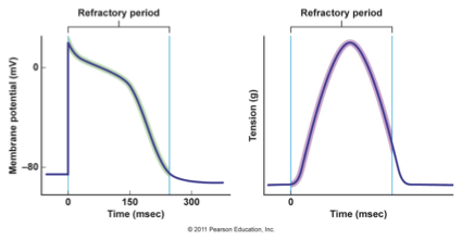
Why must contractile cell (muscle cells of the heart) action potentials be so long?
so the contractile cell will ALWAYS have enough time to contract AND relax before it receives another action potential; this prevents tetanus which can kill you if the heart experiences this (one action potential = one contraction)
how does an individual experience tetanus?
in skeletal muscle, if the muscle keeps getting action potentials telling it to contract, before it has had a time to relax, the muscle will contract harder and harder until it reaches maximal contraction (charlie horse)
how is tetanus prevented in the heart?
At the peak of the action potential, the L-type Ca channels close slowly, resulting in a very slow repolarization, and a long refractory period. During absolute refractory, no new action potentials can be produced
how is current spread throughout the heart in Excitation-Contraction Coupling in Cardiac Muscle?
gap junction connecting two different cells in the heart to contractile cell
what is the main difference between the muscle contraction mechanism between a skeletal muscle and a cardiac muscle?
In skeletal, this Ca comes from inside the cell, from the SR. In smooth muscle, the Ca comes from outside the cell. In cardiac, it comes from both. Ca entering results in Ca being released from the SR through the mechanism Calcium Induced Calcium Release (CICR
what happens to Ca following a contraction in Excitation-Contraction Coupling in Cardiac Muscle?
Calcium gets pumped back into the SR, and it also gets pumped out of the cell through the plasma membrane Ca ATPase. Both of these use ATP
what is another method to pump Ca out of the cells in Excitation-Contraction Coupling in Cardiac Muscle?
the Na / Ca exchanger, which is an antiporter, uses secondary active transport to move calcium out of the cell
what is the muscle action potential triggered by in skeletal muscle Excitation-Contraction Coupling?
by motor neuron ACh release
what happens to the DHP voltage sensor of skeletal muscle Excitation-Contraction Coupling?
DHP voltage sensors on plasma membrane open ryanodine receptors on SR
where does Ca come from in skeletal muscle Excitation-Contraction Coupling?
Ca released from SR
what removes Ca in skeletal muscle Excitation-Contraction Coupling?
Ca removed from cytosol by SERCA pumps on SR
whats triggers muscle action potential in cardiac muscle Excitation-Contraction Coupling?
by positive current spread through gap junctions
whats happens to the DHP voltage sensors in cardiac muscle Excitation-Contraction Coupling?
DHP voltage sensors open ryanodine receptors on SR
what happens to the voltage-gated Ca channels in cardiac muscle Excitation-Contraction Coupling?
Voltage-gated Ca channels on plasma membrane also open