biomechanics mmm quiz 4- dpt
1/19
There's no tags or description
Looks like no tags are added yet.
Name | Mastery | Learn | Test | Matching | Spaced | Call with Kai |
|---|
No analytics yet
Send a link to your students to track their progress
20 Terms
What muscle attaches to the medial border of the tibia:
no muscle attaches to the medial border of the tibia
What nerve typically passes around the neck of the fibula:
common peroneal (fibular)
The distal tibia and fibula form the ankle mortise. These structures articulate with which tarsal bone:
talus
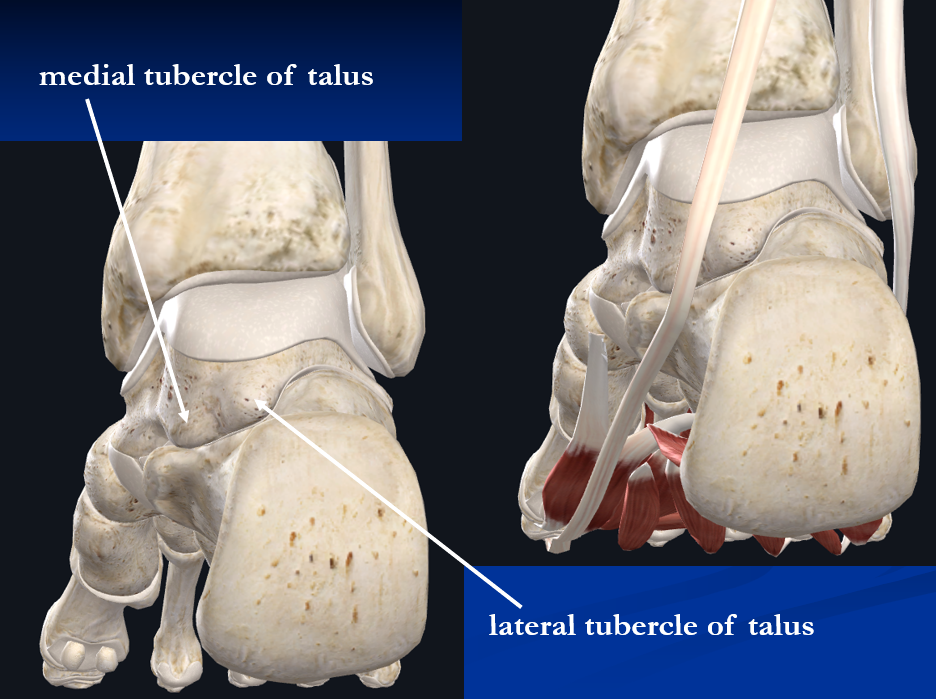
The medial and lateral tubercles of the talus are pictured below. Together they form a small channel for the passage of one of the tendons of the foot. What is the name of the tendon that passes between these tubercles:
flexor hallucis longus
The subtalar joint involves the articulation between the talus and the:
calcaneus
The medial process of the calcaneus pictured below is the attachment of what ligamentous structure:
plantar fascia
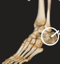
What is the bony landmark of the foot circled in the following picture:
peroneal tubercle
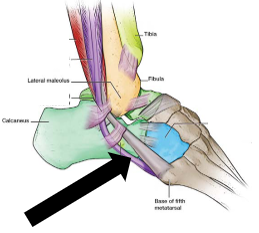
One tendon passes superior to the landmark circled above in Question #7, and one tendon passes inferior to this landmark. Which tendon, pointed to by the arrow in the following picture, passes inferior to this landmark:
peroneus longus
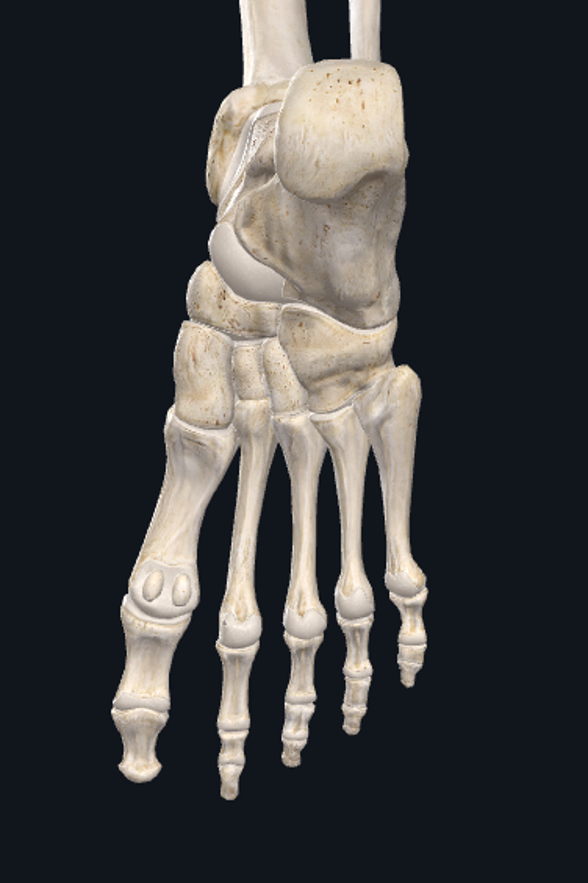
The most prominent tuberosity on the medial aspect of the foot is pictured below. This tuberosity is part of the bone that functions as the keystone of the medial longitudinal arch. What is the name of this tuberosity:
navicular tuberosity
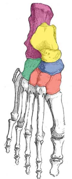
The foot consists of both longitudinal and transverse arches. The keystone of the transverse arch that runs across the midfoot is this bone. This bone is also the high point of the dorsum of the foot:
middle cuneiform
Two of these muscles have distal attachments around the base of the 5th metatarsal. Which one of these muscles does NOT have an attachment to the 5th metatarsal:
peroneus longus
Which of the following statements about the tibialis anterior is NOT correct:
the proximal attachment of the tibialis anterior is the medial tibia and interosseous membrane
Which of the following muscles could still plantarflex the ankle if the Achilles tendon were completely torn:
tibialis posterior
Which of the following muscles is both a talocrural dorsiflexor and a subtalar everter:
extensor digitorum longus
Which of the following muscles is both a talocrural plantarflexer and a subtalar inverter:
flexor hallucis longus
There is only one foot intrinsic muscle located on the dorsum of the foot. This muscle is the:
extensor digitorum brevis
Which of the following is a 2 joint muscle:
gastrocnemius
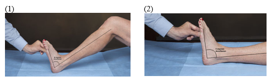
Your patient is lying on a table with her knee flexed. She maximally dorsiflexes her ankle and is able to dorsiflex her ankle 20 degrees (#1 in picture below). She then extends her knee and again maximally dorsiflexes her ankle. This time she is only able to dorsiflex her ankle 5 degrees (#2 in picture below). What is causing this change in available ROM:
the gastrocnemius becomes passively insufficient when the knee is extended and does not allow as much dorsiflexion
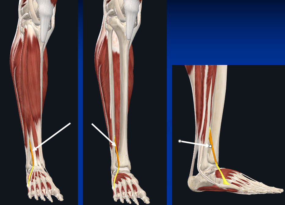
There are 3 different "peroneal" muscles. To which peroneal muscle are the arrows in the following picture pointing:
peroneus tertius
What artery lies between the extensor hallucis longus tendon and the extensor digitorum longus tendons:
dorsalis pedis