respiratory system anatomy and histology
1/22
There's no tags or description
Looks like no tags are added yet.
Name | Mastery | Learn | Test | Matching | Spaced | Call with Kai |
|---|
No analytics yet
Send a link to your students to track their progress
23 Terms
upper respiratory tract is part of what system?
conducting system
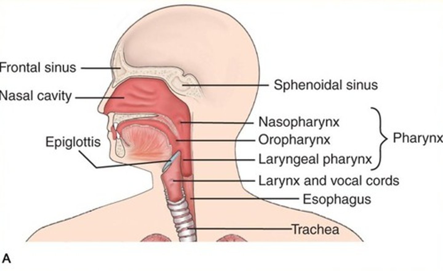
upper respiratory structures
nasal cavity
pharynx
larynx
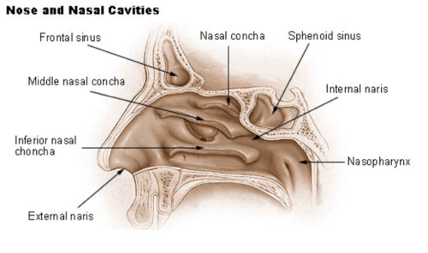
nasal cavity
nares (nostrils)
nasal conchae/turbinate bones- inferior, middle, superior
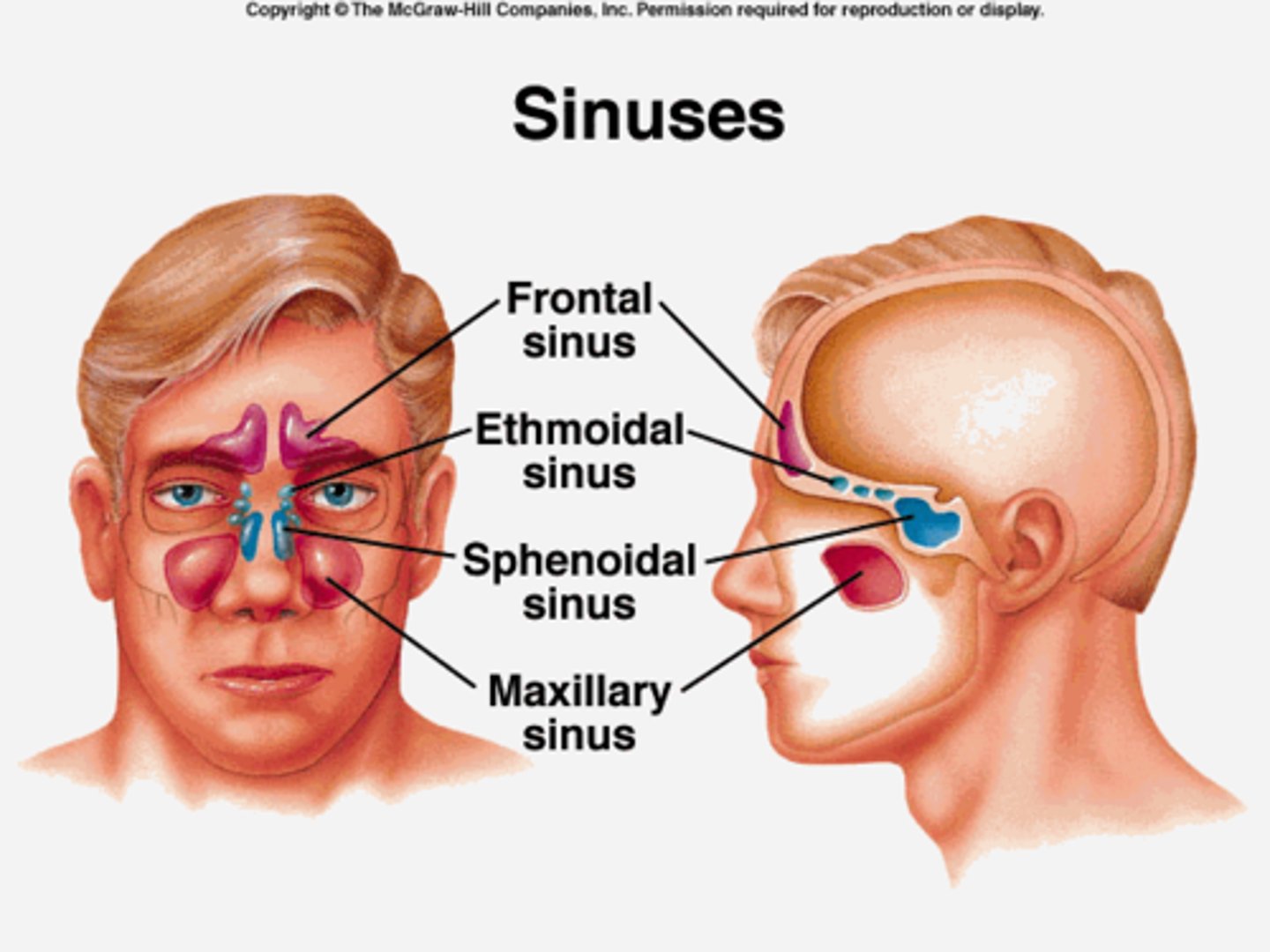
paranasal air sinuses- frontal, ethmoidal, sphenoidal, maxillary
paranasal air sinuses diagram
pharynx structures
nasopharynx- posterior to nasal cavity; sniff sound
oropharynx- posterior to oral cavities; hock sound
laryngopharynx- passageway for air and food (splits into larynx and esophagus); swallow sound
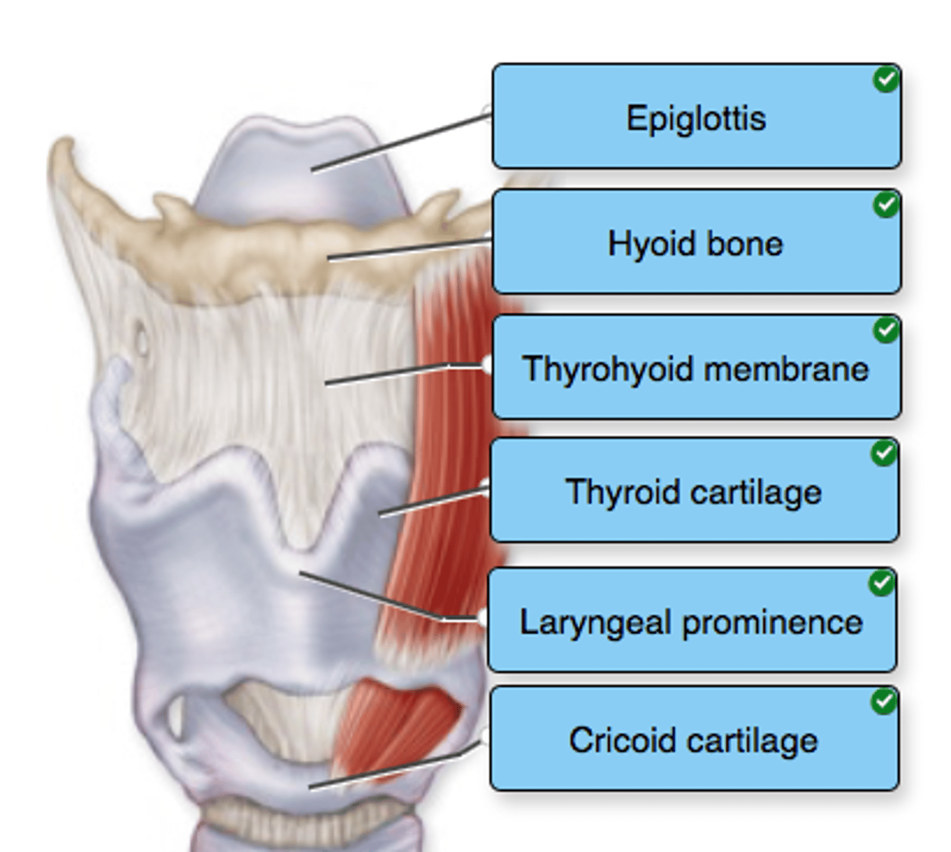
larynx cartilages
epiglottis- leaf shaped; blocks food from entering into lower respiratory tract (covers the glottis)
thyroid- shield shaped- joins at prominence "adams apple"; cricoid cartilage- ring shaped
larynx vocal folds
folds in mucous membrane
upper (false
lower (true)- vibrate as air passes
larynx diagram
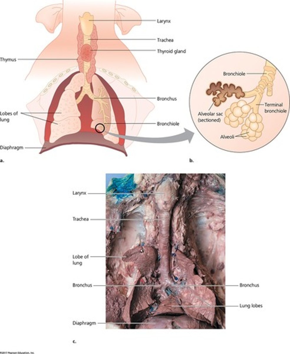
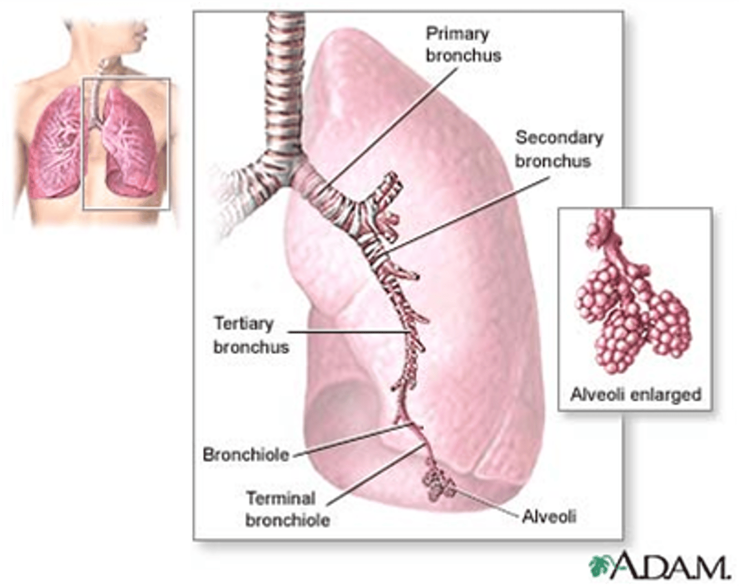
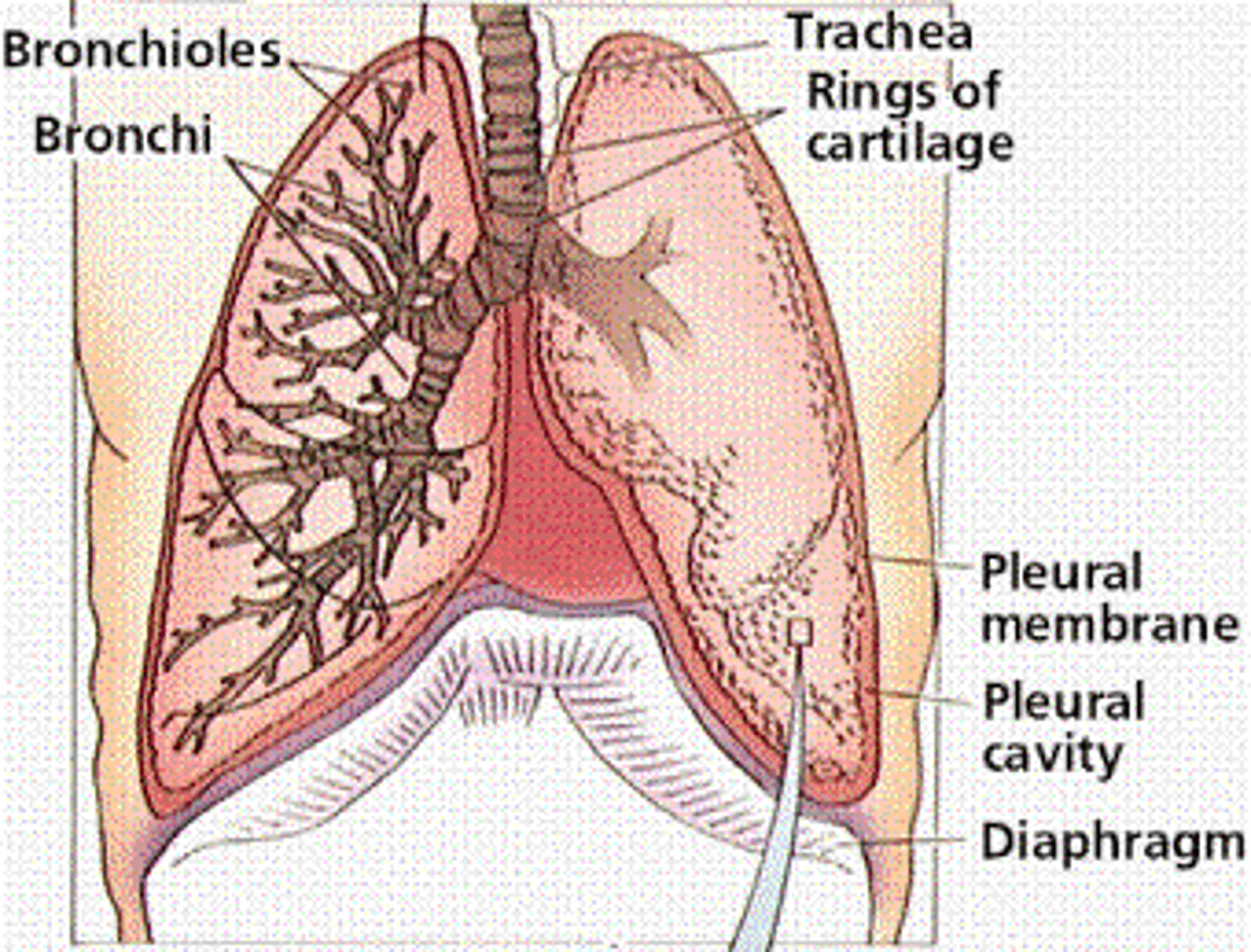
lower respiratory system includes
trachea
within lungs- bronchi (primary, secondary, tertiary); bronchioles (terminal and respiratory); alveoli (alveolar sacs)
lower respiratory tract includes what systems
conducting system- trachea terminal bronchioles
exchange system- respiratory bronchioles and alveoli
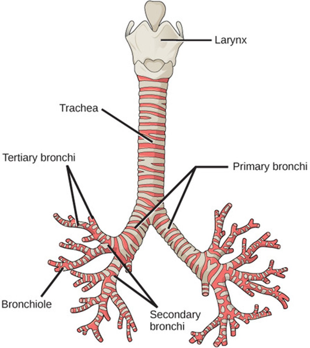
lower respiratory system diagram
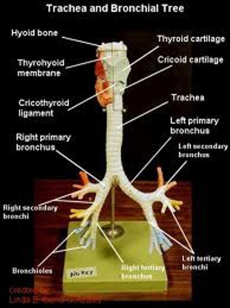
trachea: anatomy
posterior to hear
made of C shaped rings of cartilage- not complete rings to allow for esophagus to expand; structure and support to prevent collapse
branches into primary bronchi
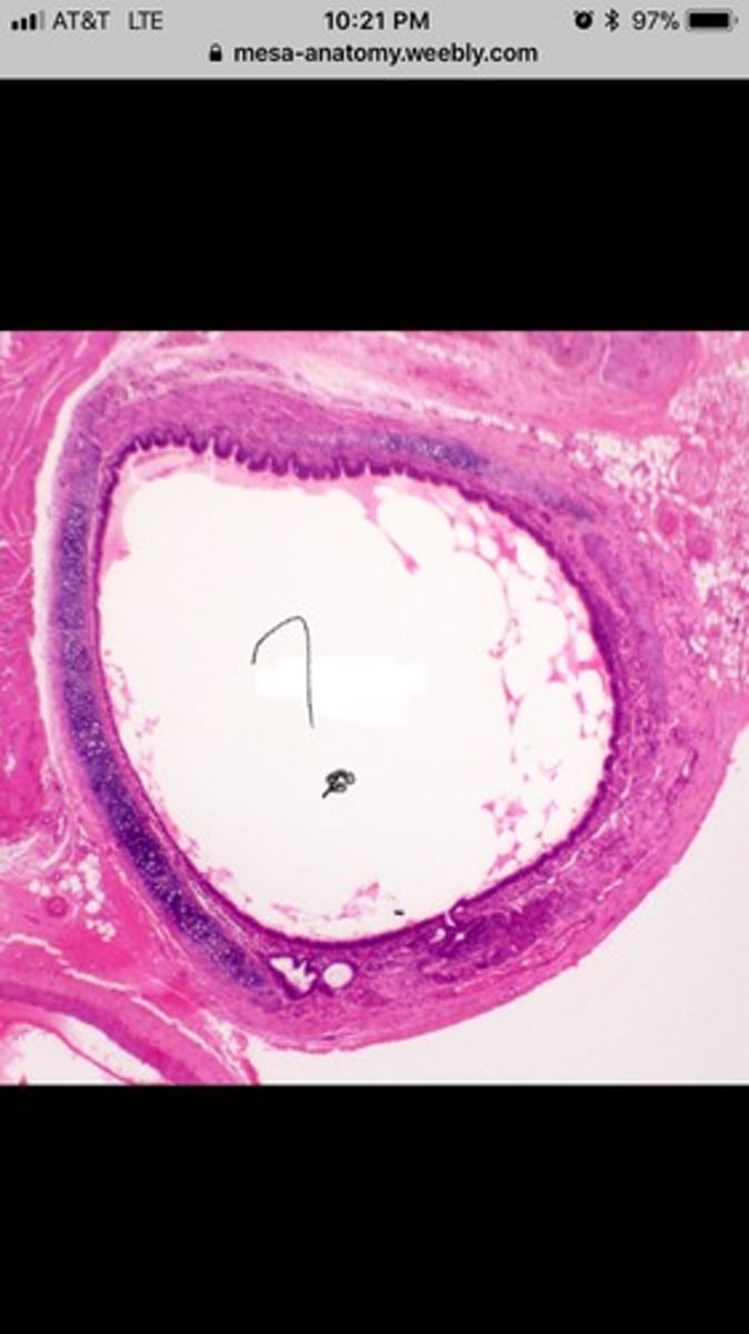
trachea: histology
layers- mucosa- ciliated pseudostratified columnar epithelium with goblet cells (mucus producing cells); mucus sticks to dirt/dust particles and is swept out of respiratory tract by cilia
submucosa
cartilage- hyaline cartilage
adventitia- external layer
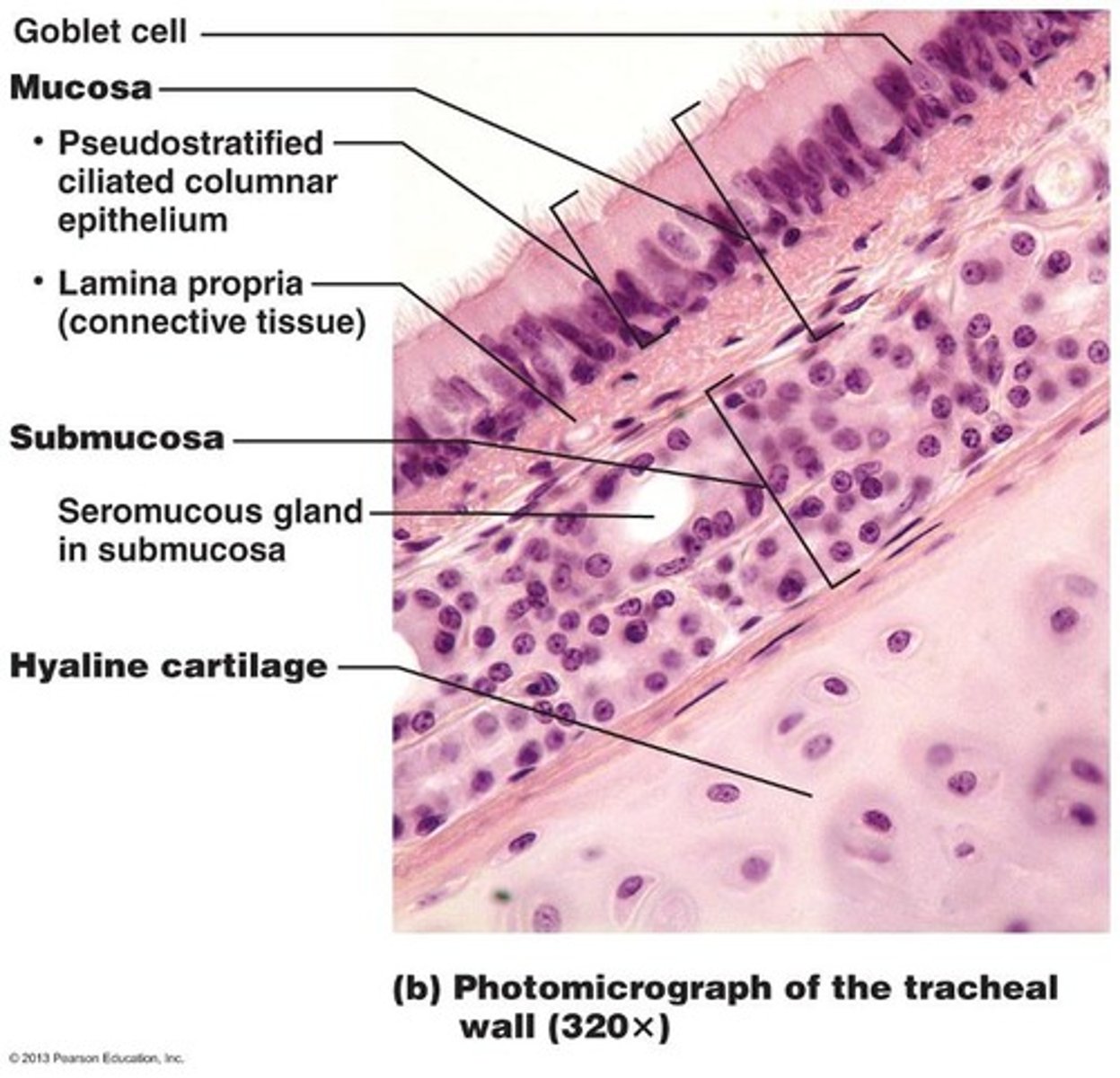
trachea histology ex.
bronchi: anatomy
primary- right and left; divides further once inside lungs; full ring of cartilage
secondary and tertiary (aka intrapulmonary bronchi)- in humans 3 lobes in right lung and 2 lobes in left lung (bc of heart); patches of cartilage
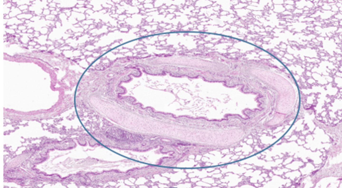
intrapulmonary bronchi: histology
ciliated pseudostratified columnar epithelium- goblet cells still producing mucus
plates of hyaline cartilage- no rings
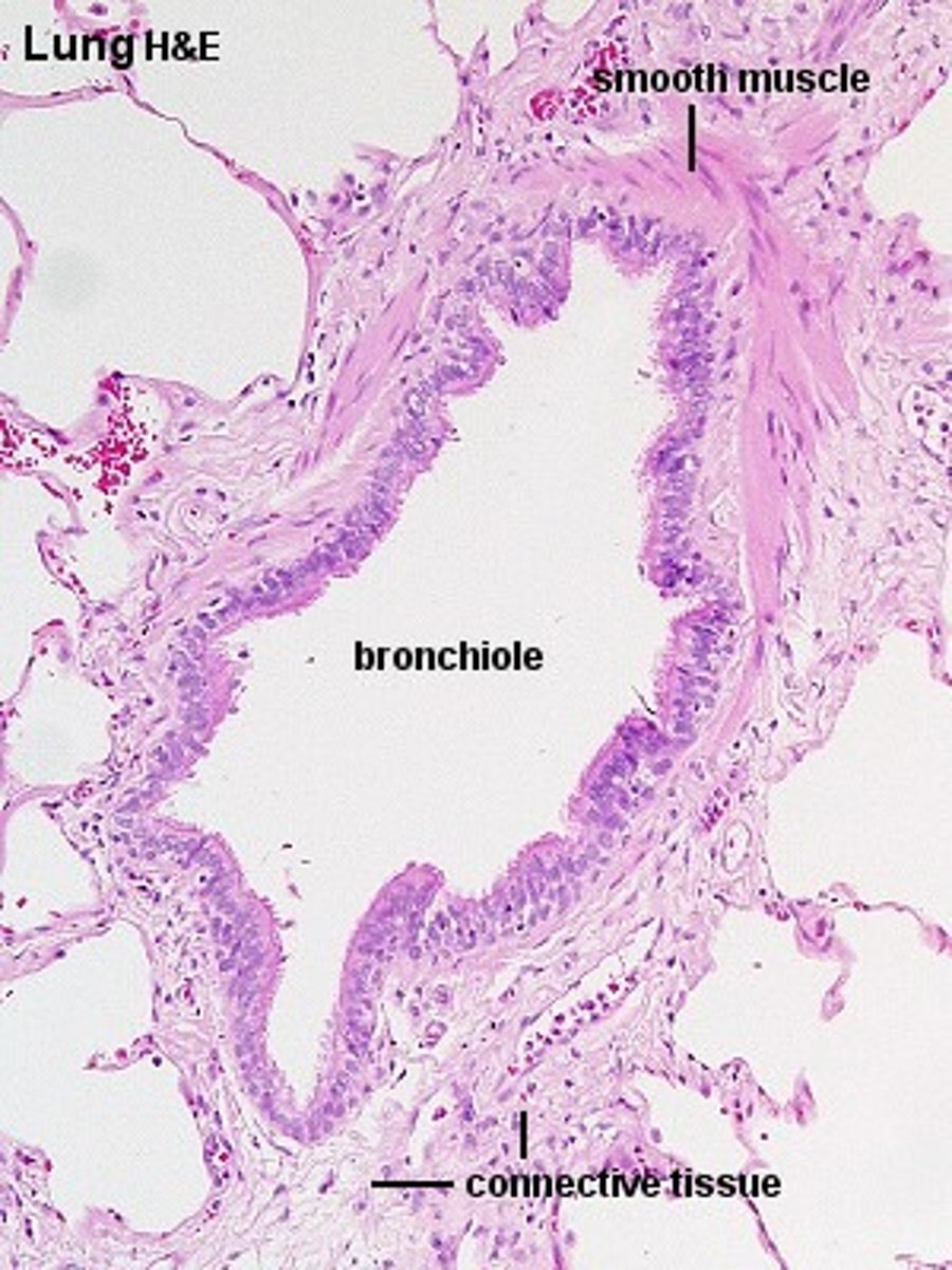
bronchioles: anatomy
no cartilage just smooth muscle- bronchoconstriction (constriction of bronchiole, response to cold/allergens); bronchodilation (dilation of bronchioles, response to heat/caffeine)
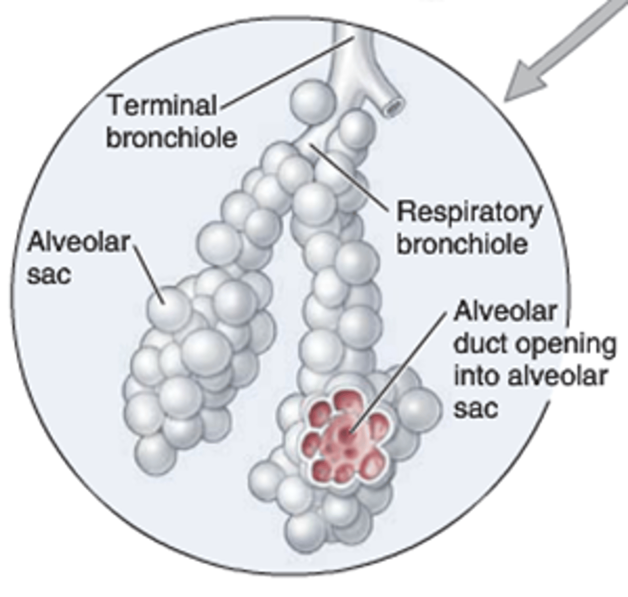
terminal bronchioles
terminal bronchioles- smallest part of conducting system; last part of respiratory system that doesn't participate in gas exchange
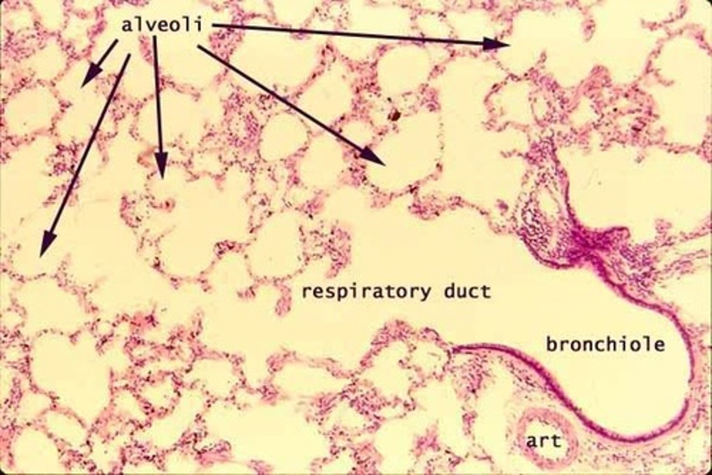
respiratory bronchioles: beginning of exchange system
give rise to alveoli
bronchioles: histology
terminal- transition from ciliated simple columnar to nonciliated simple cuboidal; goblet cells decreasing
lose goblet cells before cilia
respiratory- simple cuboidal; no cilia; no goblet cells
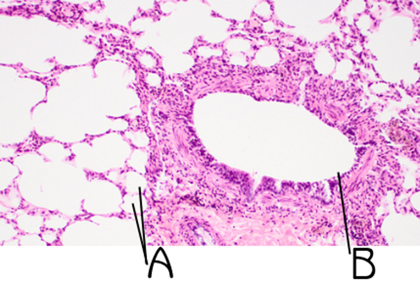
respiratory bronchiole histology
difficult to find- only able to distinguish if there are alveoli leading directly off them
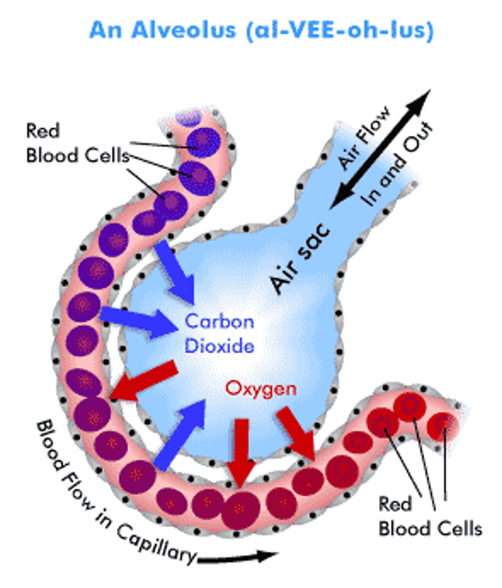
alveoli
clustered in alveolar sacs
last part of respiratory system and exchange system
lots of surface area allow for lots of diffusion
surrounded by capillaries- share a basement membrane for gas exchange
alveoli: histology
simple squamos- very thin and great for diffusion
respiratory membrane
shared membrane between alveoli and capillary
Co2 (waste/deoxygenated) moves into the alveoli to be exhaled
O2 (nutrients/oxygenated) moves from alveoli into capillaries to go to rest of body
dissection of pig respiratory system