BIS 102 Midterm 2
1/80
Earn XP
Description and Tags
Cheng M2 Spring 2026
Name | Mastery | Learn | Test | Matching | Spaced | Call with Kai |
|---|
No analytics yet
Send a link to your students to track their progress
81 Terms
Sanger’s Nobel Prize
Sanger’s breakthrough in analyzing insulin protein 1° sequencing (1958)
Pauling’s Nobel Prize
Pauling’s interpretation of chemical bonds in protein 2° structure (1954)
Anfinsen’s Nobel Prize
Anfinsen’s dogma in recognizing protein RNase 3° folding (1972)
Merrifield’s Nobel Prize
Merrifield’s “peptide synthesis” of a full-function 3° RNase (1984)
Levinthal’s Nobel Prize
Levinthal’s resolving the paradox of 3° folding kinetics
Perutz’s Nobel Prize
Discovered first tertiary structure - 3° myoglobin proteins (1962)
Klug’s Nobel Prize
Virus assembly - 4° quaternary structures (1982)
Agents affecting protein structures: Urea
disrupts hydrogen bonds
Agents affecting protein structures: SDS
disrupts hydrophobic cores of proteins
Agents affecting protein structures: Heat/High Temps
disrupts weak forces
Agents affecting protein structures: Beta-Mercaptoethanol (MCE)
disrupts disulfide bonds
Agents affecting protein structures: Distilled Water
disrupts ionic interactions
Agents affecting protein structures: Organic Solvents
interact with hydrophobic amino acid side chains
Primary (1°) Level
the sequence of amino acids in the polypeptide chain dictated by covalent bonds
Secondary (2°) Level
local structures - H-bonds within the main chain (alpha helix and beta sheets)
Tertiary (3°) Level
overall three-dimensional shape involving side chain in all weak-force interactions (globular proteins)
Quaternary (4°) Level
subunit organization based on weak-force interactions between polypeptide chains (Hemoglobin)
Sanger Sequencing: Step 1
Separate Polypeptide chains
subunits are associated by weak force interactions
expose to pH extremes, 8M Urea, Guanidinium Hydrochloride or high salt concentrations disrupts the polar interactions (H bonds)
Sanger Sequencing: Step 2
Cleave (reduce) disulfide bridges
Use performic acid or BME or DTT coupled with iodoacetate (or 3-bromopropylamine)
Sanger Sequencing: Step 3
Determine N- and C-terminal analysis
N-terminal analysis: Edman’s reagent (PTC), recovered as PTH derivatives
C-terminal analysis
Carboxypeptidase A: any residues except PRO, ASP, GLU, ARG, LYS
Carboxypeptidase B: only work on ARG or LYS
Carboxypeptidase Y: on any C-terminal residue
Sanger Sequencing: Step 4
Cleave each chain into smaller fragments and determine the composition & sequence of each chain
Trypsin → Cleaves on the C-terminus of ARG and LYS
Chymotrypsin → Cleaves at Carboxyl groups of Aromatic residues: PHE, THR, TRP, LEU
Sanger Sequencing: Step 5
Repeat step 4 - using a different cleavage procedure to generate a different & overlapping set of fragments
CNBR (cyanogen bromide) → MET
Sanger Sequencing: Step 6
Reconstruct the sequence of the protein from the sequences of overlapping fragments (Use sequencing strategy table)
Sequencing Strategy Table
Enzyme | Cleavage Site |
|---|---|
Trypsin | R, K (not before P) |
Lys-C | K (not before P) |
Arg-C | R |
Glu-C | E, D |
Asp-N | D, E (N-terminal) |
Chymotrypsin | F, W, Y, L |
CNBr | M (Chemical) |
Cleavage Procedure Method (ENZYMES): Trypsin
Susceptible Residue(s): ARG or LYS
Cleavage Procedure Method (ENZYMES): Chymotrypsin
Susceptible Residue(s): PHE, TRP or TYR; LEU
Cleavage Procedure Method (ENZYMES): Clostripain
Susceptible Residue(s): ARG
Cleavage Procedure Method (ENZYMES): Staphylococcal Protease
Susceptible Residue(s): ASP or GLU
Cleavage Procedure Method (ENZYMES): Endopeptidase Lys-C
Susceptible Residue(s): LYS
Cleavage Procedure Method (CHEMICAL METHODS): Cyanogen bromide
Susceptible Residue(s): MET
Basic Structure: α-Helix
Residues per turn: 3.6 amino acids
Rise per residue: 1.5 Angstroms
Rise per turn (pitch): 3.6 × 1.5 Å = 5.4 Angstroms
Hydrogen Bonding in α-Helix
Hydrogen bonds form between every i+4 residue in the helical backbone
Carbonyl oxygen (H-bond acceptor): bonds with the i+4 amide hydrogen
Amide hydrogen (H-bond donor): bonds with the i-4 carbonyl oxygen
Rules about α-Helix: Why are proline and glycine not included in α-Helix?
Both are helix breakers!
Proline has no backbone NH/amid to form an H-bond
Glycine only has a hydrogen as a side chain so the rotation around φ and ψ is so unconstrained -- too flexible
Rules about α-Helix: Role of Serine in α-Helix Stability
Serine’s hydroxyl group can form hydrogen bonds with water or other polar groups
If a polar or H-bond-forming residue is 3-4 positions away, transient H-bonding can destabilize the helix, leading to breaks
Rules about α-Helix: Steric Hindrance and Electrostatic Repulsions in α-Helix
Bulky residues = W-W, F-F, Y-Y
cause steric hindrance
Electrostatic repulsions: Like charges = +/+ or -/- repel each other, destabilizing the helix
Basic Structure: β strands
Rise per residue (pleated sheets): 3.25 å for parallel sheets and 3.47 å for antiparallel sheets
Parallel sheets composed of fewer than 5 strands, Antiparallel sheets composted of at least 2 strands
Beta (Reverse) turns
Reverse turns involve 4 amino acids (sharp turn) and allows the peptide chain to reverse direction for forming the antiparallel β-sheet
Carbonyl Oxygen (acceptor) of one residue is H-bonded to the amide Hydrogen (donor) of the other residue -- in sequence, three residues away from each other (i+3, the 4th residue)
Anfinsen’s Tertiary (3°) structure discoveries: Uniqueness
Uniqueness of protein folding: the sequence has no other configuration with comparable free energy → free energy minimum must be unchallenged
Anfinsen’s Tertiary (3°) structure discoveries: Kinetic Accessibility
Kinetic accessibility: the path from unfolded to folded state must be smooth → avoid highly complex changes like knots or high-order conformations
Anfinsen’s Tertiary (3°) structure discoveries: Stability
Stability: small changes in the surrounding environment cannot change the minimum energy configuration → protein remains stable in its folded state despite minor environmental fluctuations
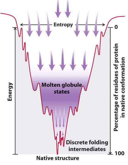
Free Energy Surface - Funnel Model
The free energy surface resembles a funnel.
The native state is at the bottom of the funnel, representing the most stable conformation.
This is not like a soup plate, which would imply multiple low-energy states.
Steep free energy surface ensures stability by making it difficult for the protein to unfold or adopt alternative conformations (protects native state)
Weak Force Interactions Types
Weak chemical forces restrict organisms to a small window of environmental conditions and fitness (pH, temp, salt concentration) which includes:
van der Waals interactions
Hydrogen bonding
Ionic interactions
Hydrophobic interactions
Van der Waals interactions
Induced electric interactions occur when atoms are close together
Significant when many contacts form complementary surfaces
Hydrogen bonding
Donor and acceptor pair: Direction dependent
The donor is hydrogen covalently bonded to an electronegative O or N
The acceptor is a lone pair on O or N
Ionic interactions
Stronger than H bonds/Not directional
Strength can be influenced by solvent properties
Ionization of amino acids
Hydrophobic interactions
Occur when nonpolar groups are added to water
Water molecules H-bonding
Nonpolar groups interfere with water H bonding and aggregate/need to minimize exposure
Cryo-Electron Microscopy 2017: Structural Preservation
Structural preservation is achieved by rapid freezing methods that generate vitrified water for Cryo-EM imaging
Cryo-Electron Microscopy 2017: Low-Dose Imaging
Low-dose imaging addresses challenges and breakthroughs associated with imaging biological structures using EM techniques
Cryo-Electron Microscopy 2017: Recording with Direct Electron Detectors
Direct Electron Detectors improve upon CCD cameras by enhancing signals through reduced noise in vitrified water
Cryo-Electron Microscopy 2017: Data Processing - Signal Enhancement
Data processing involves mathematical functions that determine cryoEM structures, where superimposing 2D images enhances signal clarity for higher resolution
Chaperons
Protein complexes that adjust hydrophobic interactions by providing a 2nd chance at protein folding → subvert kinetic trapping
Protein Life Cycle: Synthesis
Step 1 - The life cycle begins with the synthesis of a ribosome of a polypeptide chain, whose primary structure is dictated by an mRNA.
Protein Life Cycle: Folding
Step 2 - As synthesis proceeds, the polypeptide begins to fold into its native conformation
Protein Life Cycle: Processing
Step 3 - Folding may be accompanied by processing events such as
proteolytic cleavage of an N-terminal leader sequence (the most common form of post-translational processing)
formation of disulfide bonds (S—S).
Protein Life Cycle: PTM
Step 4 - Post-Translational Modifications found in proteins that may, e.g. attach a fatty acid molecule
Protein Life Cycle: Translocation
Step 5 - the modified peptide to a membrane.
Protein Life Cycle: Activation
Step 6 - If needed, binding an allosteric effector may trigger the adoption of a catalytically active conformation.
Protein Life Cycle: Aging
Step 7 - Proteins become damaged by chemical attacks, deamidation, or denaturation over time.
Protein Life Cycle: Ubiquitination
Step 8 - Damaged proteins may be “marked" by the covalent attachment of several ubiquitin molecules (Ub).
Protein Life Cycle: Recycling
Step 9 - The ubiquitinated protein is subsequently degraded to its component amino acids, which become available for the synthesis of new proteins
Human diseases linked to abnormalities of protein folding
Alzheimer’s disease
Familial amyloidotic polyneuropathy
Cancer
Creutzfeldt-Jakob Disease
Hereditary emphysema
Cystic fibrosis
Current theory of protein folding (in resolving Levinthal’s paradox)
Protein folding is a cooperative and sequential process which may involve intermediate states, including transition states, and is often guided by the protein’s primary structure. Specific proteins may require helpers like enzymes or chaperones to fold properly.
How long does most protein folding take, and what does this imply about the process?
Most folding is completed in about a second, implies that the process is highly efficient and not random. This rapid folding supports the idea of a guided, cooperative mechanism rather than a trial-and-error search through all possible conformations.
What is a “hydrophobic collapse,” and how does it contribute to protein folding?
A hydrophobic collapse is a process where nonpolar residues aggregate or coalesce. This collapse helps drive the folding process by reducing the exposure of nonpolar regions to water, leading to a more stable and compact structure.
What are the subsequent steps in protein folding after secondary structures form?
After secondary structures form, the next steps involve:
The cooperative aggregation of folding nuclei to form domains.
Long-range interactions between secondary structures or other hydrophobic regions.
The formation of a “molten globule” state, where assembled domains take on a more defined structure
Thermodynamics of protein folding
Protein folding is driven by enthalpy (H) being predominant over entropy (S) in the Gibbs free energy equation. This means that heat is released as the polypeptide folds into a more ordered state. The energy landscape of folding can be visualized as a 3D approach involving:
Conformational space (all possible shapes the protein can take).
Reaction coordinate (the pathway of folding).
Free-energy minimum (the most stable, lowest-energy state).
How to increase protein stability
Increased H-bonds
Fewer hydrophobic residues are exposed to the solvent
Charged residues on the surface
Ion-pair networks on the surface
Electrostatic optimization
Shorter loops
Clustering of aromatic residues
Improved packing of the protein core
Characteristics of the core of a globular protein structure
The helices and sheets in the core of a globular protein are typically constant and conserved in sequence and structure
Characteristics of the surface of a globular protein structure
Surface is composted of loops/coils and tight turns that connect helices and sheets of the core
Surface elements can interact with small molecules or other proteins
Basis for enzyme-substrate interactions, cell signaling, and immune responses
X-ray Crystallography
Intense X-rays are directed at a pure protein crystal, causing diffraction patterns that are processed to create electron density maps.
Best for high-resolution atomic structures of stable crystallizable proteins
X-ray Crystallography Pros and Cons
Pros: most productive method, representing the majority of. the PDB entries
Cons: requires large amounts of pure protein, crystallization is slow and difficult, flexible proteins are challenging