Anatomy Exam 2
1/25
Earn XP
Description and Tags
Muscle name, origin, insertion, innervation, function. Muscle borders.
Name | Mastery | Learn | Test | Matching | Spaced | Call with Kai |
|---|
No analytics yet
Send a link to your students to track their progress
26 Terms
What are the 9 regions of abdominal wall?
Epigastric
Umbilical/Periumbilical
Hypogastric/Pubic
Left & Right Hypochondriac Regions
Left & Right Lateral/Lumbar Regions
Left & Right Inguinal/Iliac Regions
Landmark: Umbilicus
“belly-button”
Lies at L3-L4 level in someone who is lean
Landmark: Pubic Symphysis
The point at which two pubic bones join
Landmark: Pubic Crest
A bony ridge running laterally from the pubic symphysis to pubic tubercle
Landmark: Pubic Tubercle
A small, rounded elevation to which inguinal ligament attaches
Landmark: Iliac Crest
A bony ridge that extends posteriorly from anterior superior iliac spine
Landmark: Anterior Superior Iliac Spine (ASIS)
The most anterior end of iliac crest
Landmark: Iliac Tubercle
A thickened portion of the iliac spine located approximately 6 cm posterior to ASIS
Landmark: Inguinal Groove
Marks division between abdomen and thigh.
Site of inguinal ligament which is palpable throughout its length extending from ASIS to pubic tubercle
Landmark: Epigastric Fossa
“Pit of the stomach”
A depression just inferior to xiphoid process
Landmark: Linea Alba
The “White Line” is formed by the fusion of fascial planes that surround the abdominal muscles. Extends from xiphoid process to pubic symphysis, and marks the location of the medial border of the rectus abdominis muscles.
Landmark: Linea Semilunaris
Curved groove (convex laterally) that extends from 9th costal cartilage to pubic tubercle and marks the location of the lateral borders of the rectus abdominis muscles.
Landmark: Midinguinal Point
Midpoint of line extending from ASIS to pubic symphysis
Borders of Abdominal Wall
Superior: Right & Left Costal Margins
Inferior: Horizontal line connecting anterior superior iliac spines
Lateral: Vertical line extending through anterior superior iliac spines
Transpyloric Plane
Horizontal plane at L1 level
Found midway between the suprasternal notch and pubic symphysis; it passes through the pylorus of the stomach
Subcostal Plane
Horizontal plane at L3 level
Passes through the most inferior point of ribcage
Transumbilical Plane
Horizontal plane at L3/L4 level
Passes through the umbilicus
Transtubercular Plane
Horizontal plane L5 level
Passes through the iliac tubercles
What are the layers of abdominal wall?
Skin
Superficial Fascial: Camper’s Fascia and Scarpa’s Fascia
Muscles
Transversalis Fascia
Extraperitoneal Fat
Parietal Peritoneum
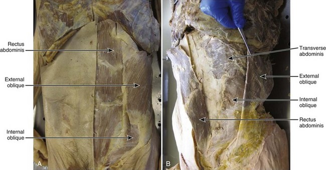
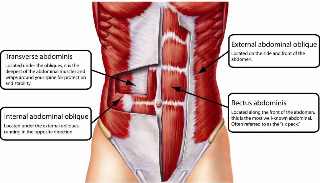
What are the 5 muscles of the anterior abdominal wall?
External Oblique Muscle
Internal Oblique Muscle
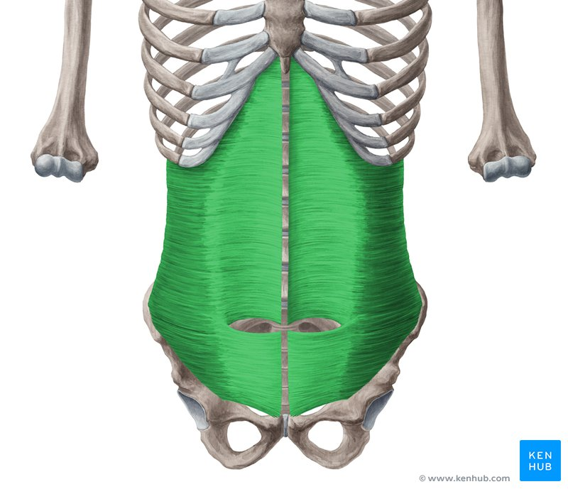
Transversus Abdominis Muscle
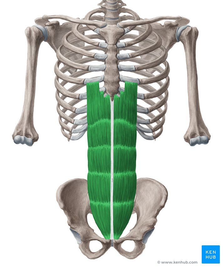
Rectus Abdominis Muscle
Pyramidalis Muscle
Name muscle, origin, insertion, innervation and function.
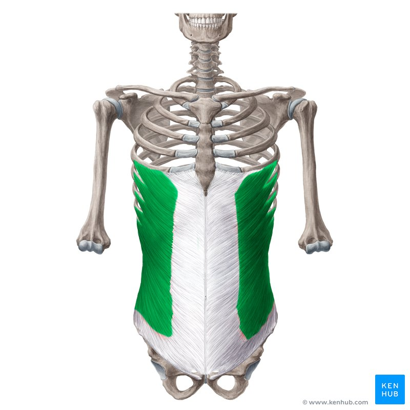
External Abdominal Oblique Muscle
Origin: Ribs 5-12
Insertion: Via aponeurosis to linea alba, pubic crest, iliac crest
Innervation: Ventral rami of lower 6 thoracic spinal nerves, iliohypogastric nerve and ilioinguinal nerve
Function: Support and compress abdominal contents, assist flexion and rotation of trunk

Name muscle, origin, insertion, innervation and function.
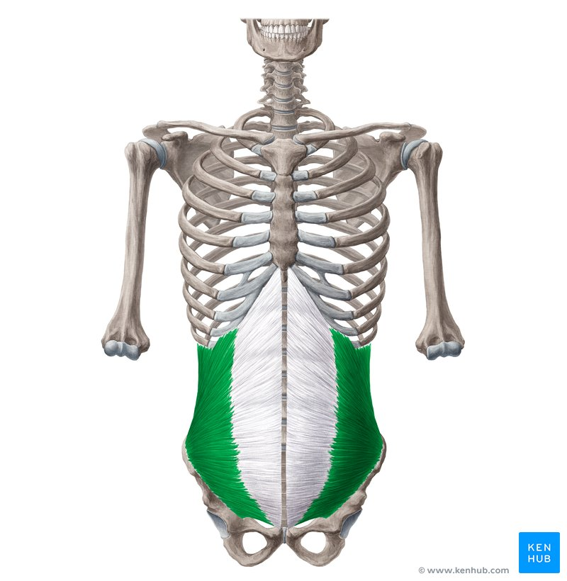
Internal Abdominal Oblique Muscle
Origin: Lumbar fascia, iliac crest, lateral 2/3 inguinal ligament
Insertion: Via aponeurosis onto ribs 10-12, xiphoid process, linea alba and pubic symphysis
Innervation: Ventral rami of lower 6 thoracic spinal nerves, iliohypogastric nerve, ilioinguinal nerve
Function: Support and compress abdominal contents, assist flexion and rotation of trunk
Name muscle, origin, insertion, innervation and function.
Transversus Abdominis Muscle
Origin: Ribs 7-12, lumbar fascia, iliac crest, lateral 1/3rd inguinal ligament
Insertion: Xiphoid process, linea alba, pubic symphysis
Innervation: Ventral rami of lower 6 thoracic spinal nerves, iliohypogastric nerve, ilioinguinal nerve
Function: Support and compress abdominal contents. Acts as a girdle for abdominal cavity.
Name muscle, origin, insertion, innervation and function.
Rectus Abdominis Muscle
Origin: Pubic symphysis, pubic crest
Insertion: Xiphoid process and costal cartilage of ribs 5-7. (3 tendinous intersections within the muscle of the belly)
Innervation: Ventral rami of lower 6 thoracic spinal nerves
Function: Flex trunk, support and compress abdominal contents
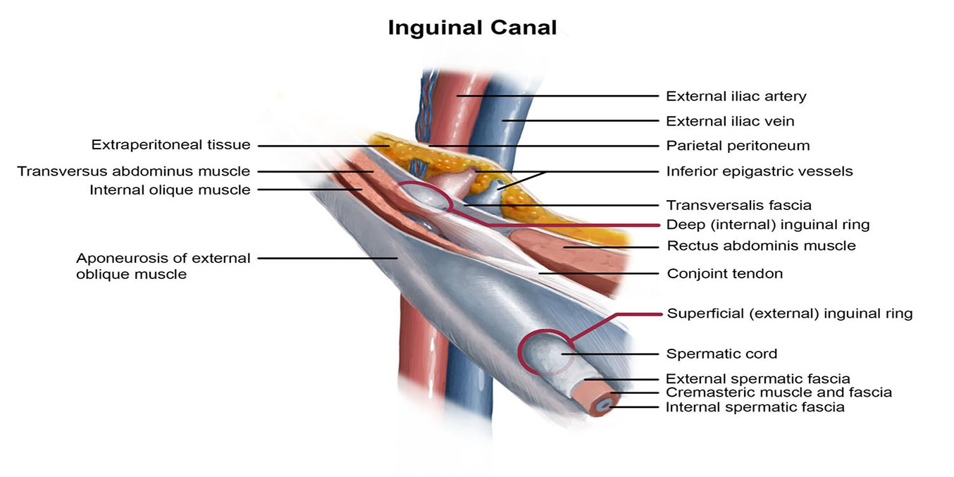
Boundaries of Inguinal Canal
Anterior Wall
Posterior Wall
Inferior and Superior boundaries
Anterior wall: aponeurosis of external abdominal oblique muscle
Posterior wall: transversalis fascia and conjoint tendon
Inferior boundary (floor): inguinal and lacunar ligaments
Superior boundary (roof): fibers of internal abdominal oblique and transversus abdominis muscles
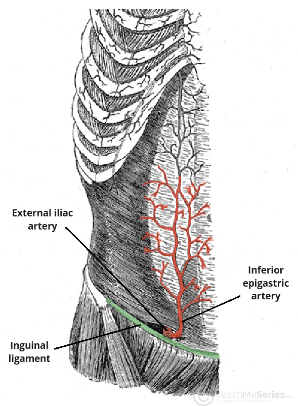
What is Hasslebach’s Triangle and what are its borders?
Site of direct inguinal hernias.
Lateral: inferior epigastric artery
Inferior: Inguinal ligament
Medial: rectus abdominis muscle