CH426 - Carmel Long Q
1/17
There's no tags or description
Looks like no tags are added yet.
Name | Mastery | Learn | Test | Matching | Spaced | Call with Kai |
|---|
No analytics yet
Send a link to your students to track their progress
18 Terms
Explain the physical principle upon which the scanning tunnelling microscopy (STM) technique is based.
STM is based on the physical principle of quantummechanical tunnelling. This tunnelling event occurs as a result of the overlap of the wave-functions of the tip and surface atoms, enabling electrons to tunnel through the energy barrier. This gives rise to a tunnelling current, It which depends on the bias voltage, VB, and the tip-tosample distance, d.
It is proportional to VB, increasing in a linear manner as VB is increased, while It is related to the tip-tosample separation through an exponential term. This makes It very sensitive to d. As a result small changes in d give relatively high It values. Typical values of d are 1 nm (which is equivalent to 10 angstroms).
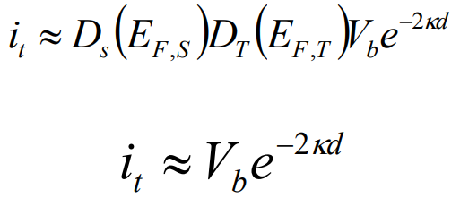
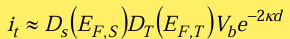
The tunnelling current equation is given by the expression,
Define the parameters in this expression and show how this equation is central in enabling nm resolution in the STM technique.
It is the tunnelling current. Ds and DT are the density of states of electrons in the Fermi levels of the sample and the tip, VB is the bias voltage. This is the difference between the potential of the tip and sample:
VB = VS - VT
If VB = 0, no current flows. A bias voltage is needed to create an energy difference between the tip and sample, allowing electrons to tunnel.
The tunnelling current depends exponentially on two key factors: k, which relates to the probability of tunnelling, and d, the tip–sample distance.
Distance d is the most important factor because the current changes extremely quickly with it. For every 0.1 nm increase in d, the current drops by about a factor of 10.
This strong exponential dependence makes STM extremely sensitive to distance and allows it to achieve ~0.1 nm resolution.
Describe how the STM tip is moved in atomic steps in the x- and y- directions.
The STM tip is moved in atomic steps using a piezoelectric scanner. Small voltages applied to the piezo electrodes make the tube bend by extremely tiny amounts (picometres to ångströms). This allows very precise movement in the x and y directions. Because the movement per volt is so small, the tip can be positioned with sub-ångström accuracy, enabling atomic-resolution imaging.
The STM technique can be used in a constant current mode or a constant height mode. Describe the difference between these two approaches.
Constant current mode: The tip scans across the surface while the tunnelling current is kept constant. The height (z) is continuously adjusted to maintain this current, producing a 3D map of (x, y, z). The z-values represent surface height and show atomic or molecular features.
Constant height mode: The tip height is kept fixed as it scans across the surface. The tunnelling current is measured instead, giving a (x, y, It) map. Changes in surface height cause changes in current.
Differences: constant current measures height changes by moving the tip, while constant height measures current changes with a fixed tip position.
Can the STM technique be used to analyse insulating materials? Provide a clear explanation to account for your answer.
No, STM cannot be used for insulating materials.
This is because STM relies on a tunnelling current between the tip and the sample. Insulators have a large band gap and do not have свобод electrons available at the surface, so no current can flow. Without a tunnelling current, the STM cannot function or form an image.
Briefly describe one surface analytical technique that can be used to provide chemical information.
SEM can provide chemical information because the electron beam interacting with the surface produces signals that depend on the elements present.
When a focused beam of electrons hits the sample, it generates secondary electrons (used for surface shape), but also X-rays and Auger electrons. These arise when inner-shell electrons are knocked out and higher-energy electrons fall into lower energy levels.
The emitted X-ray energies are characteristic of specific elements, since they depend on the energy difference between electron shells. By measuring these X-rays, the composition of the sample can be identified.
Atomic force microscopy, AFM, is used widely in the field of nanotechnology.
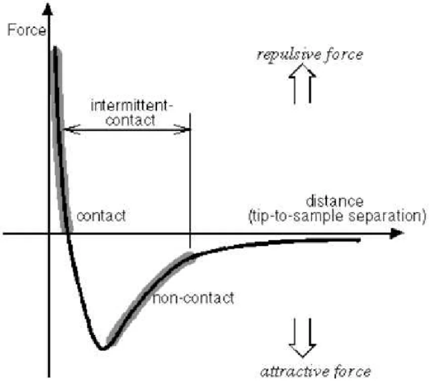
Explain the significance of the force-distance-curve and show how it is used to give the experimental techniques of contact and non-contact AFM.
The force-distance-curve gives the nature and magnitude of the forces that exist between the tip and the substrate. The force distance curve is based on the lennard-Jones potential energy curves, which give the potential energy as a function of the separation between two atoms at close distances. The repulsive force depends on (ro/d)12 while the attraction depends on (ro/d)6. The sum of these forces gives the force distance curve. Depending on the distance between the tip and the sample, the force may be attractive or repulsive. This gives the Non Contact AFM (attractive) and Contact AFM (repulsive forces) techniques.
At close distances (about 1 Ao) a large repulsive force exists giving very good resolution at the atomic scale and the measurement is less prone to moisture on the surface. This gives Contact AFM. At these close distances the tip makes intermittent contact with the surface.
At larger distances, (100 Ao) weaker attractive forces are measured to give Non-Contact AFM, making this approach suitable for biological samples that can be damaged by the intermittent contact. As the force is given as: F = -kx and k is the spring constant, then the signal from the weaker attractive forces can be made larger by changing the cantilever to give a lower k value, making x larger
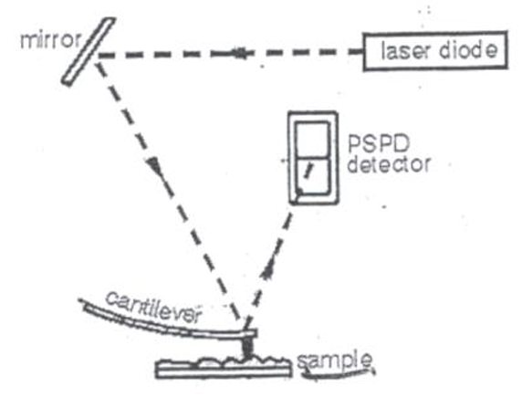
Explain and show clearly, using appropriate diagrams, how an AFM experiment is performed.
AFM is performed by scanning a sharp tip across the surface. Inter-atomic forces between the tip and sample are measured using a sensitive optical technique. The forces are measured by attaching the tip to a cantilever. A laser beam is focussed onto the tip of the cantilever and bounced back to a position-sensitive photo-detector (PSPD). As a force is registered the cantilver bends. This alters the position of the laser beam on the PSPD and this is translated using the force constant, k, to give the inter-atomic forces.
Forces are measured as the tip is scanned across the surface to give a map of the topography of the surface with atomic resolution.
Estimate the force required to deflect the cantilever by 2 nm. The force constant of the cantilever is 10 pN/nm.
F = -kx = pN
F = force, k = spring constant, x = distance.
Assuming that you need to increase the deflection to 4 nm, how would you achieve this increase? Clearly explain your answer.
Because the cantilever obeys Hooke’s law, the deflection is directly proportional to the applied force. Must double the applied force. In practice, this is done by bringing the tip closer to the surface / increasing the loading force via the z‑piezo setpoint in contact AFM. The feedback loop then allows a larger repulsive tip–sample force, which produces the larger (4 nm) cantilever deflection.
In an experiment, single walled carbon nanotubes (SWCNTs) were functionalized with –COOH groups.
What two surface analytical techniques would you use to characterize these materials?
Two suitable surface analytical techniques are X‑ray Photoelectron Spectroscopy (XPS) and Fourier Transform Infrared Spectroscopy (FTIR). XPS provides elemental and chemical‑state information, allowing detection of C=O and O–C=O species characteristic of –COOH groups. FTIR identifies functional groups through vibrational signatures, confirming the presence of carboxyl groups via their characteristic C=O and O–H stretching bands.
Describe clearly the information that you would expect to obtain using the two selected techniques.
XPS would provide elemental composition, chemical‑state information, and quantification of –COOH functionalisation through shifts in the C 1s and O 1s peaks. FTIR would provide vibrational signatures of the carboxyl groups (C=O, O–H, C–O), confirming their presence and chemical attachment to the SWCNT surface.
Explain the physical principles on which the following techniques are based. In your answer ensure that you discuss the significance of the tunnelling current equation and the force-distance curve.
Atomic Force Microscopy (AFM)
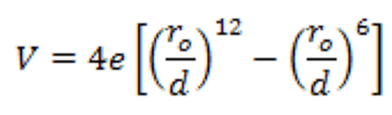
The AFM technique is based on the principle of interatomic forces, which exist between atoms at close distances. These forces can be computed using the Lennard-Jones potential energy curves. The LennardJones potential curve is a model that approximates the interactions between two atoms.
V is the potential energy, d is the distance between the two atoms, ro is the separation at which the potential energy is zero, V = 0, and e is the depth of the potential well. The variation of the repulsive force on the distance between the atoms depends on (ro/d)12 while the attraction depends on (ro/d)6.
The sum of the attractive and repulsive forces is measured to give the force distance curve.
In Contact AFM, the distance between the tip and sample is approximately 1 Ao , and a large repulsive force is measured, giving good resolution and making the measurement less prone to water or moisture on the surface of the sample.
Attractive forces are measured in non-contact AFM, where the distance, is 100 Ao , making this approach suitable for biological samples that can be damaged by the intermittent contact between the sample and the tip.
In an experiment, Fe-doped CuO2 nanostructures were synthesised. Describe the surface techniques that you would use to characterise these materials. Provide clear reasons for selecting a technique and describe the type of information that you would hope to obtain.
Two suitable techniques are XPS and STM.
XPS gives chemical information such as which elements are present, their oxidation states (e.g. Fe²⁺/Fe³⁺, Cu⁺/Cu²⁺), and bonding. It confirms that Fe has been successfully doped and shows how much is present.
STM shows the surface at atomic scale and reveals how Fe doping changes the structure and electronic behaviour, including defects and changes in local electronic states.
Together, XPS provides chemical composition, while STM shows atomic structure and electronic effects of doping.
Briefly, describe how the STM experiment is performed.
In STM, a sharp tip (< 1 mm in diameter) made of an inert but conducting material (e.g., W, Pt, Pt-Ir) is brought sufficiently close to the sample to enable the tunnelling process, typically d is about 1 nm.
The tip is scanned across the surface, to give a map of the tunnelling current as a function of the position. A piezo electric material is used to move the tip in atomically small increments. A bias voltage, Vb, is applied between the tip and the sample so that there is a difference in the energy of the electrons on the tip and the sample.
The measurement can be made using two modes:
in the constant height mode, the tip is maintained at a constant height and It is monitored and recorded to give a three dimensional plot of (x, y, It).
Alternatively, It can be maintained constant and the height of the tip, z, is measured as a function of the position of the tip on the sample to give a map of (x, y, z).
In an experiment, Ce-doped CuO2 nanostructures were synthesised and analysed using STM.
What information would you expect to obtain from this STM experiment?
From STM analysis of Ce‑doped CuO₂ nanostructures, you would obtain atomic‑scale topographic images of the surface and detailed information about the local electronic structure. STM would reveal how Ce doping modifies the CuO₂ lattice, where dopants are located, and how the electronic properties vary across the surface. It provides structural and electronic information, but not direct chemical identification.
Name one other technique that you would use to obtain additional information on the Ce-doped CuO2 nanostructures. What information would you expect to obtain using this additional technique?
A suitable additional technique is X-ray Photoelectron Spectroscopy (XPS).
XPS identifies the elements present and their oxidation states, such as Ce³⁺/Ce⁴⁺ and Cu⁺/Cu²⁺. It also shows changes in oxygen bonding and the Ce/Cu ratio.
This confirms successful doping and shows how Ce affects the surface chemistry, which STM cannot measure.
A new nanomaterial, that has a very low electrical conductivity, was synthesised. Would you select STM to gain information of the size and shape of this new nanomaterial? Explain your answer.
No, STM would not be selected. STM requires a measurable tunnelling current, which only flows if the sample is electrically conductive. A nanomaterial with very low conductivity cannot sustain tunnelling, so the STM feedback loop cannot operate and no topographic image can be obtained. Instead, AFM should be used, as it provides size and shape information for insulating materials.