Muscle proximal/distal attachments
1/93
There's no tags or description
Looks like no tags are added yet.
Name | Mastery | Learn | Test | Matching | Spaced | Call with Kai |
|---|
No analytics yet
Send a link to your students to track their progress
94 Terms

Gluteus maximus proximal attachment
Ilium, sacrum, coccyx, sacrotuberous ligament

Gluteus maximus distal attachment
75% iliotibial band, 25% gluteal tuberosity
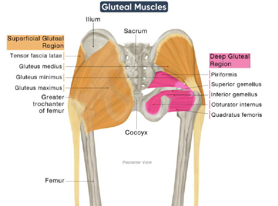
Gluteus medius proximal attachment
Gluteal surface of ilium

Gluteus medius distal attachment
Greater trochanter

Gluteus minimus proximal attachment
Anterior region of ilium
Gluteus minimus distal attachment
Greater trochanter

Tensor Fascia Lata proximal attachment
ASIS, Iliac crest

Tensor Fascia Lata distal attachment
Iliotibial band

Piriformis proximal attachment
Anterior aspect of sacrum
Piriformis distal attachment
Greater trochanter
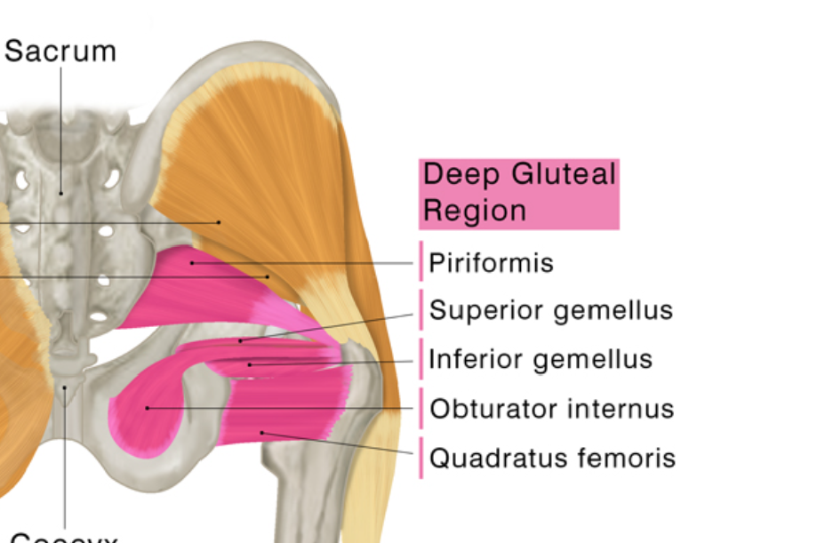
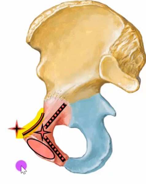
Superior gemellus proximal attachment
Ischial spine

Superior gemellus distal attachment
Trochanteric fossa

Obturator internus proximal attachment
Obturator foramen

Obturator internus distal attachment
Trochanteric fossa

Inferior gemellus proximal attachment
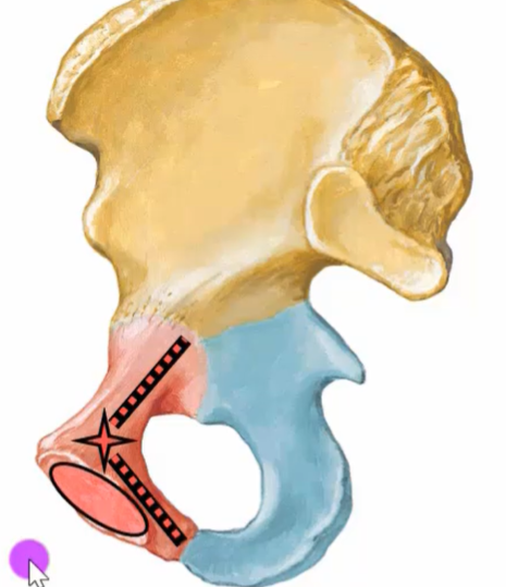
Ischial tuberosity

Inferior gemellus distal attachment
Trochanteric fossa

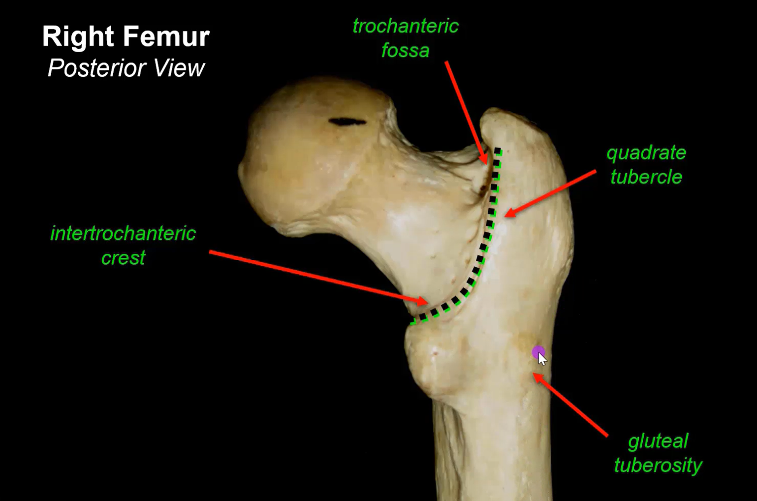
Quadratus femoris proximal attachment
Ischial tuberosity

Quadratus femoris distal attachment
Quadrate tubercle of intertrochanteric crest
What is this?
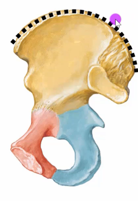
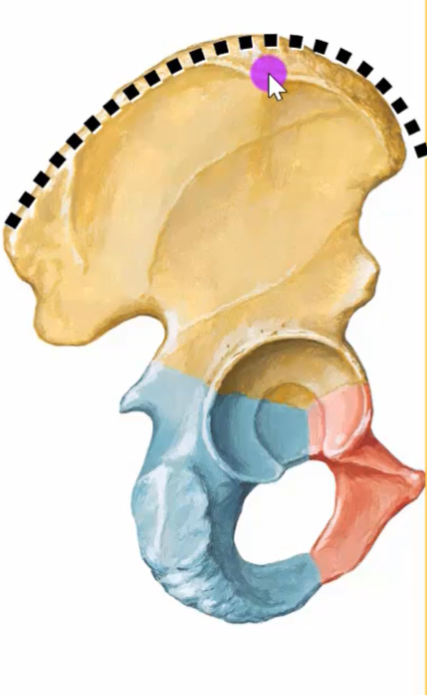
Iliac crest
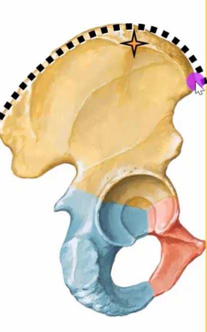
What is this?
Iliac tubercle
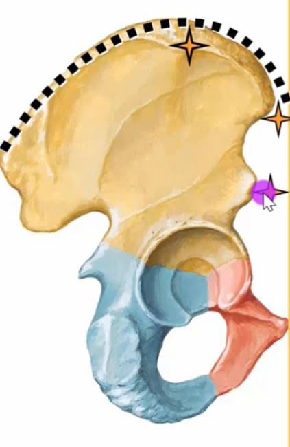
Anterior Superior Iliac Spine
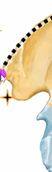
Anterior Inferior Iliac Spine
Posterior Superior Iliac Spine and Posterior Inferior Iliac Spine
Iliac tuberosity
Iliopubic eminence
Ramus
Ischial spine
Ischial tuberosity
Obturator foramen
Sciatic notches
Rami
Symphyseal surface
Pubic crest
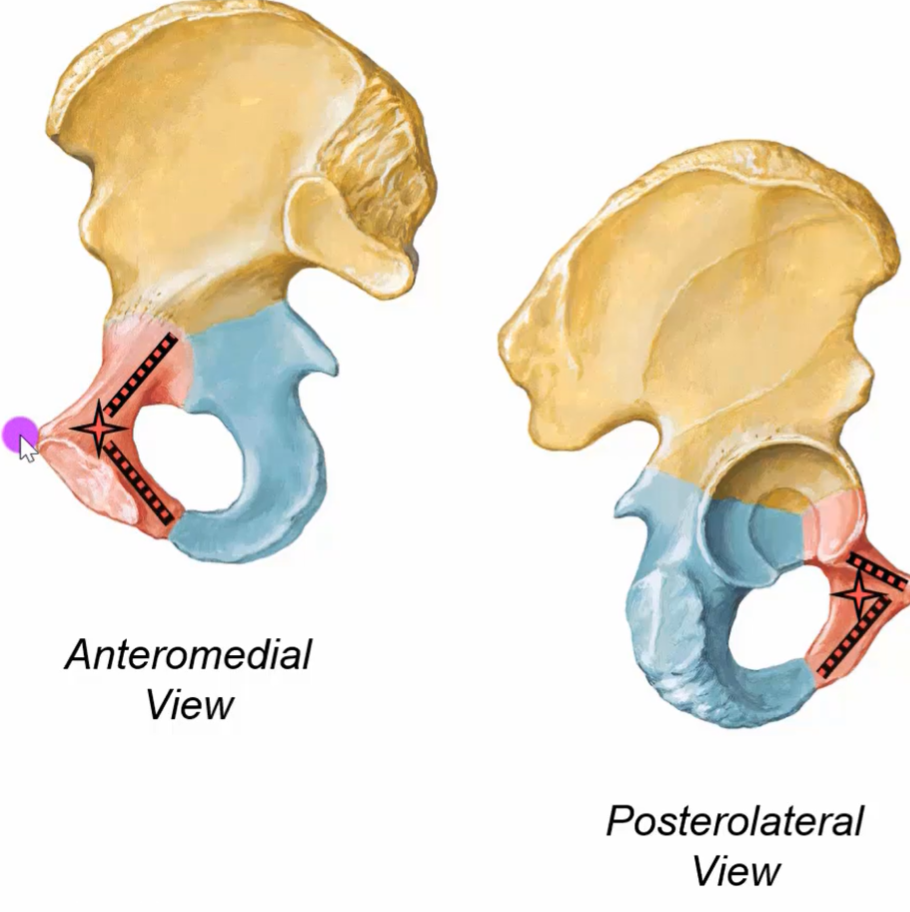
Sacrotuberous ligament attachments
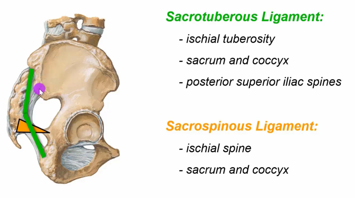
Vertically from sacrum to ischial tuberosity
Sacrospinous ligament
Sacrum to ischial spine
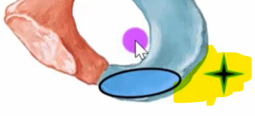
Greater sciatic foramen
Passage for piriformis, sciatic nerve, gluteal vessel and other neurovascular structures.
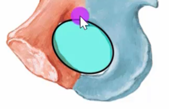
Lesser sciatic foramen
Passage for pudendal neurovascular bundle and tendon to obturator internus.
WOW
Superior gluteal nerve supplies…
Gluteus medius, gluteus minimus and tensor fascia lata
Inferior gluteal nerve supplies…
Gluteus maximus
Sciatic nerve path
Passes below the piriformis through the gluteal region to the posterior thigh before splitting into a tibial and common fibular nerve.
Lumbosacral joint structure
Anteriorly, it is a secondary cartilaginous joint AKA symphysis. Posteriorly, it is 2 synovial joints.
Ligaments of the spine/vertebral column
Anterior longitudinal ligament, posterior longitudinal ligament, interspinous and supraspinous ligament, ligamentum flavum.
Sacroiliac joint - Joins sacrum to ilium
Synovial anteriorly, fibrous posteriorly.
Sacroiliac joint ligaments
Anterior sacroiliac, posterior sacroiliac, Interosseus sacroiliac, accessory (sacrotuberous, sacrospinous)
Pubic symphysis ligaments
Superior pubic ligament, arcuate ligament
Pubic symphysis is a…
Secondary cartilaginous joint
Hip joint is a…
Synovial ball and socket joint
Iliofemoral intrinsic ligament
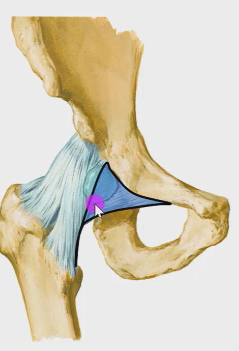
Pubofemoral intrinsic ligament
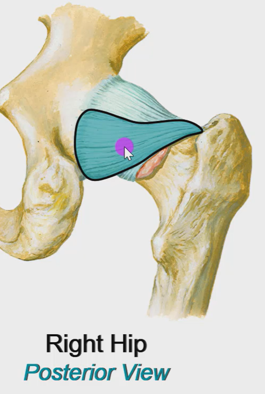
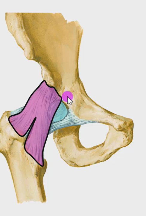
Ischiofemoral intrinsic ligament
Weakest of the intrinsic ligaments is…
Ischiofemoral ligament
Hilton’s Law
Nerves supplying a joint also innervate the muscles that move that joint and the overlying skin.
Blood supply to femoral head
Medial and lateral circumflex femoral arteries, Retinacular arteries, artery to head of femur
Blood supply to femoral neck
Medial and lateral circumflex femoral arteries
Iliopsoas is a…
HIP FLEXOR
Hip closed pack position
Extension, adduction, internal rotation
Femoral neck fracture consequences
Can disrupt retinacular arteries and the artery to the head of the femur, leading to avascular necrosis.
Function of skin
Sensation, Protection, and thermoregulation, vitamin D synthesis
Epidermis
Dead protective outerlayer. Avascular, neural. Epithelial tissue.
Dermis
Inner connective tissue layer, highly vascular and neural.
Layers of skin
Epidermis, dermis, subcutaneous tissue, deep fascia
Skin nerve supply
Somatic voluntary nerves for sensation, autonomic involuntary nerves for function.
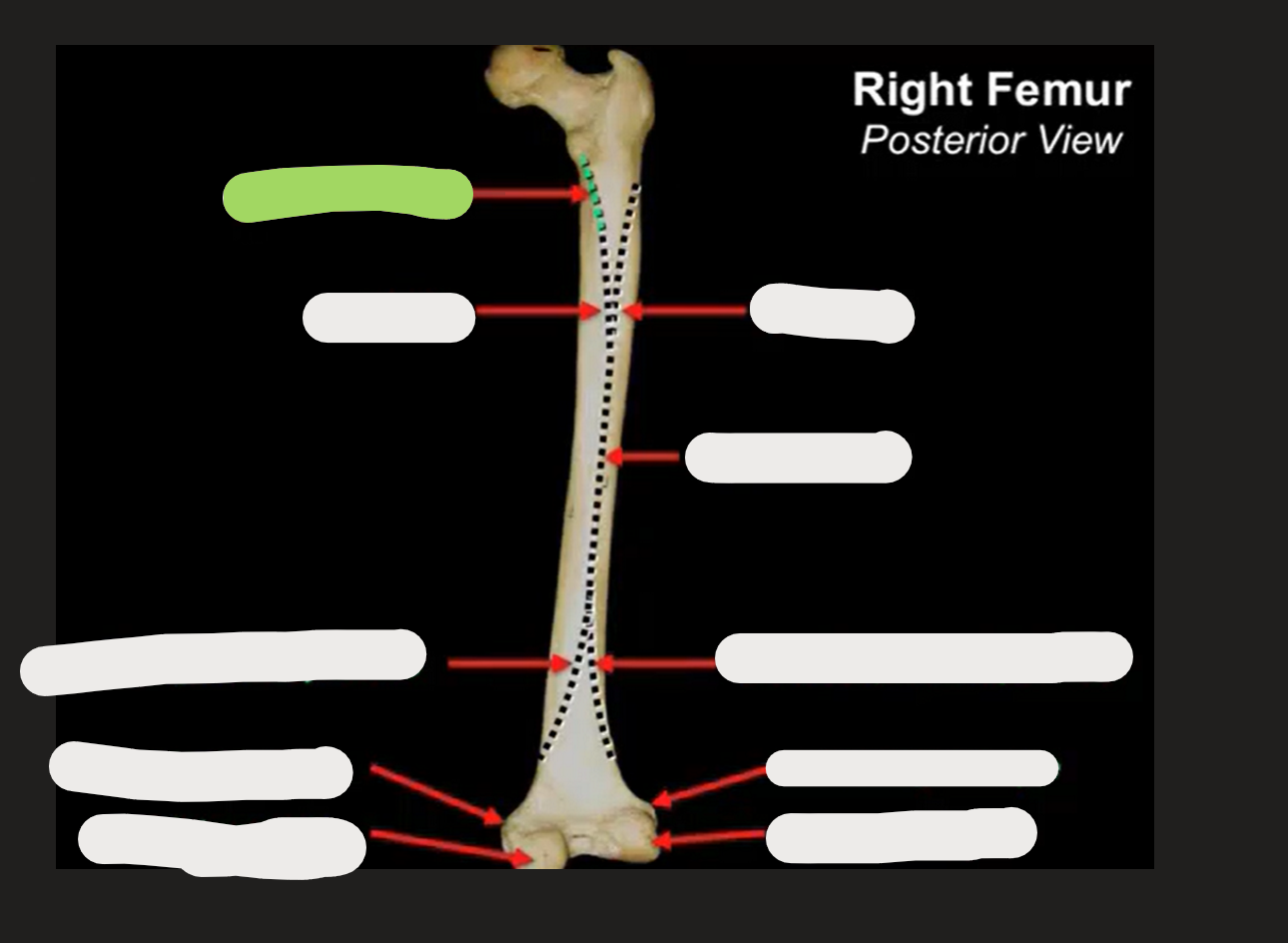
What is this?
Pectineal line
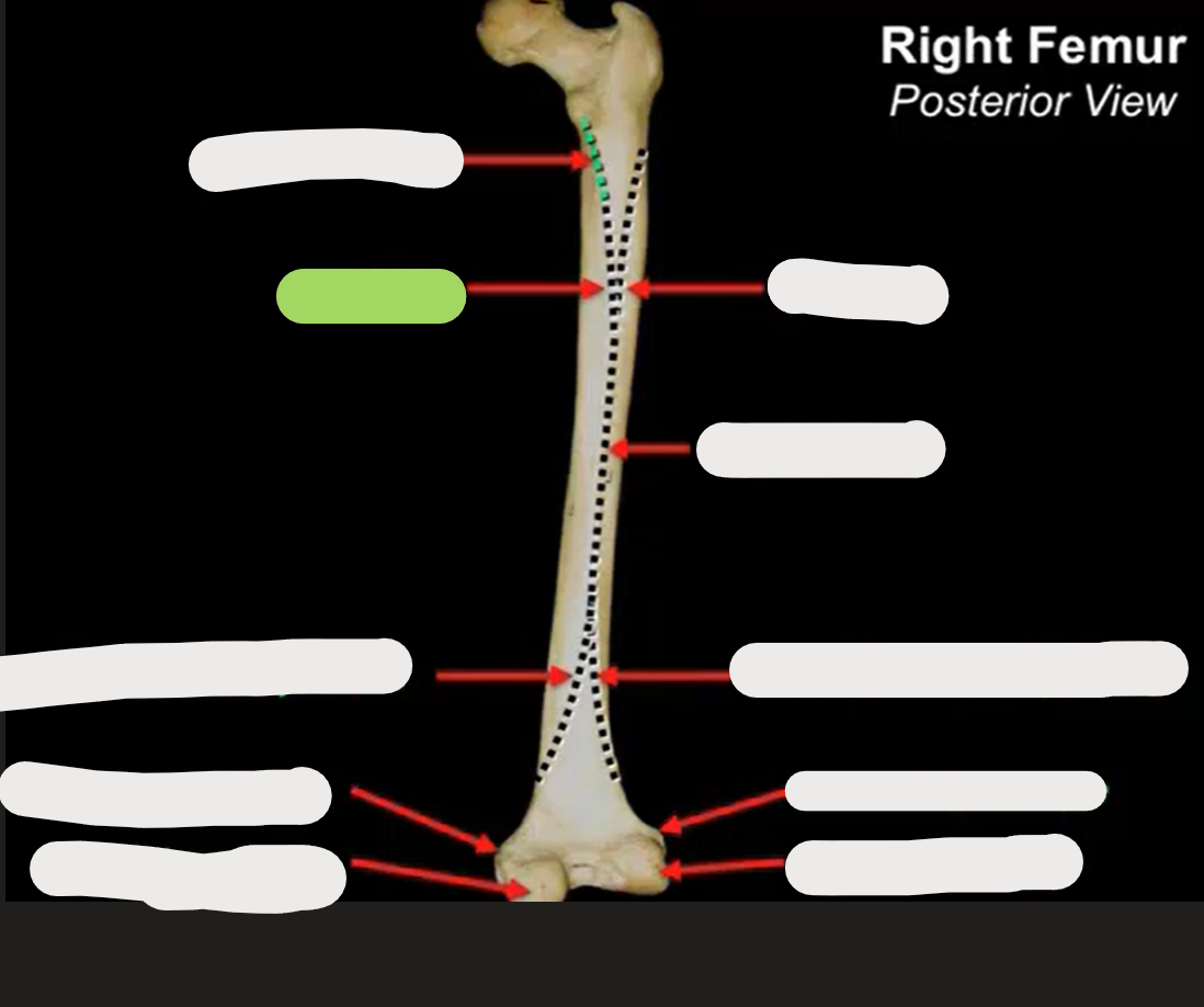
What is this?
Medial femoral lip
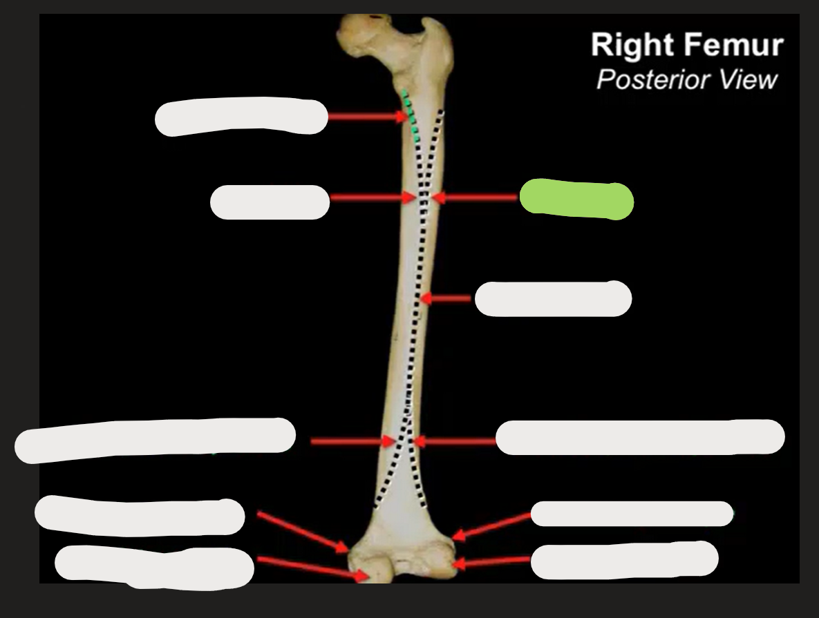
What is this?
Lateral femoral lip
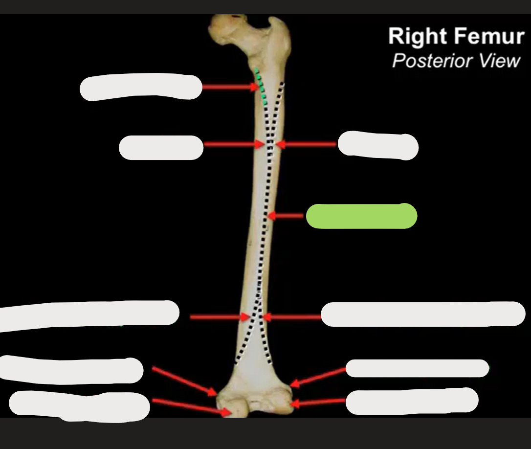
What is this?
Linea aspera
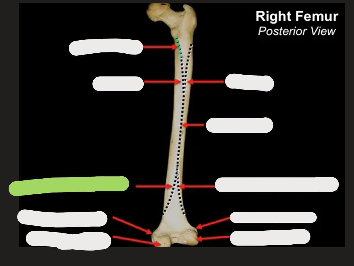
What is this?
Medial femoral supracondylar line
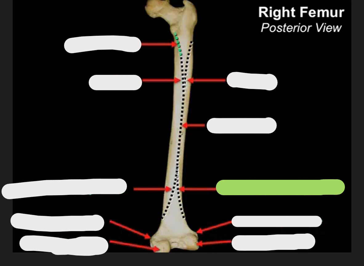
What is this?
Lateral femoral supracondylar line
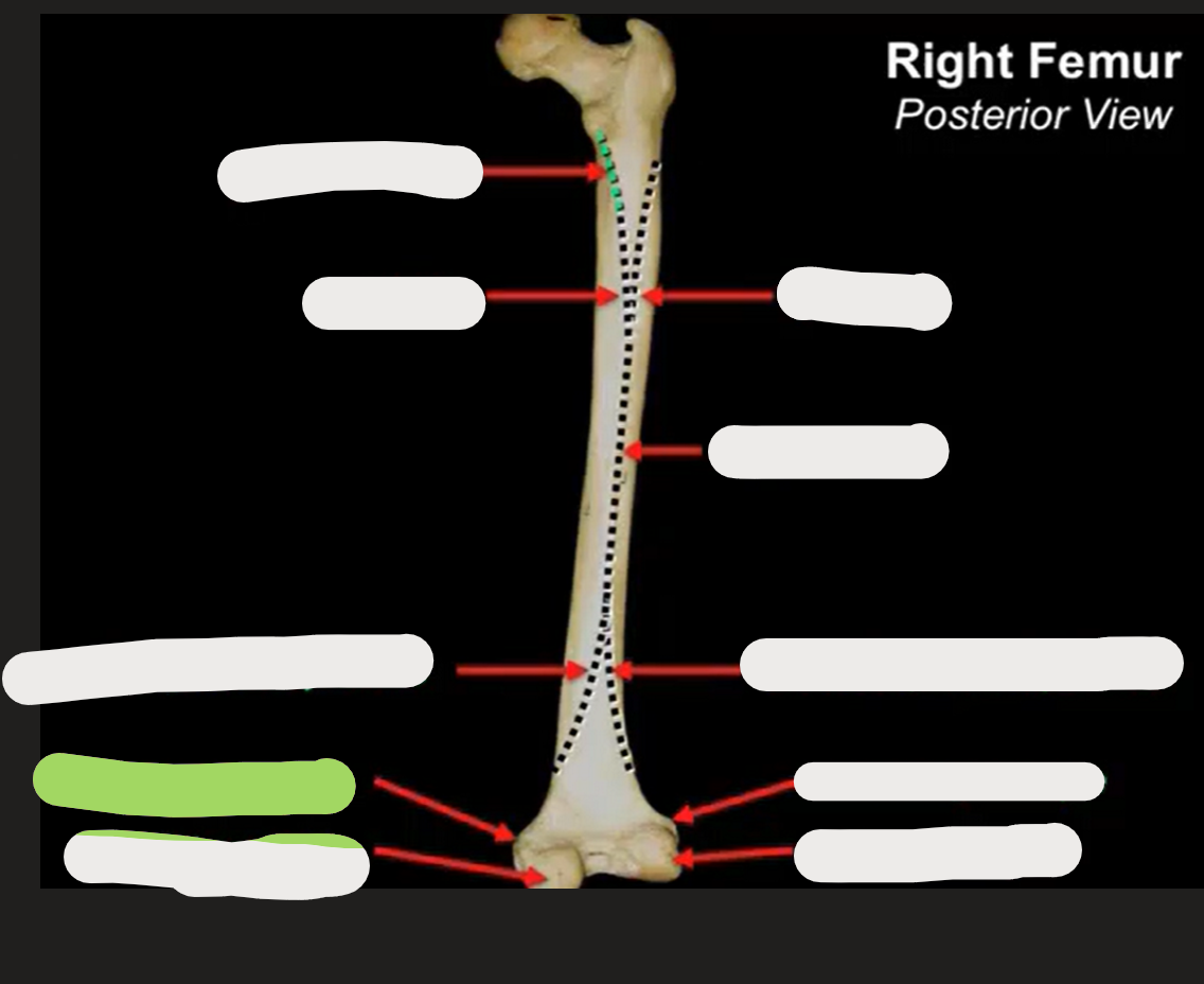
What is this?
Medial femoral epicondyle
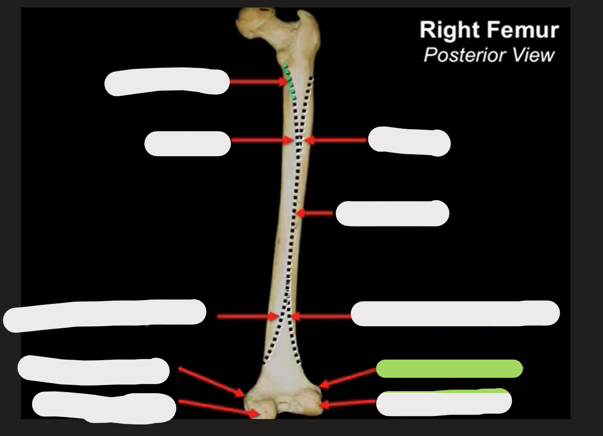
What is this?
Lateral femoral epicondyle
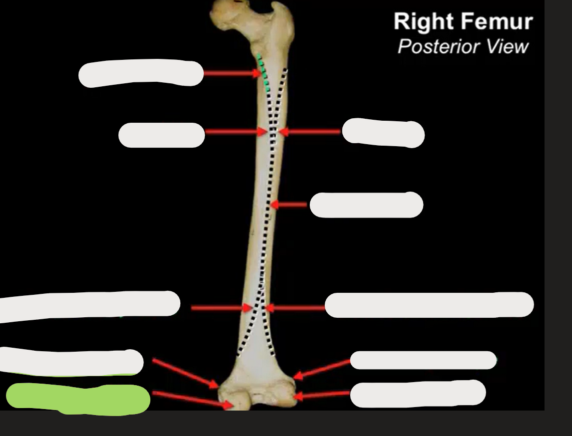
What is this?
Medial femoral condyle
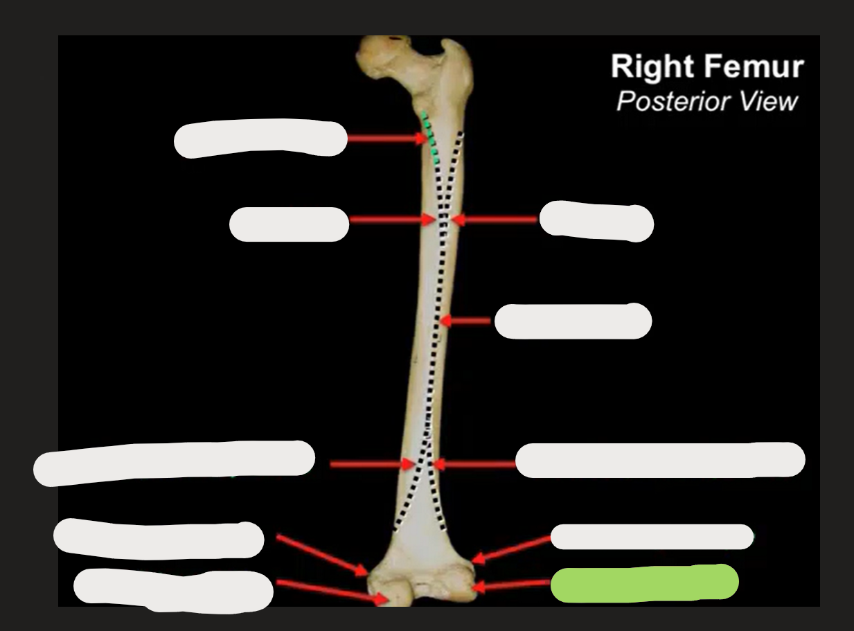
What is this?
Lateral femoral condyle
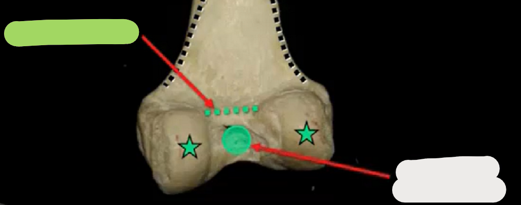
What is this?
Intercondylar line.
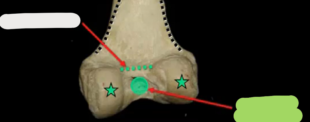
What is this?
Intercondylar fossa.
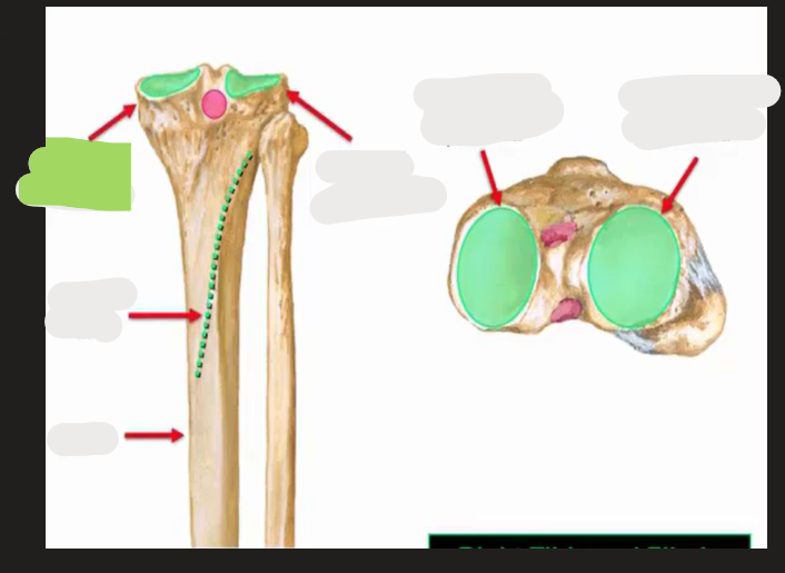
What is this?
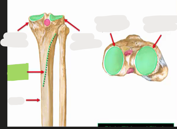
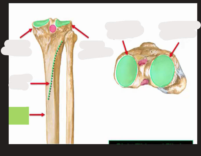
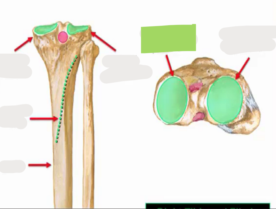
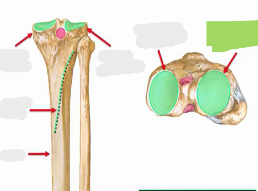
Medial tibial condyle
What is this?
Soleal line
What is this?
Tibial shaft
What is this?
Medial tibial plateau
What is this?
Lateral tibial plateau
Fibula main parts top to bottom
Apex, head, neck, shaft
Semimembranosus attachments
Proximal: Ischial tuberosity of pelvis, Distal: Posterior medial tibial condyle
Semimembranosus/Semitendinosus role
Hip extension, knee flexion, internal rotation of tibia
Semitendinosus attachments
Proximal: Ischial tuberosity, Distal: Pes Anserinus
Biceps femoris (long head) attachments
Proximal: Ischial tuberosity, distal: Head of fibula and lateral tibial condyle
Biceps femoris (long head) role
Hip extension, knee flexion, external rotation of tibia
Biceps femoris (short head) attachment
Proximal: Lateral supracondylar line, Distal: Head of fibular and lateral tibial condyle
Biceps femoris (short head) role
Knee flexion, external rotation of tibia
Semitendinosus, semimembranous, and biceps femoris long head innervation
Tibial portion of sciatic nerve
Biceps femoris short head innervation
Common fibular portion of sciatic nerve
Gastrocnemius attachments
Proximal attachment: Medial and lateral femoral condyles. distal attachment: Calcaneus
Plantaris attachment
Proximal attachment: Lateral femoral condyle, distal attachment: Calcaneus
Popliteus
Proximal attachment: Lateral femoral condyle, Distal attachment: Posterior surface of tibia.
Tarsel Tunnel contents (Tom dick and very naughty harry)
Tibialis posterior, Flexor Digitorium Longus, Arteries, Vein, Nerve, Flexor Hallucis Longus