Locomotor
1/92
There's no tags or description
Looks like no tags are added yet.
Name | Mastery | Learn | Test | Matching | Spaced | Call with Kai |
|---|
No analytics yet
Send a link to your students to track their progress
93 Terms
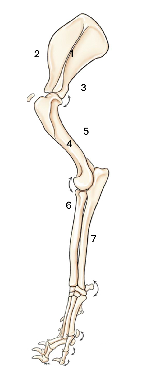
Name the labelled muscles
1 = Shoulder stabilisers. Supraspinatus, infraspinatus, subscapularis, teres minor
2 = Shoulder extensors. Brachiocephalicus, biceps brachii, supraspinatus
3 = Shoulder flexors. Deltoid, ters major, triceps brachi
4 = Elbow flexors. Biceps brachii and brachialis
5 = Elbow extensors. Triceps brachi - long head, accessory head, medial head, lateral head.
6 = Carpus and digit extensors. Extensor carpi radialis, abductor pollicic longus, common digital extensor, lateral digital extensor.
7 = Carpus and digit flexors. Flexor carpi radialis, superficial digital flexor, deep digital flexor, flexor carpi ulnaris.
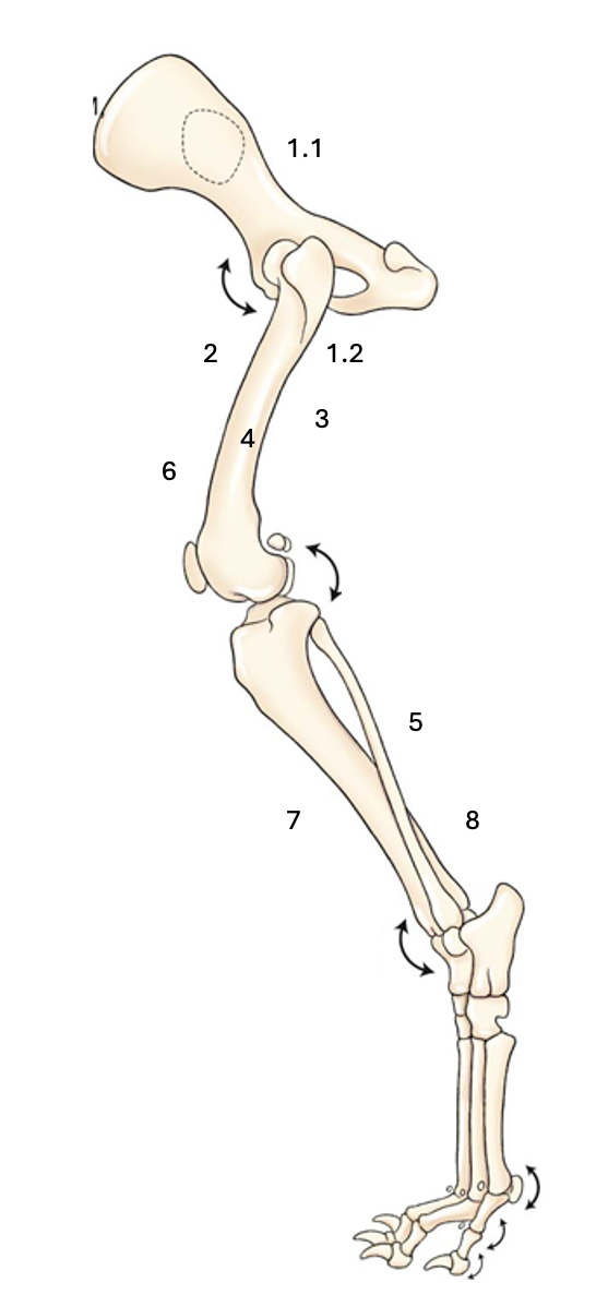
Name the labelled muscles
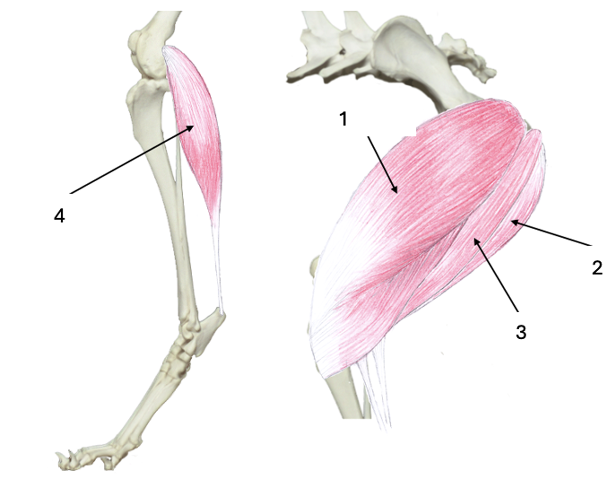
1.1 = Hip Extensors. Gluteals - deep, middle and superficial
1.2 = Hip Extensors. Hamstrings - semimembranosus, semitendinosus, biceps femoris
2 = Hip Flexors. Tensor fascia latae, Sartorius, Quadriceps femoris - rectus femoris, vastus lateralis, vastus intermedius, vastus medialis
3 = Hip Adductors. Gracilis, Adductor, Pectineus, External obturator
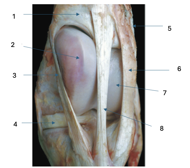
4 = Stifle Flexors. Hamstrings - semimembranosus, semitendinosus, biceps femoris
5= Stifle Flexors. Gastrocnemius.
6 = Stifle extensors. Quadriceps femoris - rectus femoris, vastus lateralis, vastus intermedius, vastus medialis.
7 = Tarsal and digit flexors. Gastrocnemius and hamstrings via the common calcaneal tendon, superficial digital flexor.
8 = Tarsal and digit flexors. Cranial tibial and peroneus tertius.
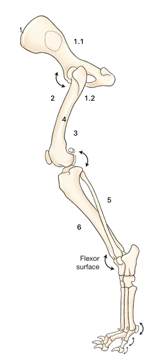
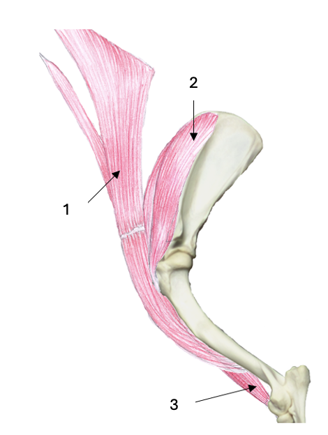
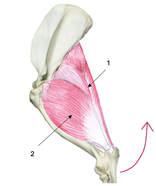
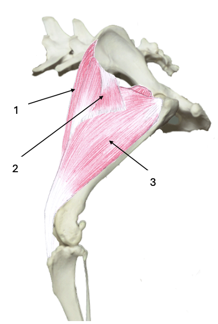
Name the labelled muscles
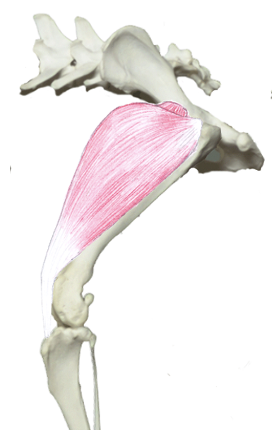
1.1 = Hip extensors. Gluteals - deep, middle and superficial
1.2 = Hip extensors. Hamstrings - semimembranosus, semitendinosus, biceps femoris
2 = Hip flexors. Sartorius, tensor fascia latae, quadriceps femoris - rectus femoris, vastus lateralis, vastus intermedius, vastus medialis,
3 = Stifle flexors
4 = Stifle extensors
5 = Tarsus and digit flexors
6 = Tarsus and digit extensors
What is the scapulothoracic joint?
A muscular attachment between the thorax and scapula.
What is the scapulohumeral joint?
The shoulder joint between the scapula and humerus.
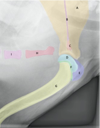
Name the structures
A = scapula
B = scapula spine
C = acromion
D = humerus head
F = epiphyseal growth plate
E = greater tubercule
G = humerus
H = manubrium
I = sternebrae
What is the role of the forelimb extrinsic muscles?
To attach the forelimb to the spine, stabilise the shoulder and move the limb during locomotion.
Which muscles are used to protract the forelimb?
Omotransverse and brachiocephalicus
What muscles are used to retract the forelimb?
Pectorals and latissimus dorsi
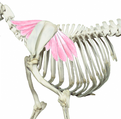
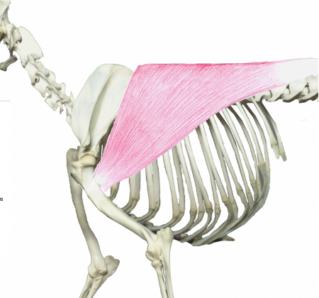
What muscle is this?
What muscle group is it in?
Serratus ventralis
Medial extrinsic muscle
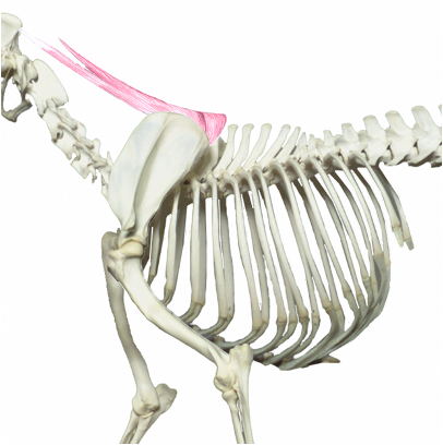
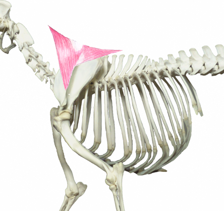
What muscle is this?
What muscle group is it in?
Rhomboid
Medial extrinsic muscle
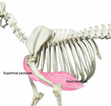
What muscles are these?
What muscle group are they in?
Superficial and deep pectoral
Medial extrinsic muscle
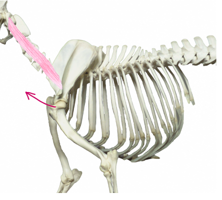
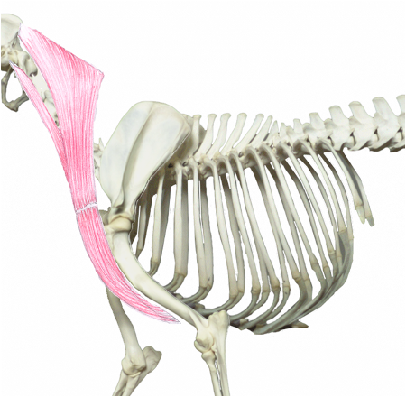
What muscle is this?
What muscle group is it in?
Omotransverse
Lateral extrinsic muscle
What muscle is this?
What muscle group is it in?
Trapezius
Lateral extrinsic muscle
What muscle is this?
What muscle group is it in?
Latissimus dorsi
Lateral extrinsic muscle
What muscle is this?
What muscle group is it in?
Brachiocephalicus
Lateral extrinsic muscle
What muscle group is this?
What muscles are these?
Shoulder stabilisers
1 = supraspinatus
2 = subscapularis
3 = teres minor
4 = infraspinatus
What muscle group is this?
What muscles are these?
Shoulder extensors
1 = brachiocephalicus
2 = supraspinatus
3 = biceps brachii
What muscle group is this?
What muscles are these?
Shoulder flexors
1 = Teres major
2 = Deltoid
3 = Triceps brachii
What three joints make up the elbow joint?
Humeroradial joint
Humeroulnar joint
Radioulnar joint
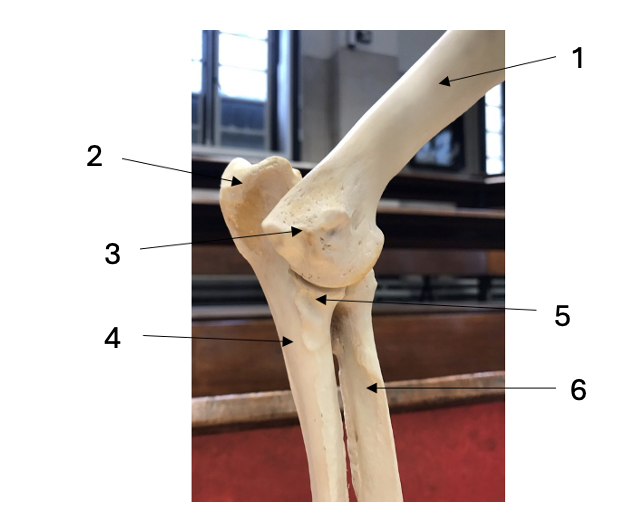
Name the structures.
1 = humerus
2 = olecranon
3 = lateral condyle
4 = ulna
5 = coronoid process
6 = radius
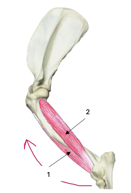
What muscle group is this?
What muscles are these?
Elbow flexors
1 = Biceps brachii
2 = Brachialis
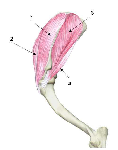
What muscle group is this?
What muscles are these?
Elbow extensors (triceps)
1 = Long head
2 = Lateral head
What bones make up the carpus?
Proximal row = ulnar carpal, intermediate carpal, radial carpal, accessory carpal
Distal row = first carpal (rarely present), second carpal, third carpal, fourth carpal
Name the structures
A = radius
B = accessory carpal
C = ulnar carpal
D = intermediate carpal
E = radial carpal
F = fourth carpal
G = third carpal
H = second carpal
I = first carpal
J = metacarpal 4
L = metacarpal 3
K = metacarpal 2
What muscle group is this?
Carpus and digit extensors
What muscle group is this?
Carpus and digit flexors.
What are the technical names for the cannon bone and splint bones in horses?
Cannon bone = metacarpal 3
Medial splint bone = metacarpal 2
Lateral splint = metacarpal 4
What bones make up the cannon bone in ruminants?
Fused metacarpal 3 and 4
What is the fetlock joint and which bones is it between?
Metacarpophalangeal joint
Cannon bone (metacarpal 3) and the long pastern (proximal phalanx)
What is the pastern joint and which bones is it between?
Proximal interphalangeal joint
Long pastern (proximal phalanx) and short pastern (middle phalanx)
What is the coffin joint and which bones is it between?
Distal interphalangeal joint
Short pastern (middle phalanx) and coffin/pedal bone (distal phalanx)
Which sesamoid bones are present in the equine forelimb?
Two proximal sesamoids on the palmar aspect of the fetlock joint
A distal sesamoid in the foot called the navicular bone
Which sesamoid bones are present in the canine forelimb?
Two proximal sesamoids at each metacarpophalangeal joint (except the dewclaw - phalanx 1)
One distal sesamoid at each distal interphalageal joint, called a navicular bone
Where is the additional sesamoid bone some dogs have?
At the tendon of the supinator muscle, near the elbow.
Where does the deep digital flexor tendon (DDFT) originate and insert, and what action does it have?
Originates at the deep digital flexor muscle on the caudal aspect of the ulna/fibula.
Inserts on the palmar/plantar aspect of the distal phalanx
Flexes the distal interphalangeal joint
What is the role of the inferior check ligament?
To attach the deep digital flexor tendon to the palmar carpal ligament in the forelimb, or the tarsus in the hindlimb
Where does the superficial digital flexor tendon (SDFT) originate and insert, and what action does it have?
Originates from the superficial digital flexor muscle on the caudal aspect of the ulna/fibula..
At the fetlock, it forms a sheath called the manica flexoria and then splits into two branches which insert on the medial and lateral aspect of the middle phalanx
Flexes the proximal interphalangeal joint
What is the role of the superior check ligament?
To attach the superficial digital flexor tendon to the distal radius
Why is there no superior check ligament in the hindlimb?
The SDFT is anchored to the calcaneus instead.
What is the manica flexoria?
A sheath formed by the SDFT which wraps around the DDFT.
Where does the suspensory ligament (interosseus muscle) originate and insert, and what action does it have?
Originates on the proximal metacarpus/tarsus and splits into two branches, which each travel over one sesamoid bone, and then cross to the cranial aspect of the long pastern, and insert on the extensor tendon.
Where does the common digital extensor tendon originate and insert, and what action does it have?
Originates from the common digital extensor muscle on the cranial aspect of the radius/tibia.
Inserts on the distal phalanx.
It extends the foot, to oppose the action of the SDFT and DDFT.
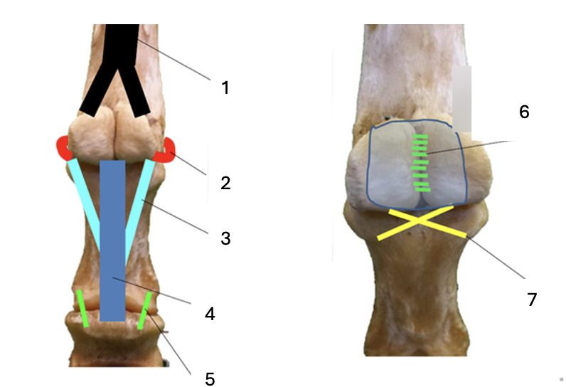
Name the structures.
1 = suspensory ligament
2 = collateral sesamoidean ligament
3 = oblique sesamoidean ligament
4 = straight sesamoidean ligament
5 = palmar ligament of the pastern
6 = intersesamoidean ligament
7 = cruciate sesamoidean ligament
How does the lacertus fibrosus contribute to the equine forelimb stay apparatus?
It connects the biceps brachii on the cranial aspect of the humerus (shoulder extensor, elbow flexor) to the extensor carpi radialis on the cranial aspect of the radius (carpus and digit extensor) so that the shoulder, carpus and digits are locked in extension while the elbow is flexed.
How does the elbow locking mechanism contribute to the equine forelimb passive stay apparatus?
Once the elbow is locked in extension, it requires minimal effort to maintain and is only unlocked when force is applied.
Why does the biceps brachii in the horse have lots of white connective tissue in in?
To allow it to store elastic energy during weight bearing which can be rapidly released during retraction.
What is the main pathway of arterial blood supply to the canine forelimb?
Ascending aorta > brachiocephalic artery > subclavian artery > axillary artery (enters forelimb) > brachial artery (after shoulder) > median artery (after elbow). it runs along the medial aspect of the limb.
Where can you take a pulse in the dog?
From the median artery, by palpating the cubital fossa - which is on the medial aspect of the elbow, or from the digital artery, by palpating the palmar aspect of the paw.
Where can you take a pulse in the horse?
From the lateral palmar artery which is part of the neurovascular bundle on the lateral aspect of the pastern (dorsal - VAN - palmar)
What is the pathway of venous blood drainage from the forelimb?
On the medial aspect of the limb, the median vein > brachial vein (above elbow) > brachial vein (above shoulder) > axillary vein (enters forelimb) > subclavian vein > brachiocephalic vein > cranial vena cava.
The cephalic vein runs along the cranial aspect of the limb and feeds into the axillary vein.
Where does autonomic cutaneous innervation to the forelimb arise?
Nerves exit the spinal cord at the cervical intumescence (C6-T2) and their ventral branches form the brachial plexus, which innervates the forelimb.
What is Sweeney in horses?
Wastage of the supraspinatus and infraspinatus muscles, which are shoulder stabilisers on the lateral aspect of the shoulder, due to damage to the suprascapular nerve as it cross over the cranial surface of the scapula.
What does the axillary nerve innervate?
The deltoid and teres major (shoulder flexors)
What does the radial nerve innervate?
The triceps brachii muscles (shoulder flexors, elbow extensors) on the caudal aspect of the humerus, and the carpus and digit flexors on the cranial aspect of the radius.
What does the musculocutaneous nerve innervate?
The biceps brachii and brachialis muscles (elbow flexors) on the cranial aspect of the humerus.
What does the median nerve innervate?
The carpus and digit extensors on the palmar aspect of the ulna.
What does the ulnar nerve innervate?
The carpus and digit extensors on the palmar aspect of the ulna.
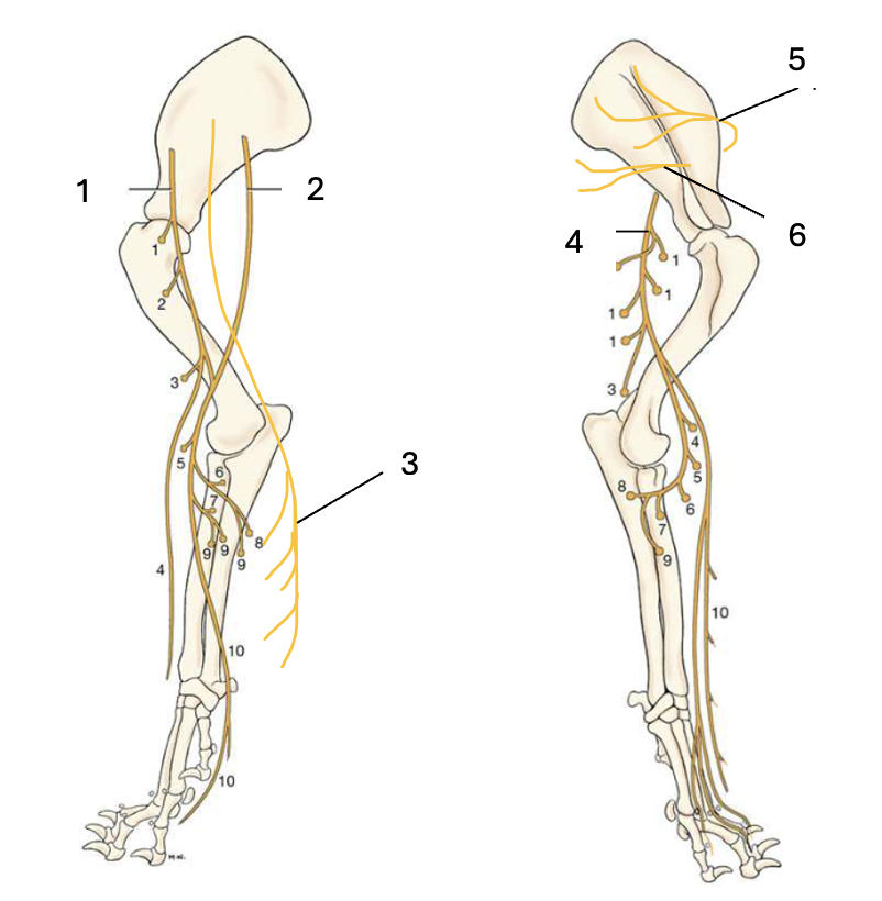
Name the labelled structures.
1 = musculocutaneous nerve
2 = median nerve
3 = ulnar nerve
4 = radial nerve
5 = subscapular nerve
6 = axillary nerve
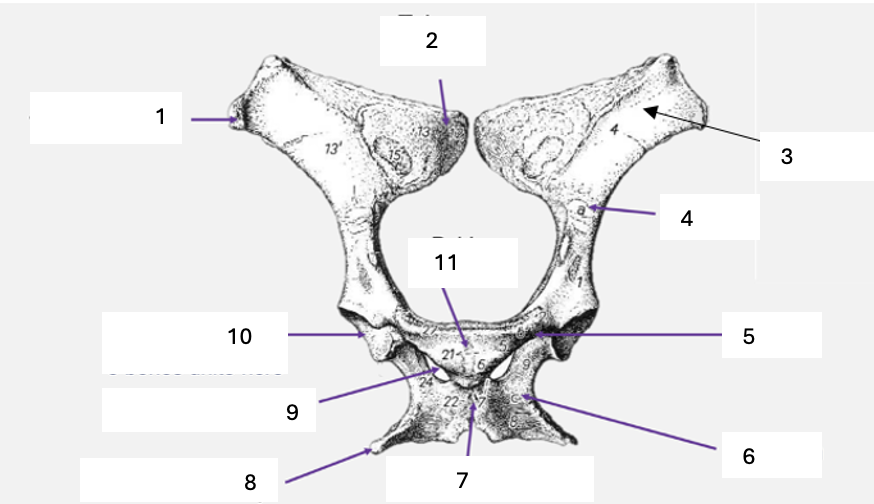
Name the labelled structures.
1 = Tuber coxae
2 = Tuber sacrale
3 = Iliac wing
4 = Ileum
5 = Pubis
6 = Ischium
7 = Ischial symphysis
8 = Ischiatic tuberosity
9 = Obturator foramen
10 = Acetabulum
11 = Pubic symphysis

Name the labelled structures
A = Ischium
B = Ischiatic tuberosity
C = Obturator foramen
D = Pubis
E = Acetabulum
F = Ileum
G = Crest of ileum
H = Ischiatic arch
I = Sacrum
J = Head of femur
K = Neck of femur
L = Greater trochanter of femur
M = Femur
What muscle inserts on the greater trochanter of the femur?
The gluteal muscles (hip extensors and hip abductors)
What additional structures do horses have at the proximal end of their femur?
A lesser trochanter on the same side as the head, which the psoas muscles (hypaxial muscles) attach to, and a third trochanter on the same side as the greater trochanter, which the superficial gluteal attaches to (hip flexor in the horse unlike in all other species where it is a hip extensor).
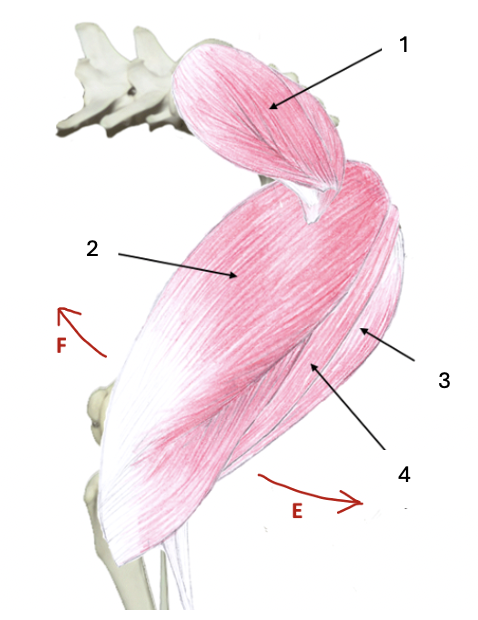
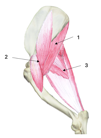
What muscle group is this?
What muscles are these?
Hip extensors
1 = Gluteal muscles (superficial, middle and deep)
2 = Biceps femoris (hamstring)
3 = Semimembranosus (hamstring)
4 = Semitendinosus (hamstring)
What muscle group is this?
What muscles are these?
Hip flexors
1 = Sartorius
2 = Tensor fascia latae
3 = Quadriceps femoris
What muscle group is this?
What muscles are these?
Hip adductors
1 = Adductor
2 = Gracilis
What is the femoral groove?
The dip between the two condyles at the distal end of the femur.
How does the patella move within the femoral groove?
It slides distally as the stifle flexes (bent leg) and proximally as the stifle extends (straight leg).
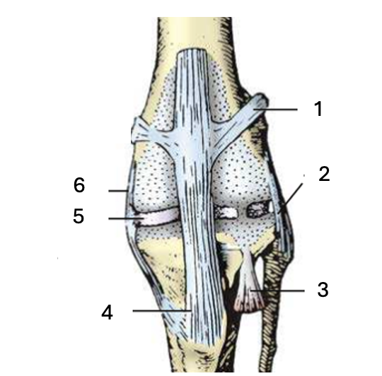
Name the labelled structures
1 = Patella femoral ligament
2 = Lateral collateral ligament
3 = Digital extensor tendon
4 = Patella ligament
5 = Menisci
6 = Medial collateral ligament
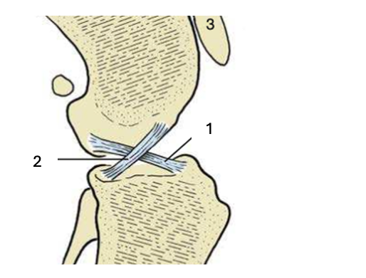
Name the labelled structures
1 = Cranial cruciate ligament
2 = Caudal cruciate ligament
What is the parapatellar cartilage in the horse?
A hook of cartilage on the medial side of the patella which is able to hook over the medial trochlear ridge of the femur.
Name the labelled structures
1 = Parapatellar cartilage
2 = Medial trochlear ridge
3 = Medial patella ligament
4 = Menisci
5 = Patella
6 = Lateral patella ligament
7 = Lateral trochlear ridge
8 = Intermediate trochlear ridge
Which species have two fabellae on the caudal aspect of the knee joint?
Dogs and cats
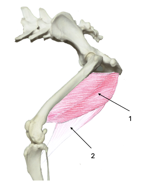
What muscle group is this?
What muscles are these?
Stifle flexors
1 = Biceps femoris
2 = Semimembranosus
3 = Semitendinosus
4 = Gastrocnemius
What muscle group is this?
What muscles are these?
Stifle extensors
Quadriceps femoris
What bones make up the tarsus?
Proximal row = talus and calcaneus
Intermediate row = central tarsal bone and 4th tarsal bone
Distal row = 1st, 2nd and 3rd and 4th tarsal bones (bridges two rows)
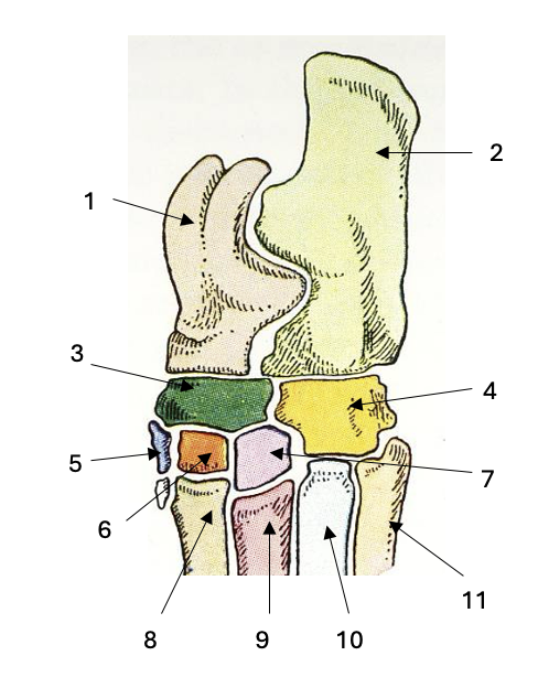
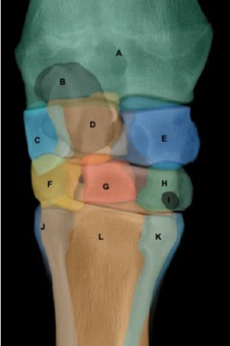
Name the labelled structures
1 = talus
2 = calcaneus
3 = central tarsal
4 = 4th tarsal
5 = 1st tarsal
6 = 2nd tarsal
7 = 3rd tarsal
8 = 2nd metacarpal
9 = 3rd metacarpal
10 = 4th metacarpal
11 = 5th metacarpal
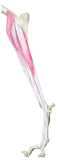
What muscles are tarsal extensors?
Gastrocnemius and hamstrings (biceps femoris, semimembranosus) via the common calcaneal tendon
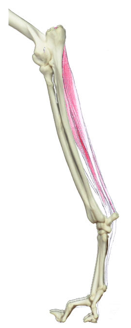
What muscles are tarsal flexors?
Cranial tibial and peroneus tertius
What contributes to the common calcaneal tendon?
The gastrocnemius tendon
The semitendinosus and biceps femoris muscles (stifle flexors)
The gracilis muscle (hip adductor)
What is the reciprocal apparatus in the horse’s hindlimb and what does it do?
The SDFT on the caudal aspect of the tibia and the peroneus tertius on the cranial aspect
It ensures the stifle and hock both flex/extend together.
What is the main pathway of arterial blood supply to the hindlimb?
Abdominal aorta > External iliac artery > Superficial femoral artery and deep femoral artery
The superficial femoral artery supplies the inguinal region
The deep femoral artery enters the hindlimb > popliteal artery (beyond stifle) > cranial tibial artery and caudal tibial artery
The cranial tibial artery supplies the dorsal distal limb and becomes the dorsal metatarsal artery
The caudal tibial artery supplies the plantar distal limb and becomes the digital artery
The saphenous artery branches off the femoral artery and runs along the medial aspect of the limb.
How is venous blood drained from the hindlimb?
Digital vein > Metatarsal vein > Tibial vein > Popliteal vein > Femoral vein > External iliac vein > Caudal vena cava
The saphenous vein also drains into the femoral vein
Where does autonomic cutaneous innervation to the hindlimb arise?
Nerves exit the spinal cord at the lumbosacral intumescence (L4 - S3) and their ventral branches form the lumbosacral plexus, which innervates the hindlimb.
What does the cranial gluteal nerve innervate?
The gluteals (hip extensors) and the tensor fascia latae (hip flexor) on the cranial lateral aspect of the pelvis.
What does the caudal gluteal nerve innervate?
The gluteals (hip extensors)
What does the sciatic nerve innervate?
The quadriceps femoris (stifle extensor) on the lateral aspect of the femur, and the stifle flexors on the caudal aspect of the femur (hamstrings - biceps femoris, semimembranosus, semitendinosus.
What does the femoral nerve innervate?
The hip flexors (sartorius, tensor fascia latae, quadriceps femoris) on the cranial aspect of the femur.
What does the obturator nerve innervate?
The hip adductors (gracilis, adductor, pectineus, obturator) on the medial aspect of the femur.
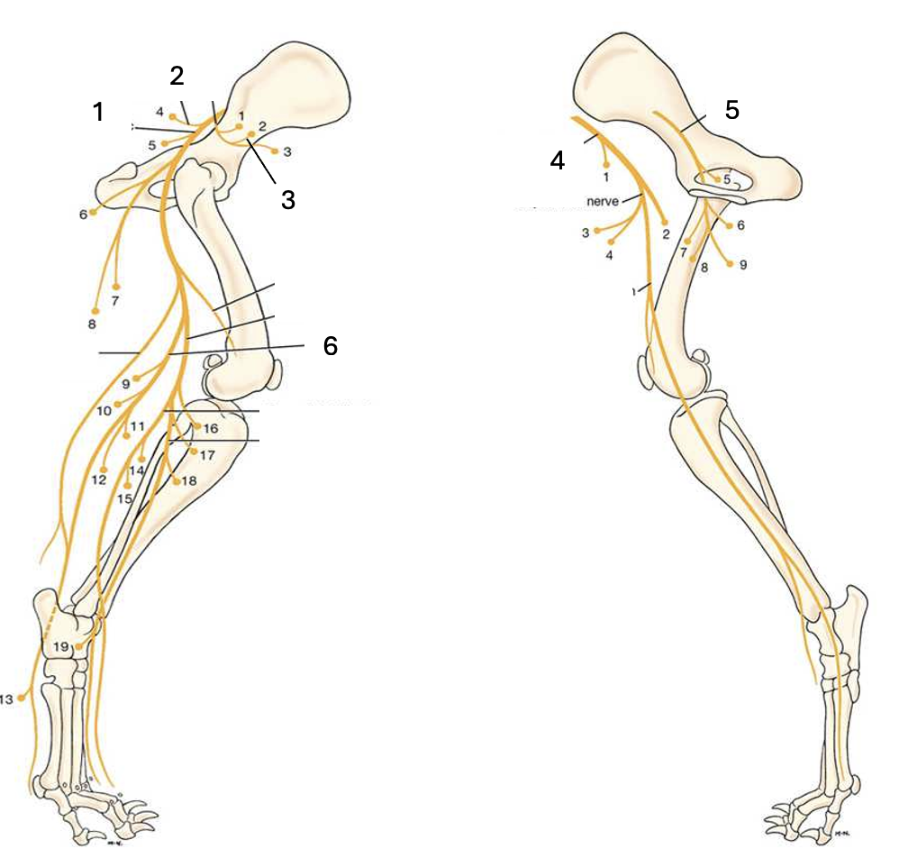
Name the labelled structures.
1 = Sciatic nerve
2 = Caudal gluteal nerve
3 = Cranial gluteal nerve
4 = Femoral nerve
5 = Obturator nerve
6 = Tibial nerve
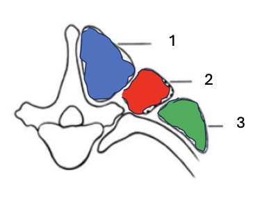
What overarching muscle group is this?
What individual muscle groups are these?
Epaxial muscles
1 = Transversospinalis system
2 = Longissimus system
3 = Iliocostalis system
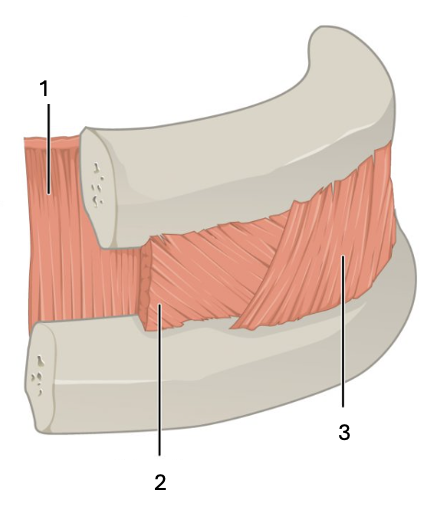
What muscle group is this?
What muscles are these?
Intercostal muscles
1 = Innermost intercostal
2 = Internal intercostal
3 = External intercostal
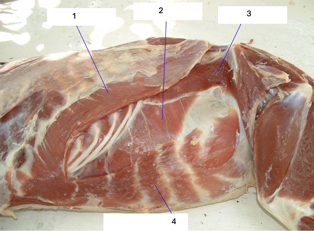
What muscle group is this?
What muscles are these?
Hypaxial muscles
1 = External abdominal obliques
2 = Transversus abdominis
3 = Internal abdominal obliques
4 = Rectus abdominis