Bio lab
1/85
There's no tags or description
Looks like no tags are added yet.
Name | Mastery | Learn | Test | Matching | Spaced | Call with Kai |
|---|
No analytics yet
Send a link to your students to track their progress
86 Terms
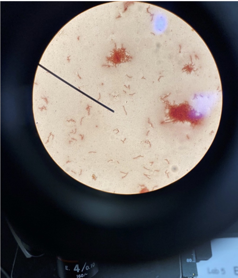
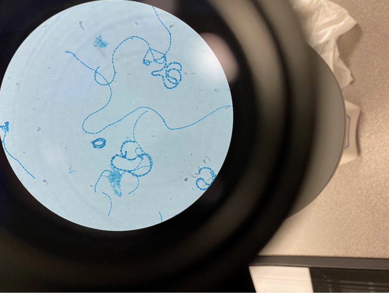
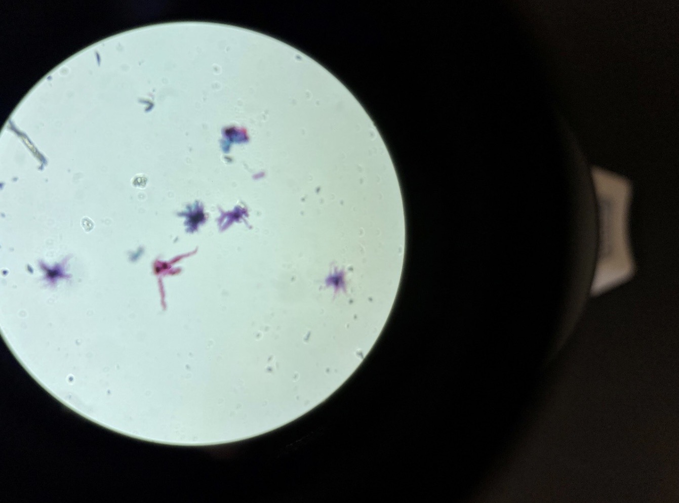
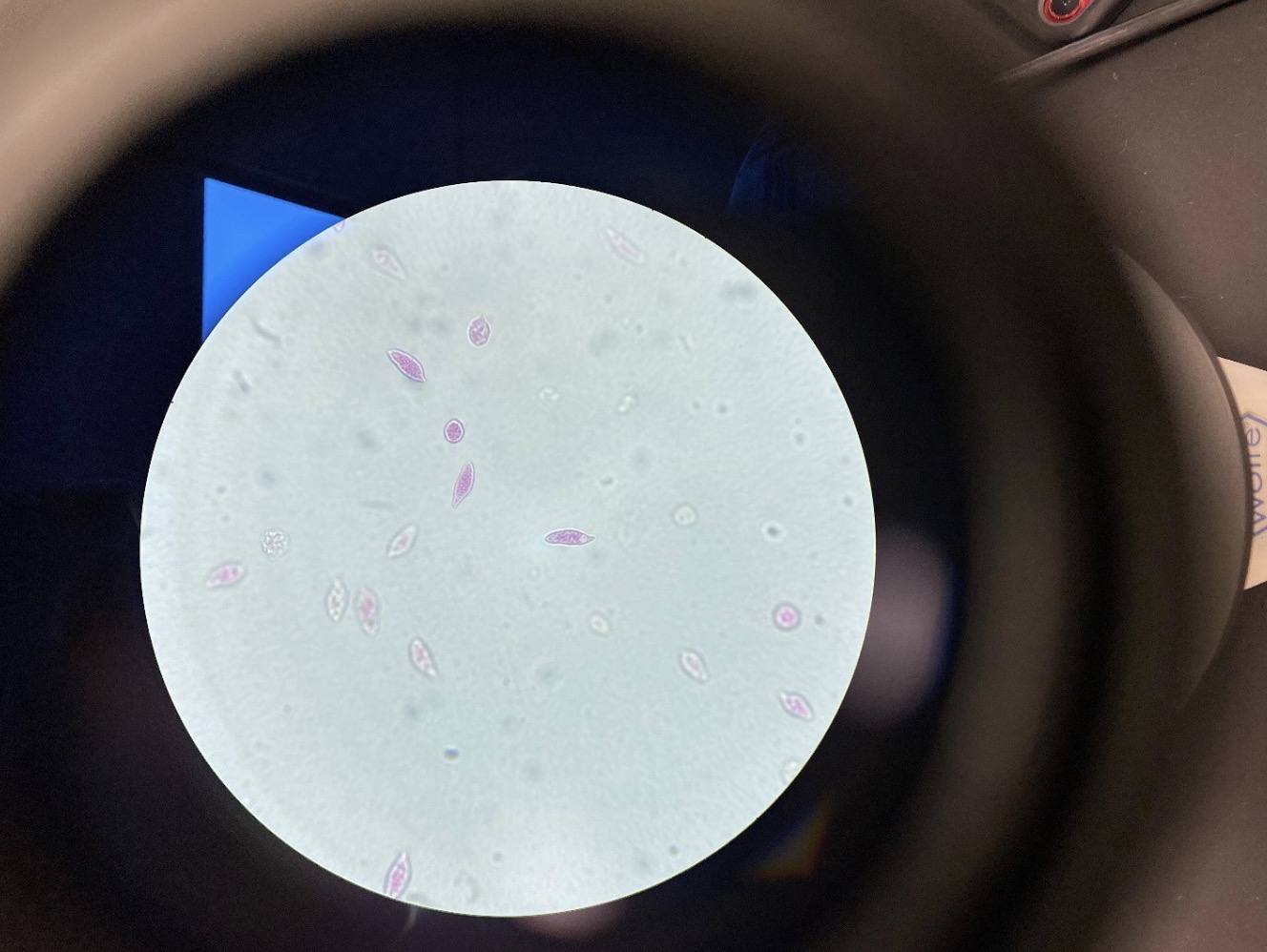
Spirillum shaped Bacteria 400x
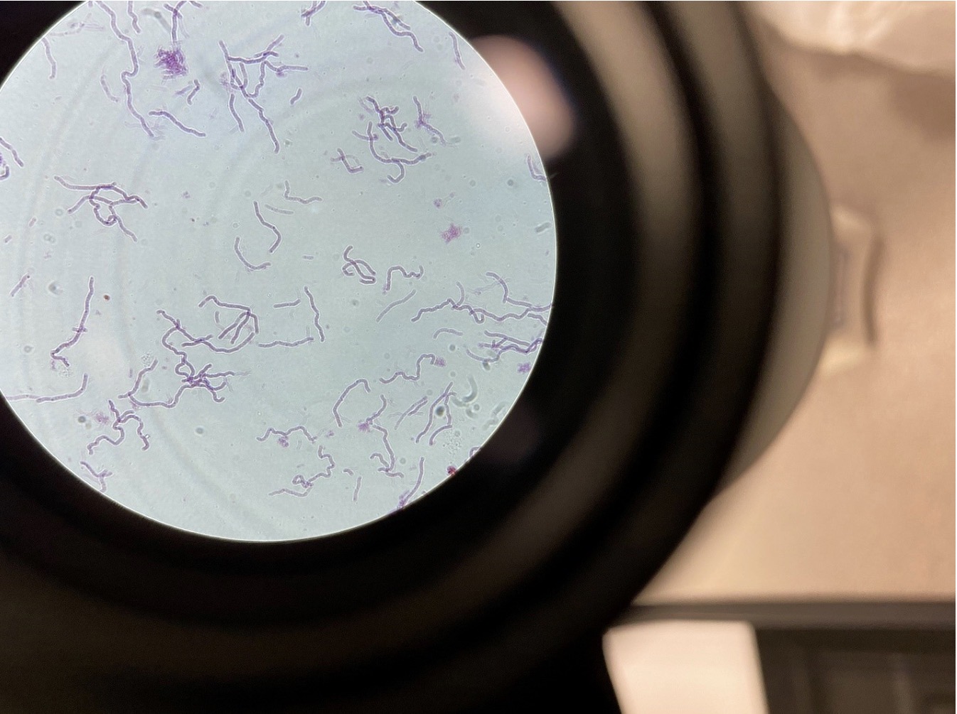
Three shapes of Gram-Positive Bacteria 400x, Spirillum (spiral, Coccus (spherical), Bacillus (rod) Gram-Positive bacteria
stain purple in a Gram Stain
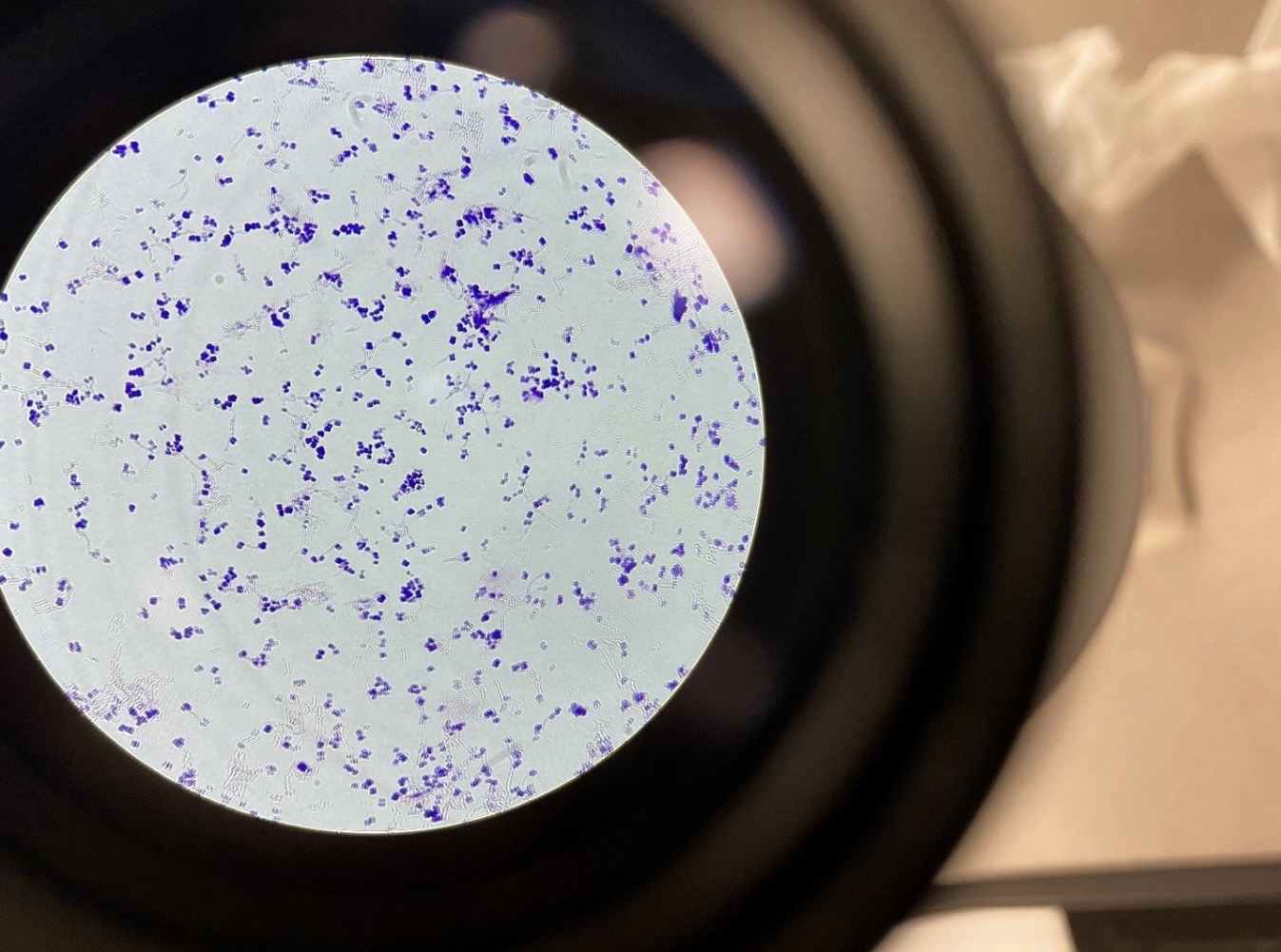
Gram-Negative Bacteria 400x. Gram-Negative bacteria stain pink in a Gram Stain
Cyanobacteria perform nitrogen fixation by breaking the triple bond in nitrogen gas and producing
nitrates, nitrites and ammonia which is usable by other organisms for proteins and nucleic acids.
Specialized cells called Heterocysts, and may be seen as enlarged cells in a filament
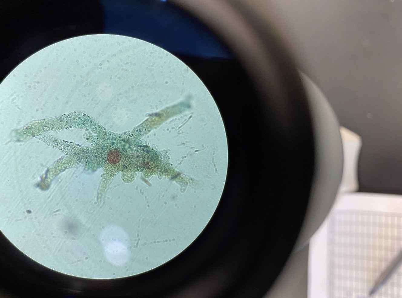
Amoeba proteus 100x, Amoeba have pseudopodia (false feet) that are used for movement and
to ingest food. Amoeba are protozoans in the Kingdom Protista. Protozoans are one-celled organisms
that ingest their food
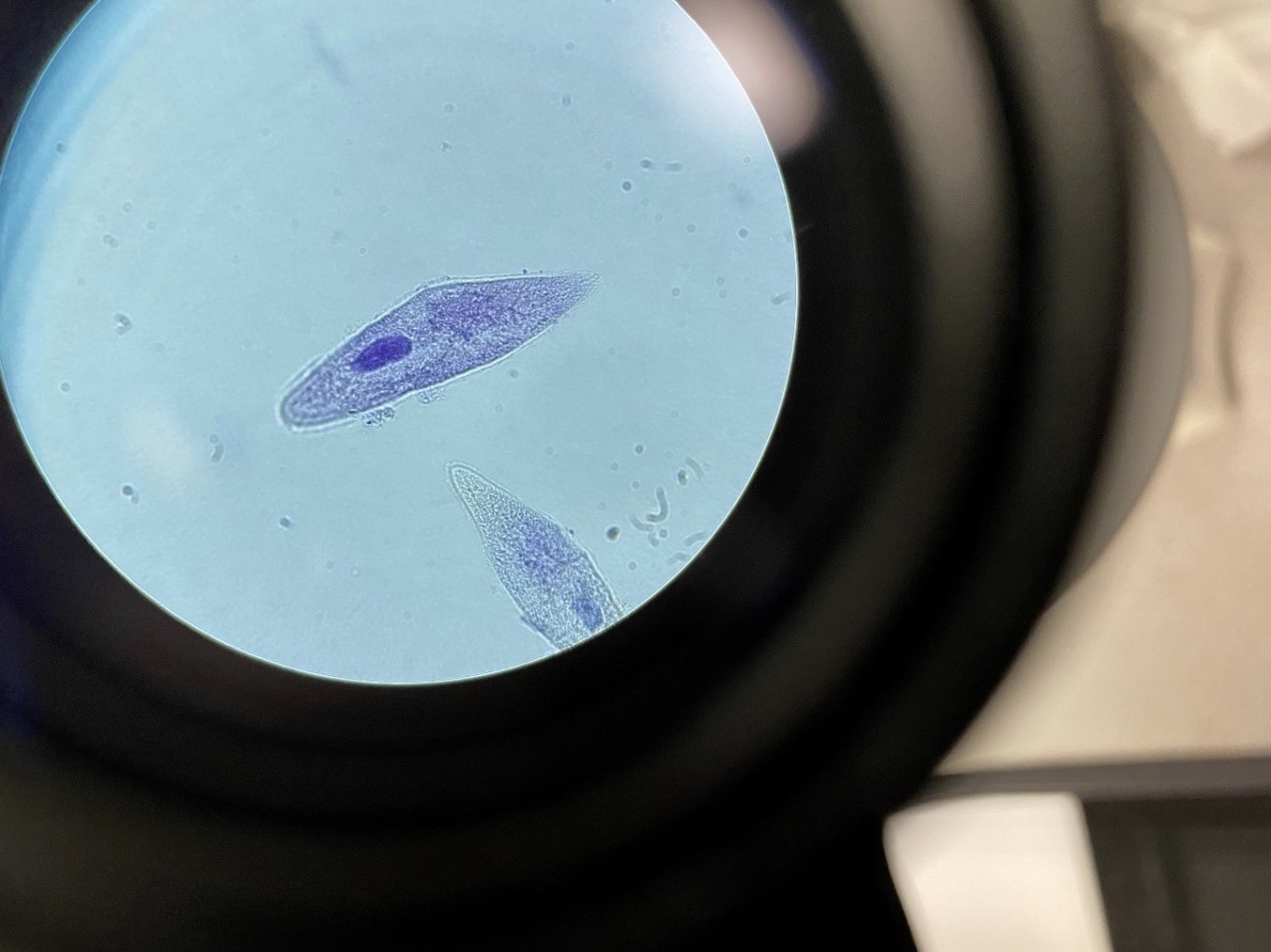
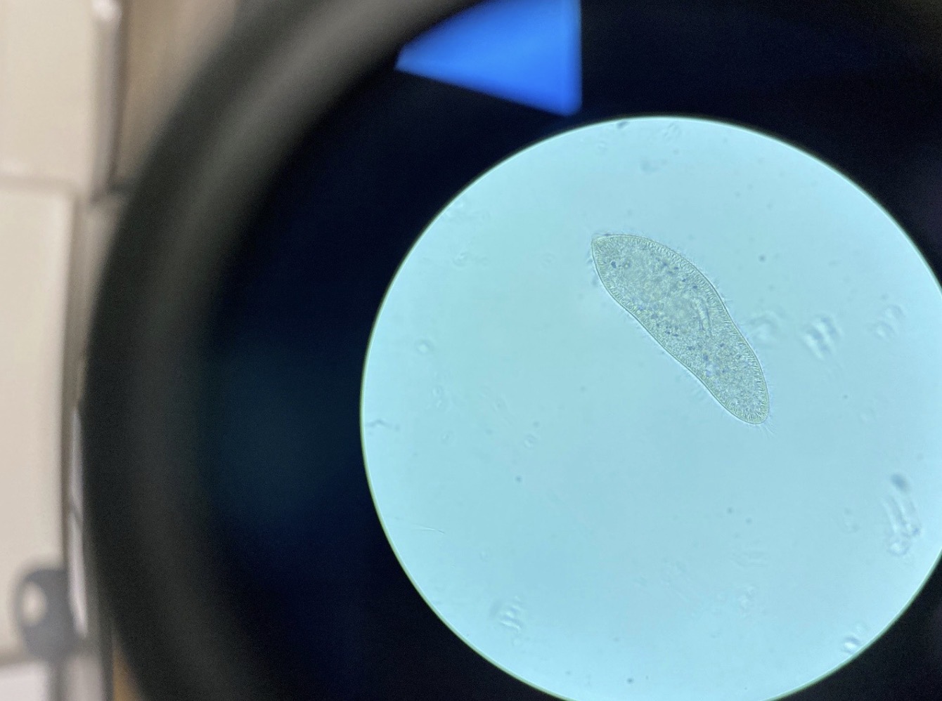
Paramecium 400x, Paramecium have cilia. Paramecium are protozoans in the Kingdom
Protista. Protozoans are one-celled organisms that ingest their food.
Euglena 400x, Euglena have a flagellum. Euglena belong to the Kingdom Protista. They have a flagellum that is used
for movement. They can be both autotrophic and heterotrophic
Chlamydomonas 400x, Chlamydomonas has two flagella. It is algae in the Volvocine line of evolution (Kingdom Protista).
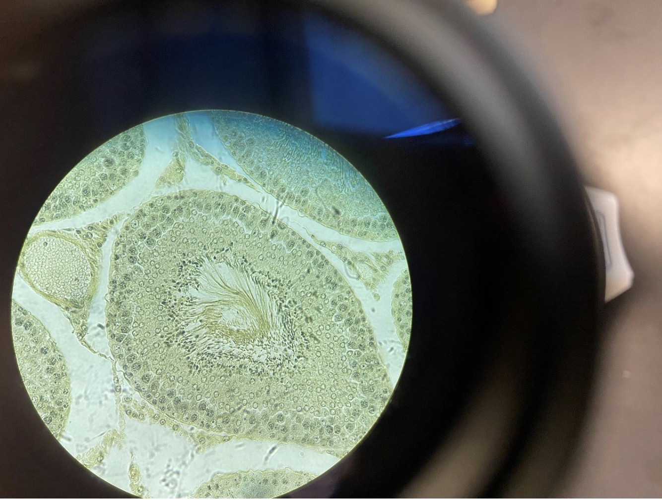
Rat Testis 400x. Flagella can be seen on sperm in the center of the seminiferous tubule. Flagella are the most primitive
structure for movement. Flagella can be found in some organisms of every kingdom of life
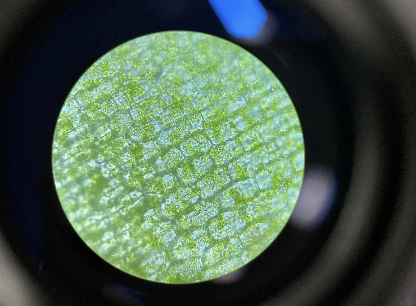
Elodea with chloroplasts 400x. Elodea is a hydrophyte (plant that lives in water). Chloroplasts are organelles that
perform photosynthesis in algae and in plants
Spirogyra with chloroplasts in spirals 400x
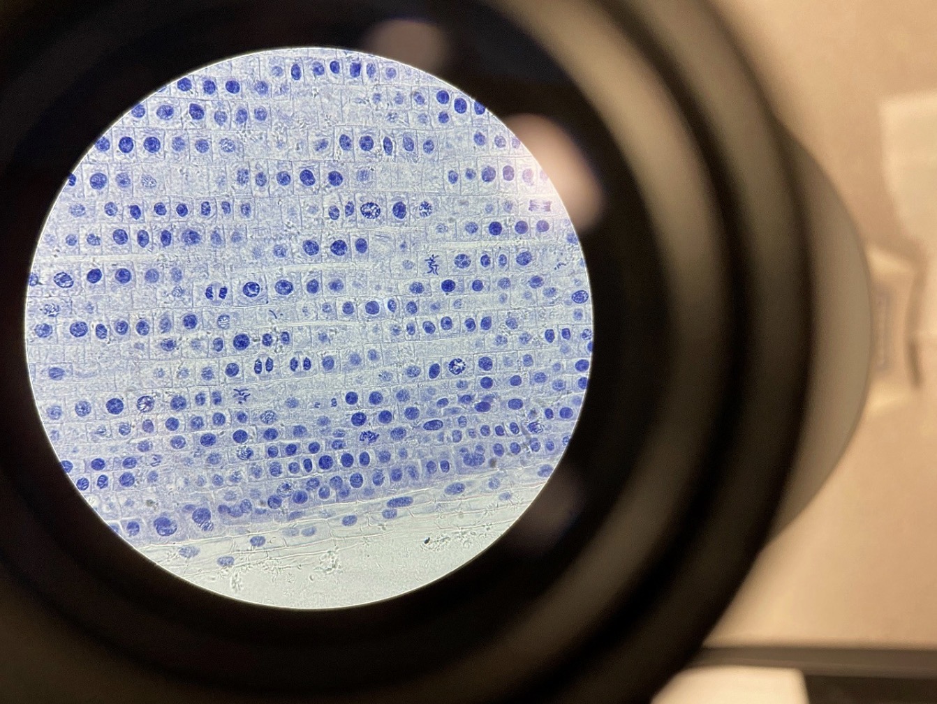
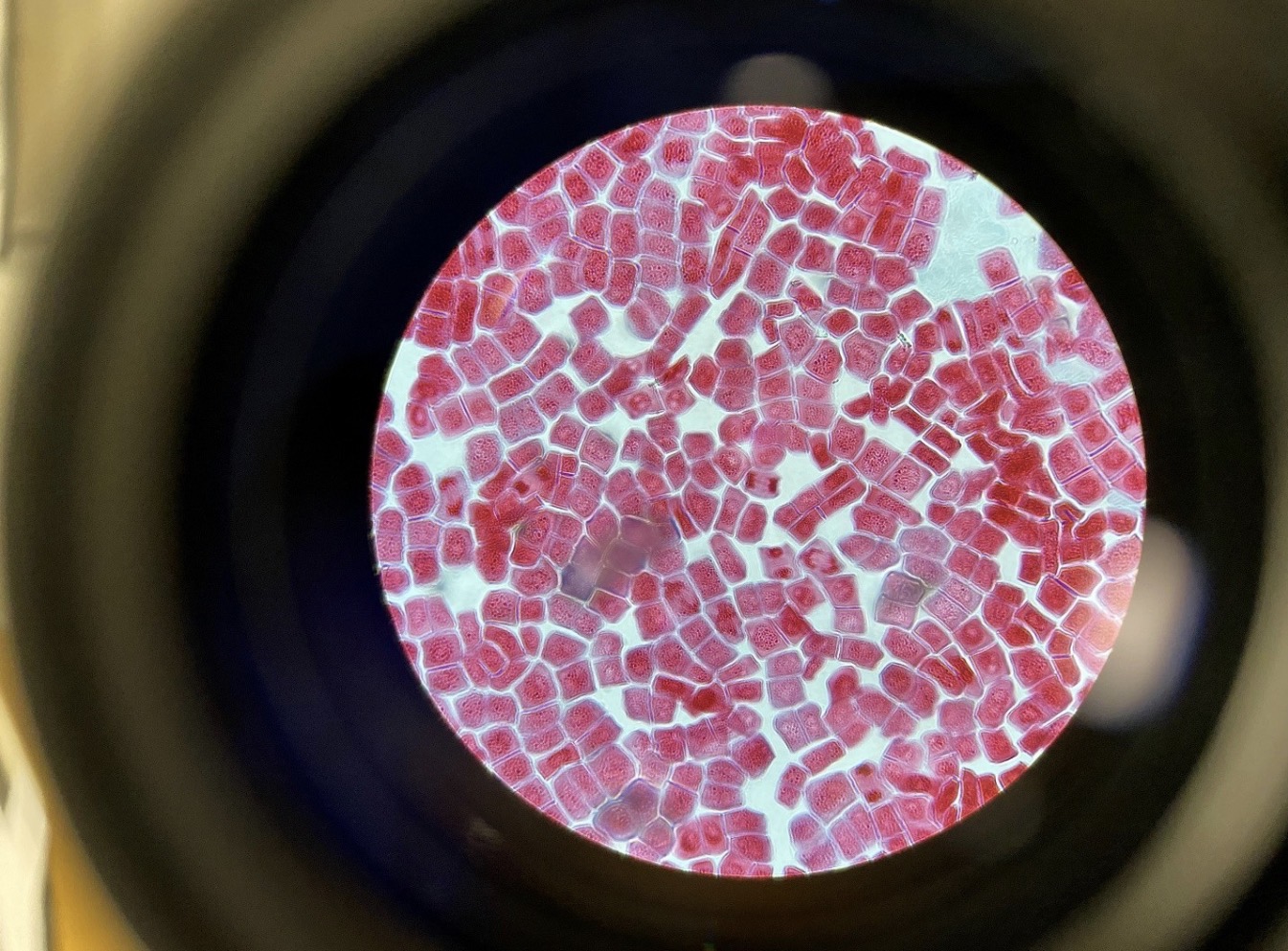
Mitosis in the Allium (onion) root tip 400x, longitudinal section. Roots grow at the apical meristem near the tip of the
root, and this is where mitosis can be found. Under the microscope condensed chromosomes can be seen in prophase;
chromosomes can be seen lined up in the middle of the cell in metaphase; chromosomes can be seen separating in anaphase,
and chromosomes can be seen at opposite poses of the cell n telophase
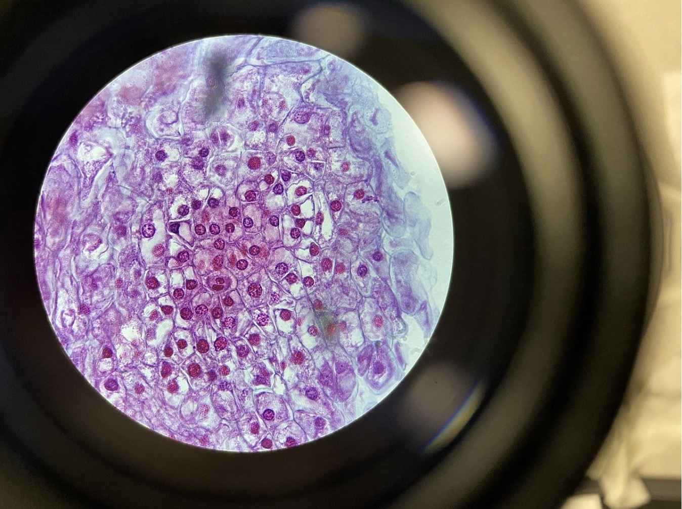
Plant Cell Mitosis 400x, cross section
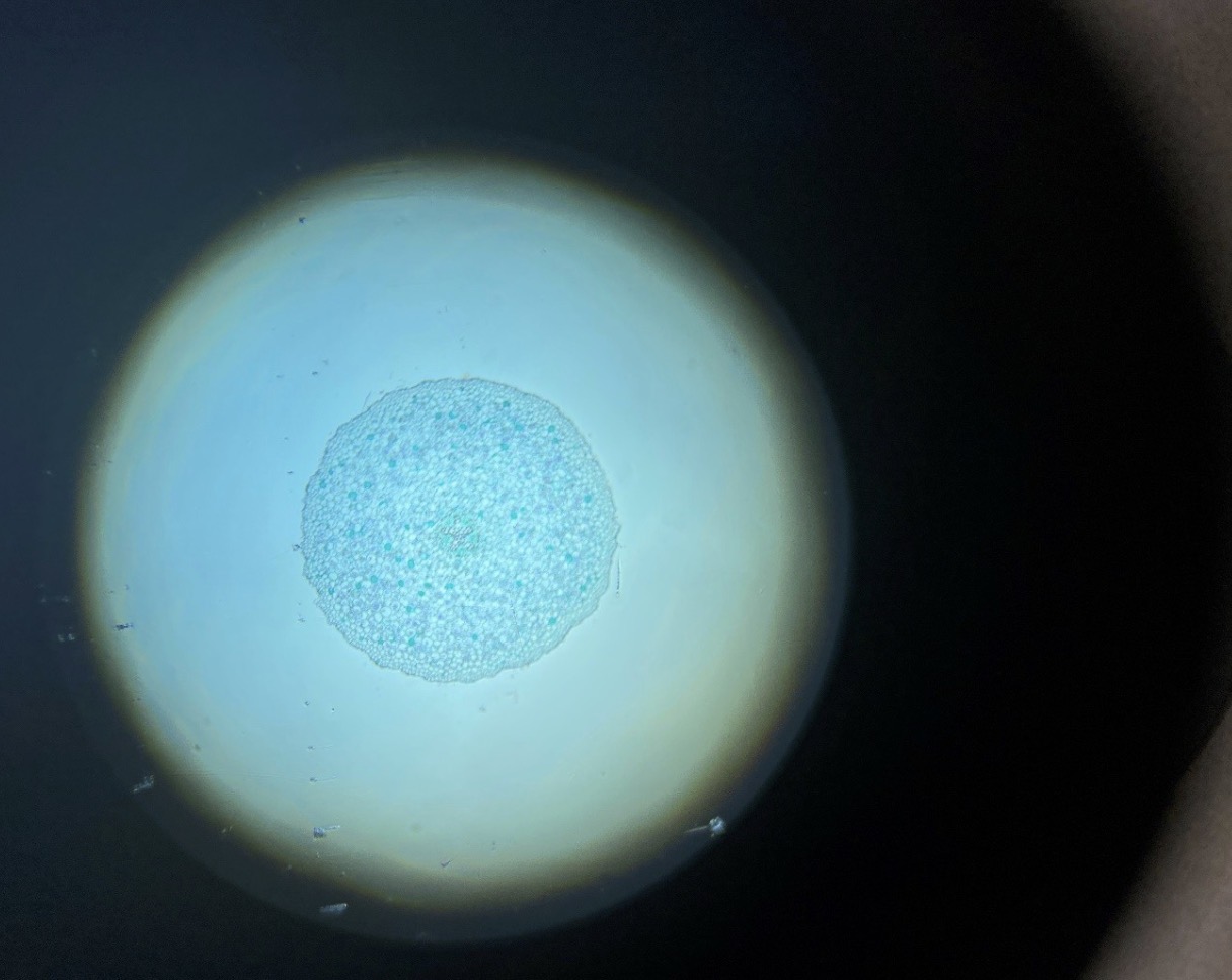
Nuclei in the Starfish Embryo 400x. The nuclei can be seen in the center of the animal cells
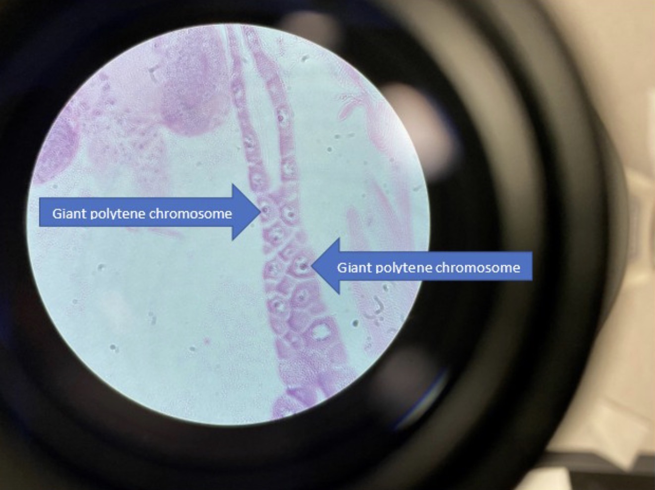
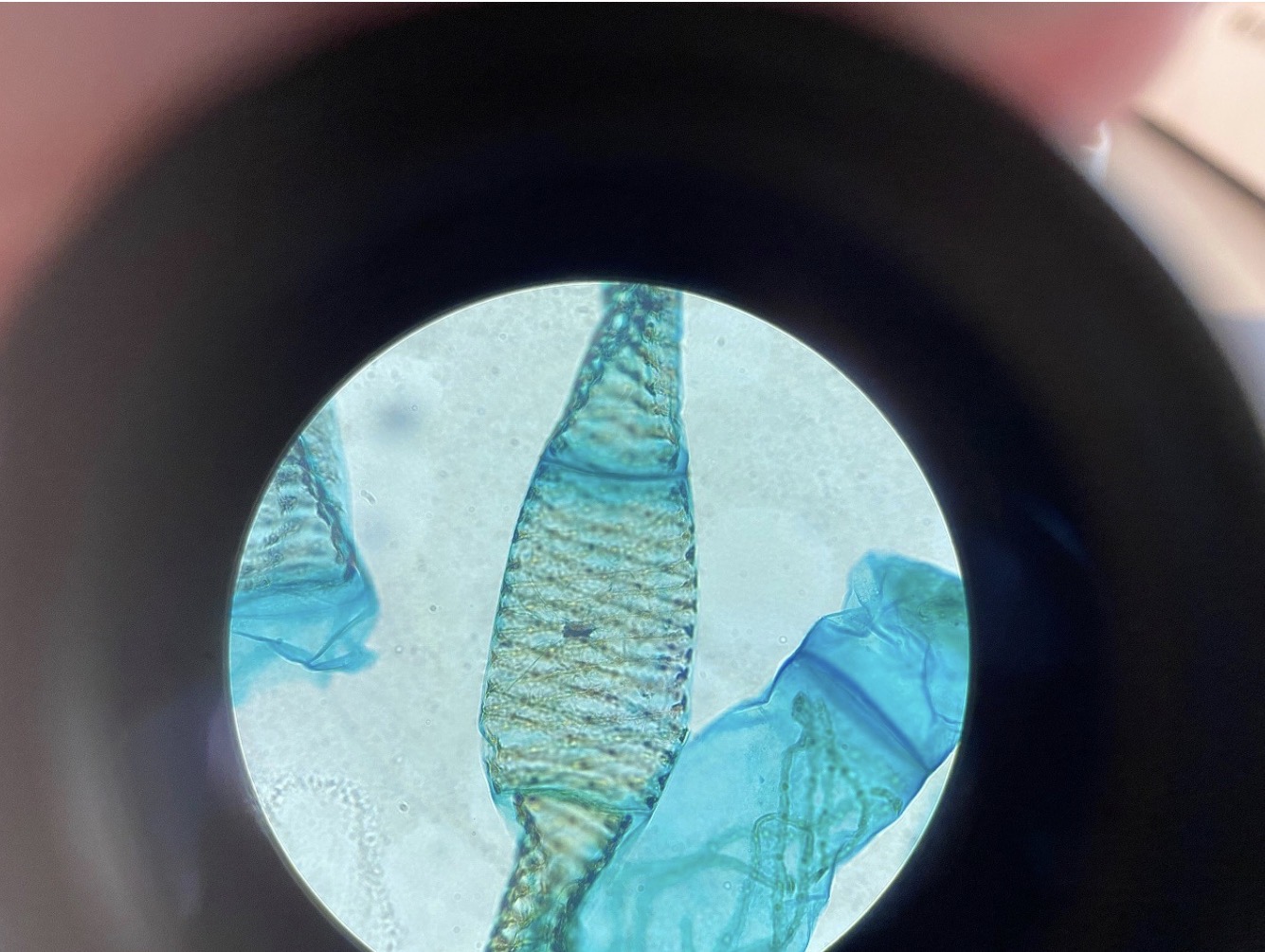
Giant Polytene Chromosomes in the Drosophila larvae 400x. Drosophila larvae must consume a great deal of food.
The chromosomes (at the tip of the blue arrows) in their salivary glands enlarge to produce proteins necessary to aid in digestion.
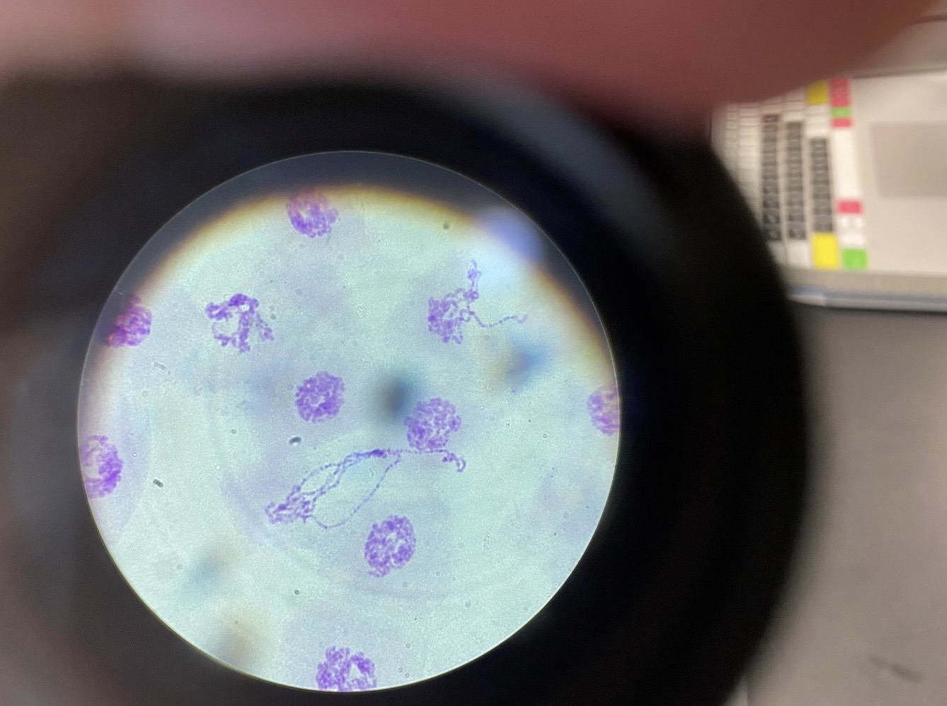
DNA from giant polytene chromosomes in Drosophila 400x.

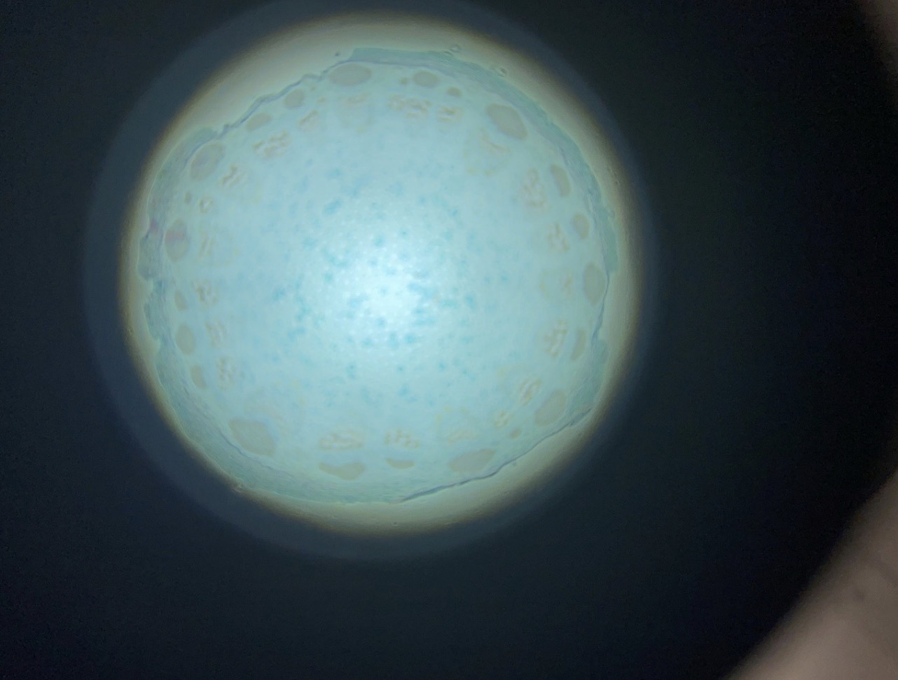
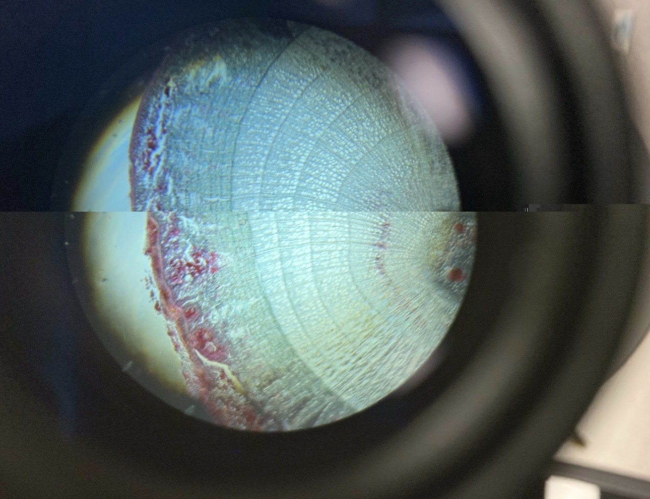
Pollen grains in a Pine Cone 400x. Nuclei can be seen in the pollen grains. Wing structures can be seen on the pollen
grains as well.
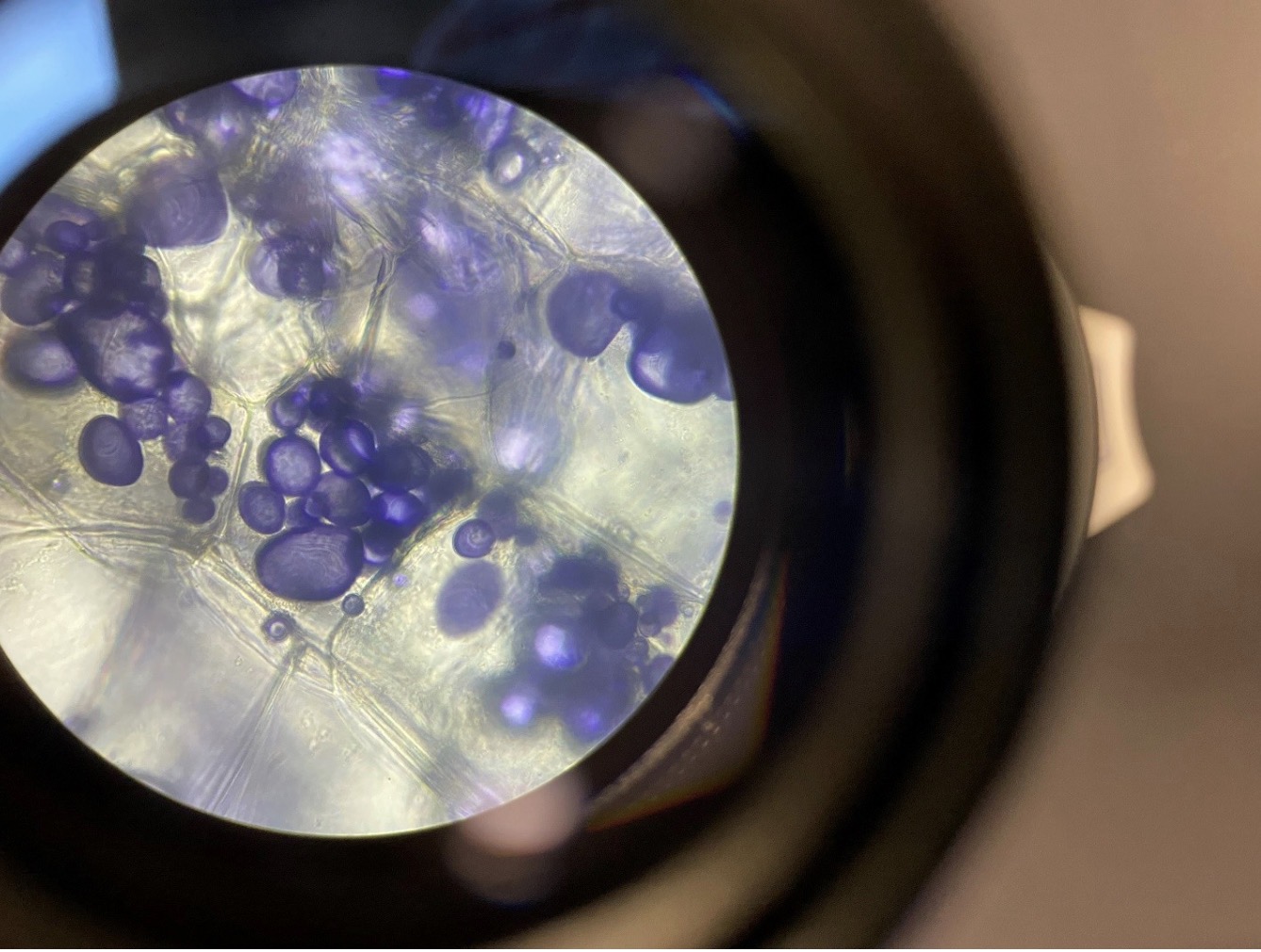
Storage Vesicles containing Starch in the Potato 400x. The starch is stained purple in the storage vesicles. With iodine
potassium iodide.
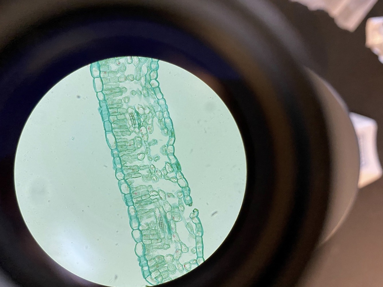
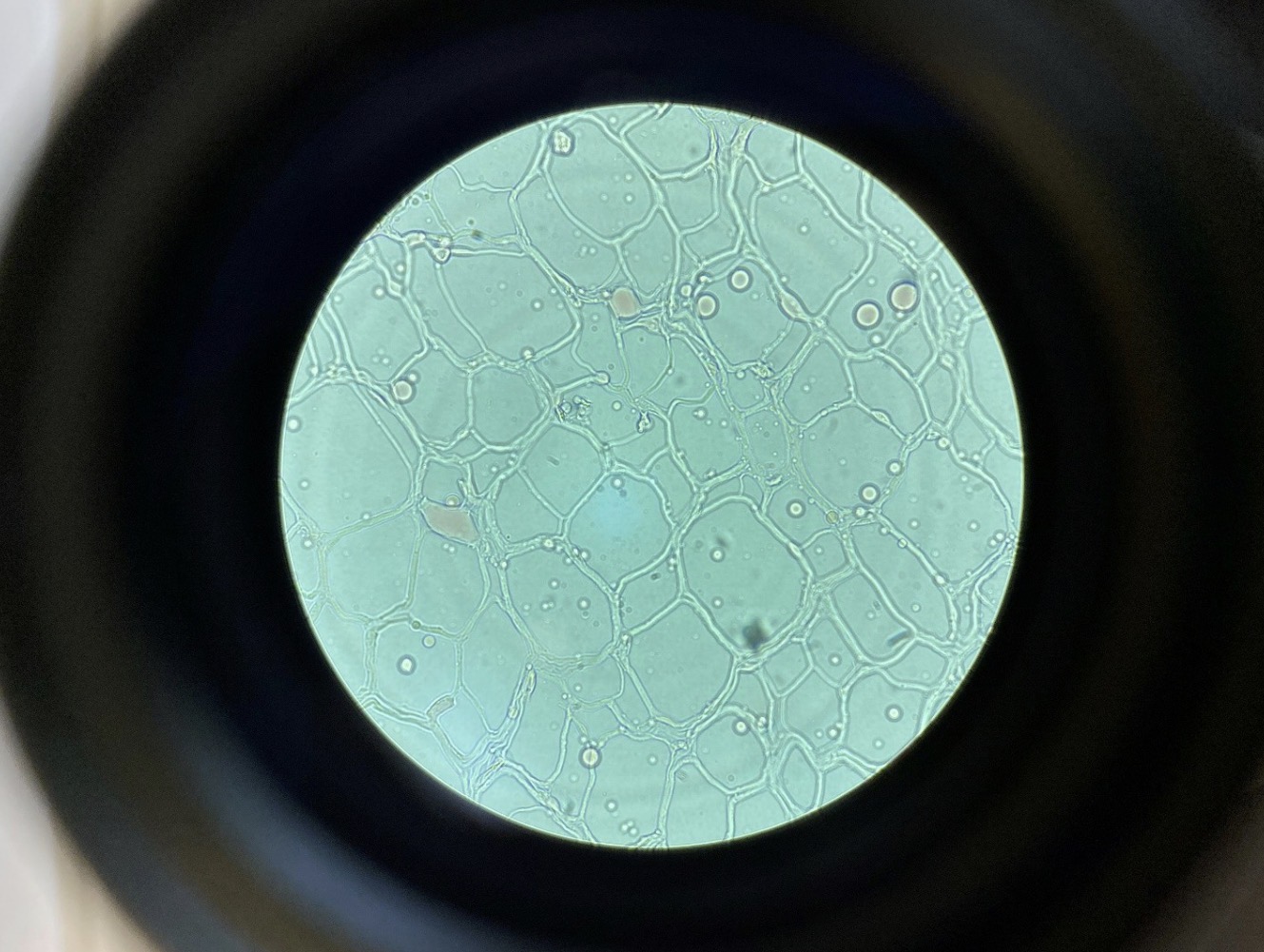
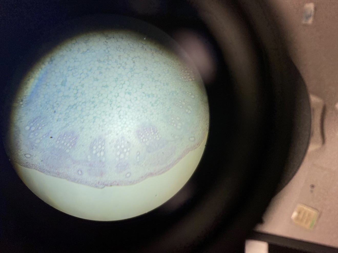
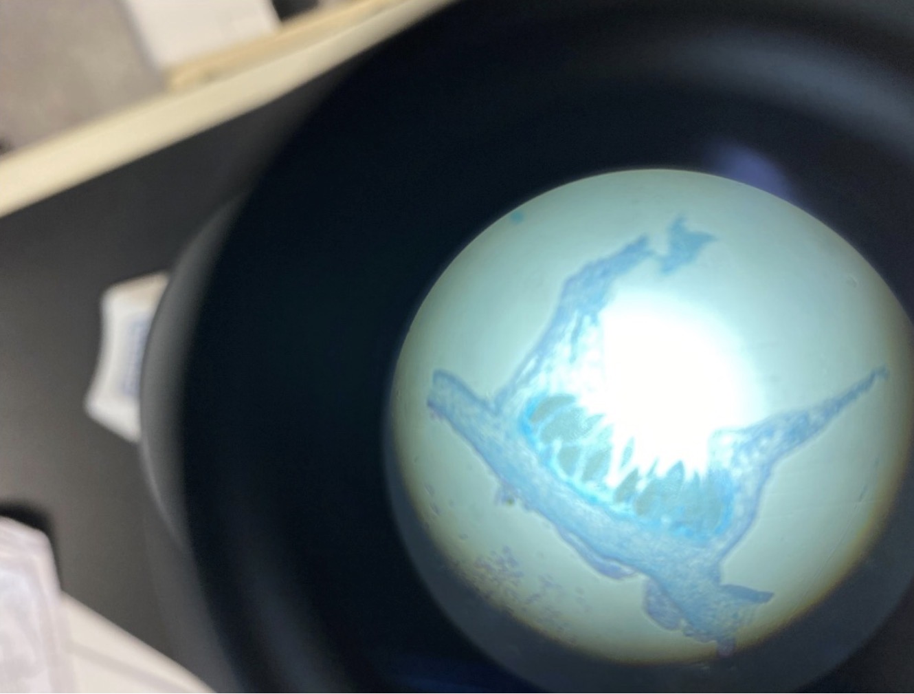
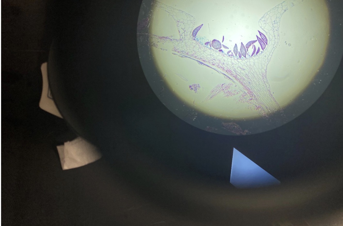
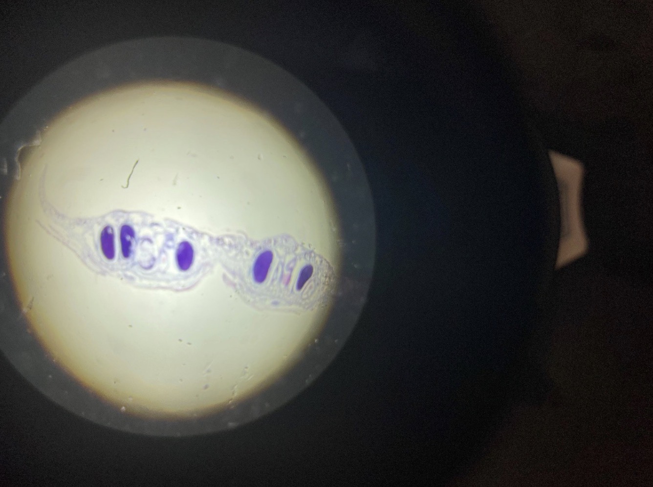
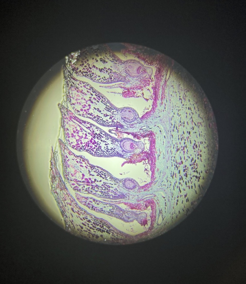
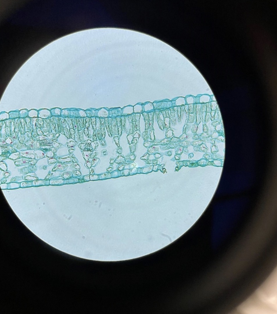
Dicot Plant Leaf 400x. Upper and Lower Epidermal Cells, Palisade Cells In a layer for photosynthesis, Spongy
Mesophyll Cells in a layer that enables gas exchange, and Guard Cells flanking a Stoma (together called a Stomata) to aid
against water loss in a Dicot Plant Leaf Upper and Lower Epidermal Cells,
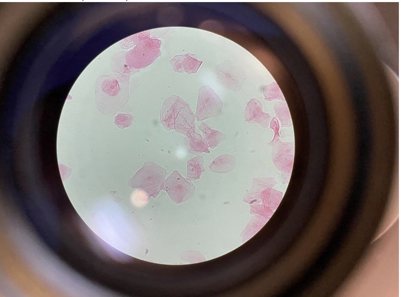
Squamous Cells from Human Cheek Epithelium 400x. Squamous cells have a flat shape. The nuclei may be seen in the
center of the cells Squamous Cells from Human Cheek Epithelium 400x. Squamous cells are flat cells that form epithelial linings.
Columnar Epithelial Cells with Microvilli from the intestines 400x. Columnar cells are column shaped cells. Here they
can be seen lining the villi in the intestine with microvilli at the apex of the cells to increase surface area for absorption of food. Columnar Epithelial Cells with The nuclei of the cells in this photograph can be seen as purple
within the pink cell.
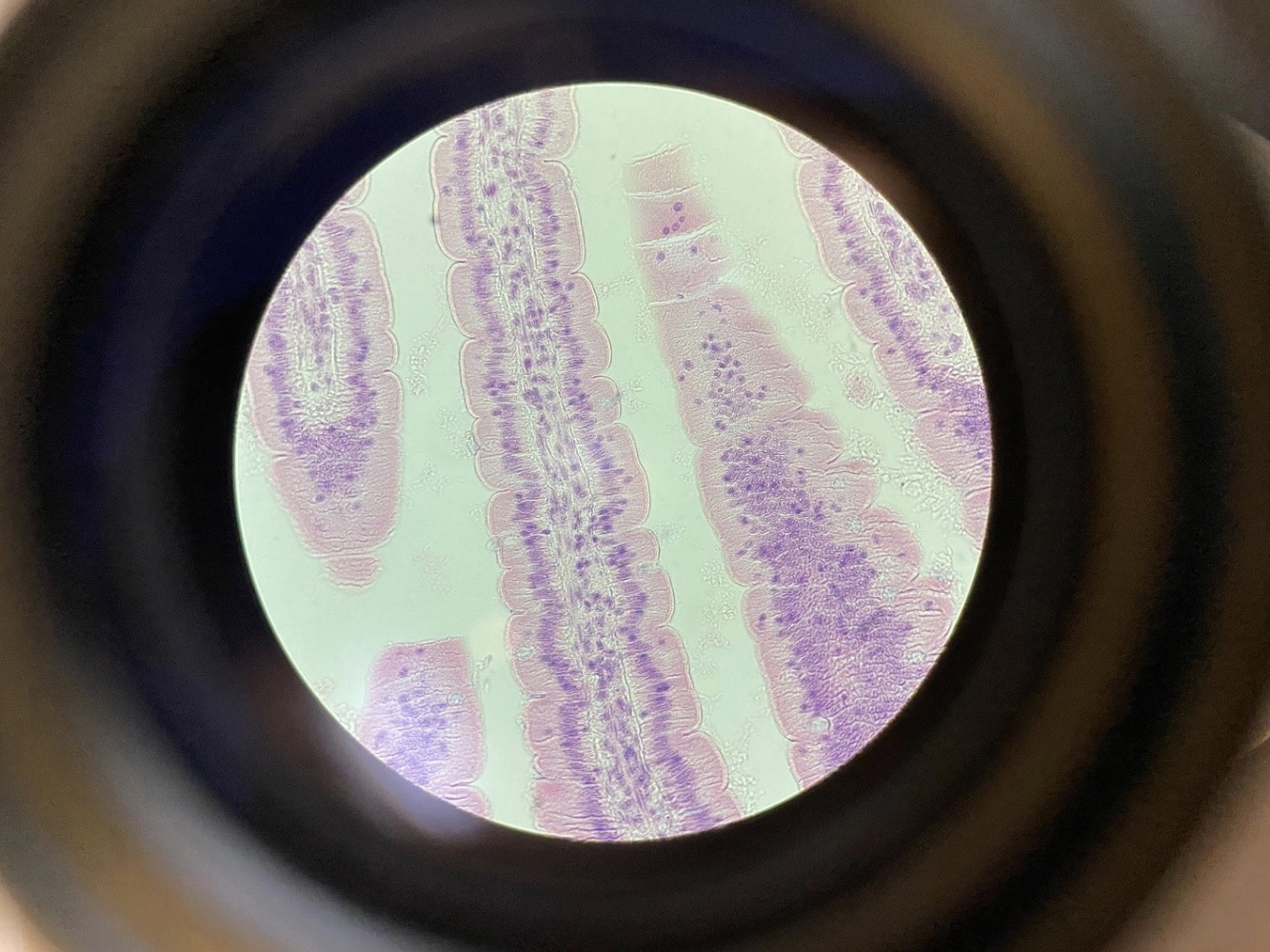
Ciliated Pseudostratified Columnar Epithelium from the Trachea 400x. Cilia in the respiratory tract help to waft up
mucus to b e swallowed. In Pseudostratified layers of cells, many of the cells on the basement membrane do not reach the
surface, so while the tissue looks stratified because thee nuclei do not line up, it is actually one cell layerCiliated Pseudostratified Columnar Epithelium from the . Pseudostratified columnar cells are column
shaped cells, and not all of the cells reach the surface. The single layer of cells looks like multiple layers. The nuclei can be seen
near the base of the cells, and cilia can be seen on the apical surface.
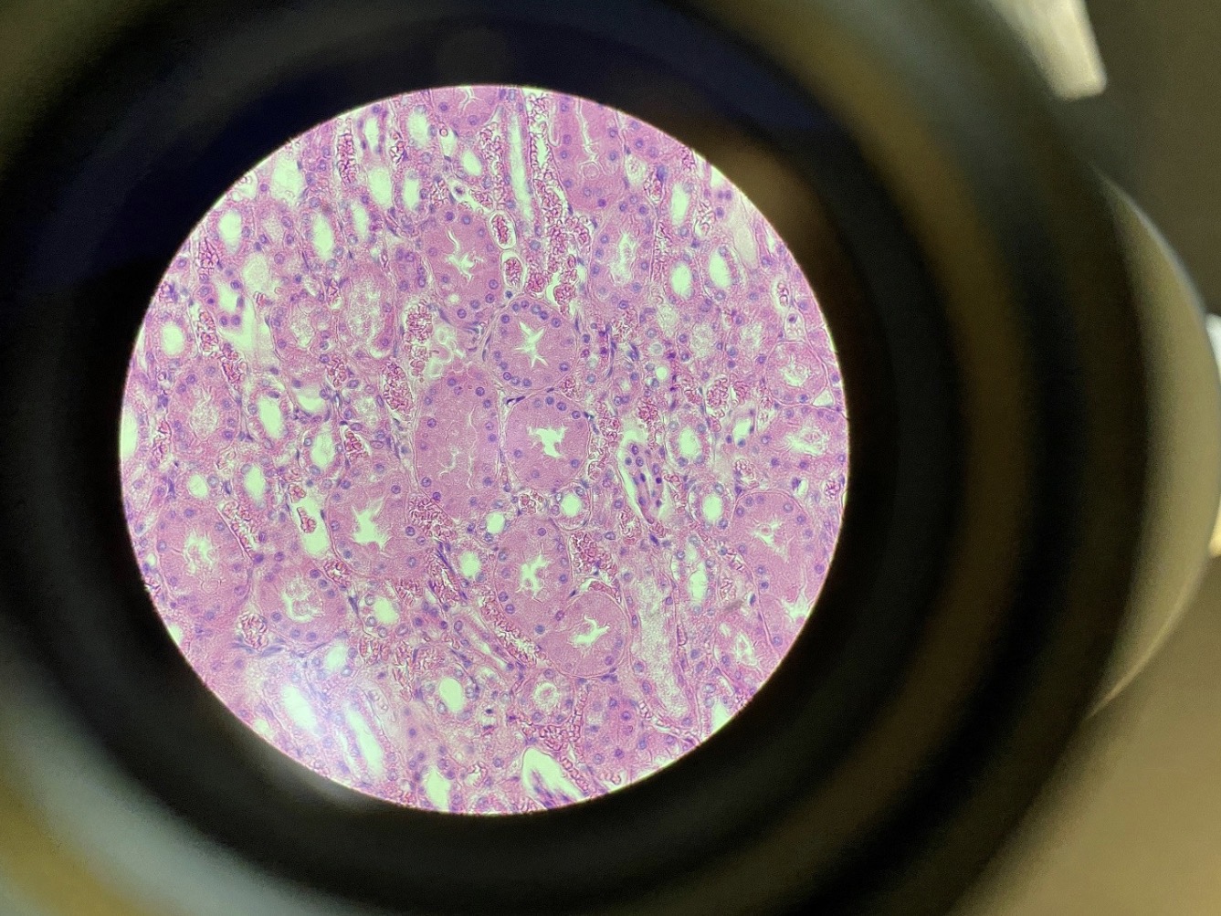
Cuboidal Cells from the Kidney 400x. The kidney contains nephrons with tubules surrounding a lumen that have
cuboidal cells to reabsorb water and salts. The cells appear to be stained pink and the nuclei stained purple in this image. Cuboidal cells are cubed shaped cells in epithelial linings. IN this
photograph they surround the lumen of tubules
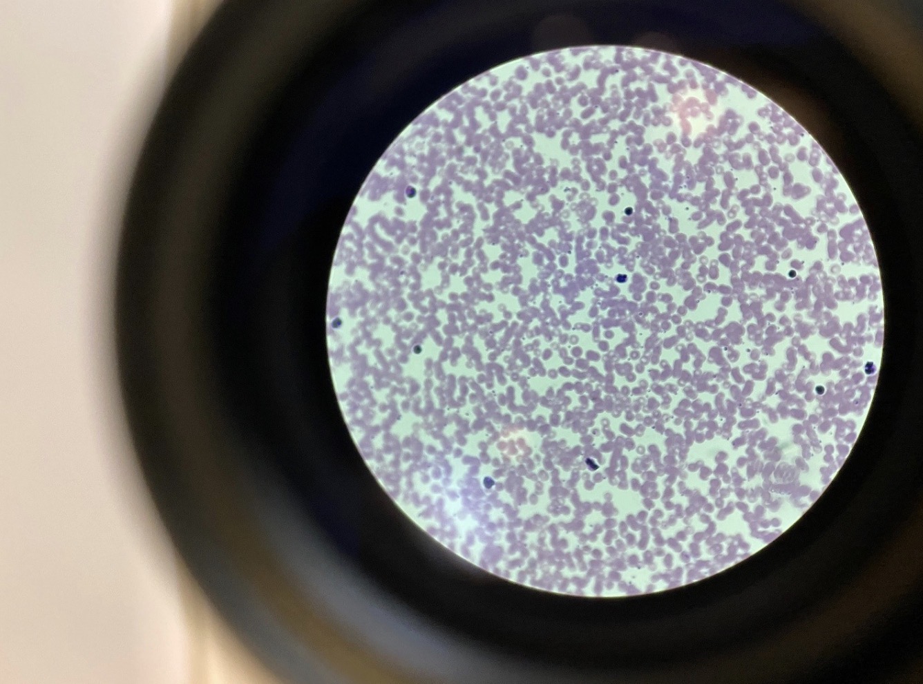
Human Blood, Connective Tissue with Liquid Matrix, 400x. Red blood cells (erythrocytes) are thinner in the center
than on the periphery. They appear pink in this image while the white blood cells (leukocytes) have purple stained nuclei Human Blood, Connective Tissue with
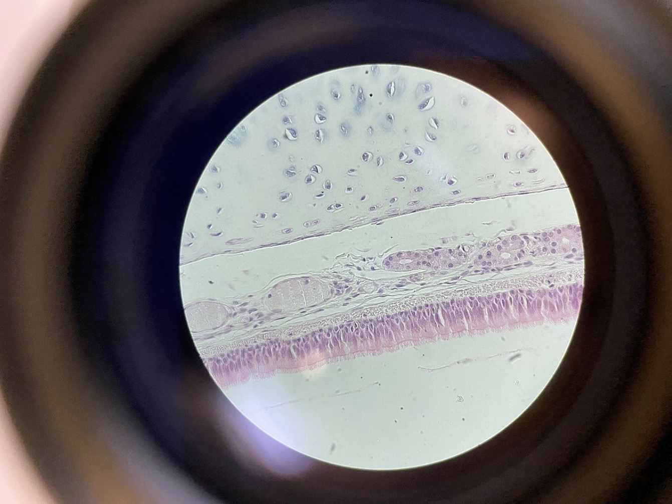
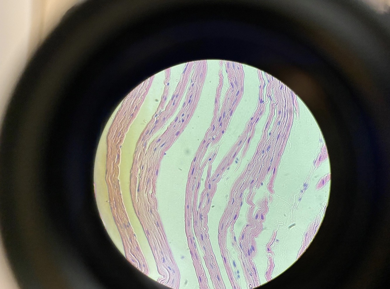
Chondrocytes in Cartilage from the Trachea 400x. This is a Connective Tissue with a Flexible Matrix Chondrocytes in Cartilage from the Trachea, Connective Tissue with a Flexible Matrix, 400x. The chondrocytes are in
little caves (lacunae) with a flexible matrix containing collagen fibers and hyaluronic acid between them
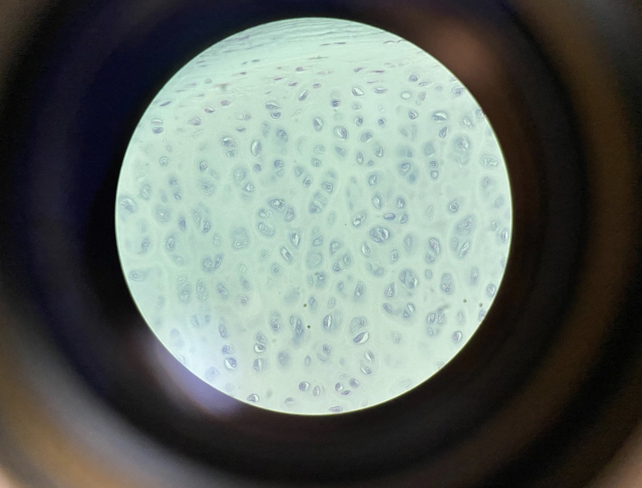
Osteocytes in Compact Bone with Haversian Systems 400x. This is a connective tissue with a solid matric containing
calcium carbonate. The Bone cells (osteocytes) are in
little caves (lacunae) with a solid matrix containing collagen fibers and calcium carbonate between them. Haversian systems
made of concentric lamellae surround a central haversian canal that contain blood vessels. Canaliculi connect the central
haversian canal to the lacunae containing the osteocytes.
Adipocytes in Adipose Connective Tissue 400x. Adipose cells have a large central vacuole containing liquid lipids. The
nucleus is forced to the periphery of the cell Adipose cells contain a large droplet of oil in a central vacuole. The
cytoplasm that contains the nucleus is forced to the periphery
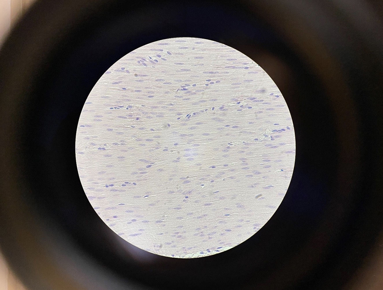
Smooth Muscle Cells 400x. Smooth muscle cells are spindle shaped cells. The nuclei of the cells can be seen as purple
and the cells in pink in this image
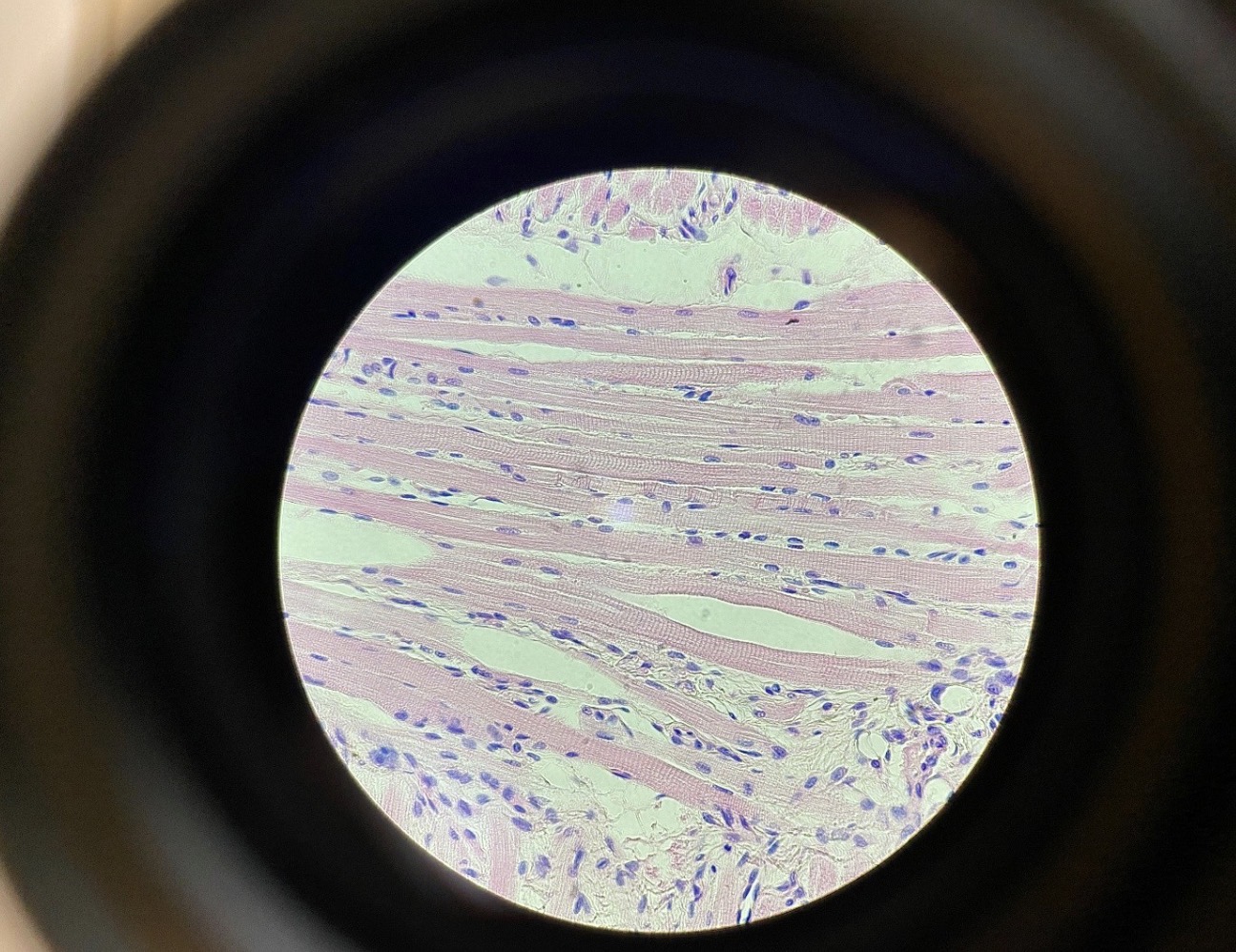
Skeletal Muscle Cells with Striations 400x. Skeletal muscle cells have lighter and darker bands. The micro-filaments
used in contractions overlap in the darker bands. The cells can be very long containing many nuclei Skeletal muscle cells are elongated with a multiple nuclei (purple) and
striations (bands of darker and lighter contractile fibers) can be seen.
cardiac Muscle Cells with Striations 400x. Cardia muscle cells are branched shaped with a single nucleus (purple) and striations can
be seen
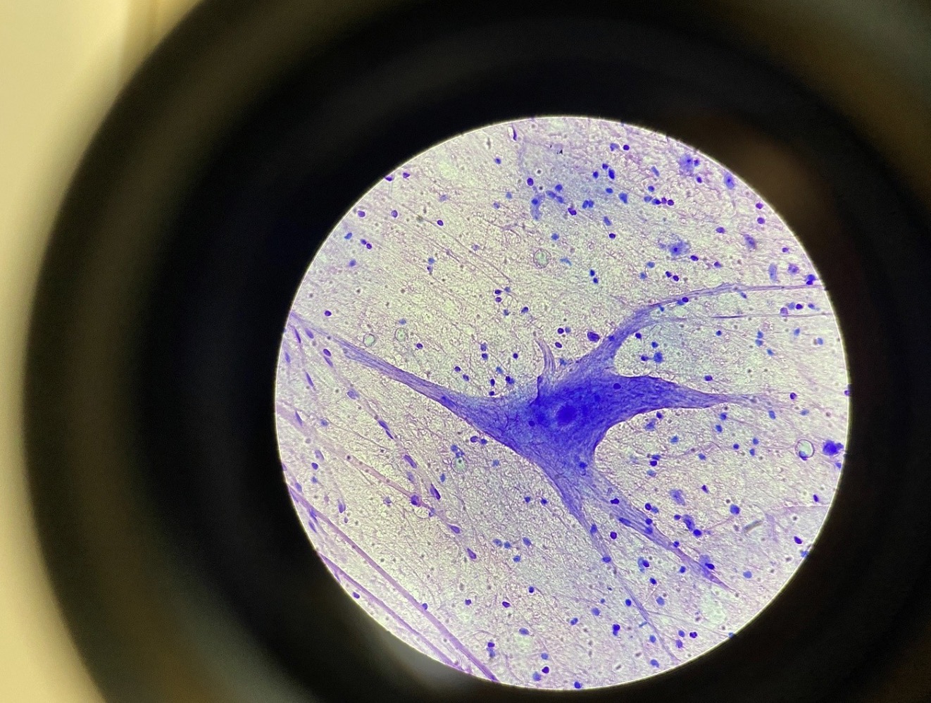
Giant Multipolar Neurons from the Spine 400x. Neurons contain dendrites that receive signals and an axon that sends
a signal. Neurons may be surrounded by cells of the microglia
Live amoeba
Live Paramecium 400x (Kingdom Protista)
Euglena stained pink 400x. The pointed end that has the flagellum and the rounded end can be distinguished in this
photograph.

Euglena stained pink 400x. The pointed end that has the flagellum and the rounded end can be distinguished in this
photograph.
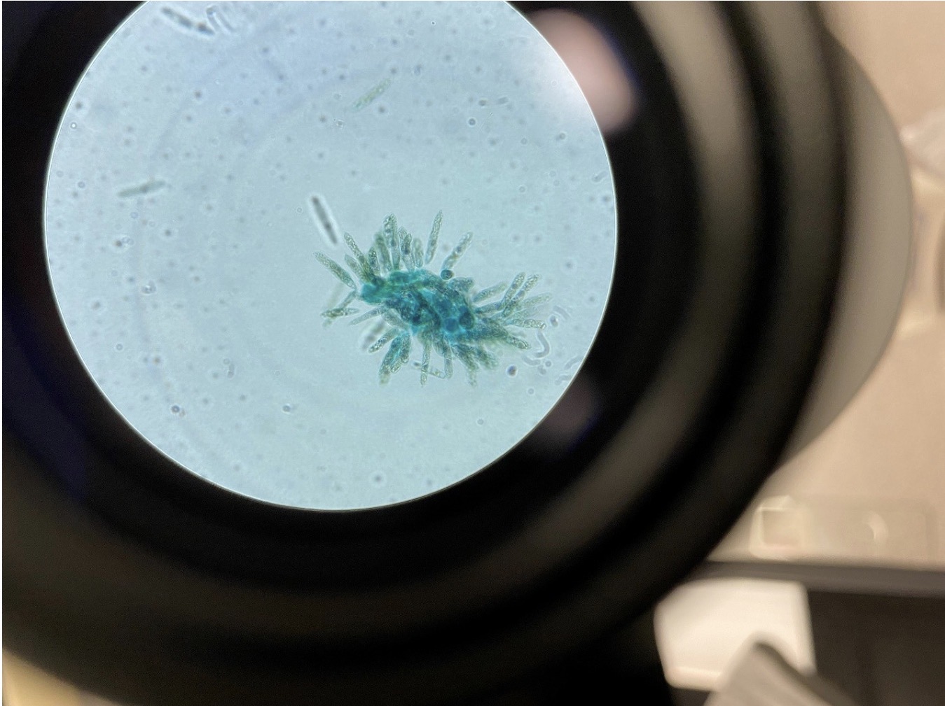
Diatom, Division Chrysophyta, Yellow and Golden Brown Algae 40x
Volvox 100x (Kingdom Protista, Algae with multicellularity and cell differentiation with two flagella per cell). This is
also algae in the Volvocine line of evolution
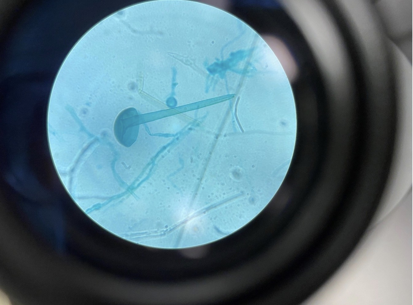
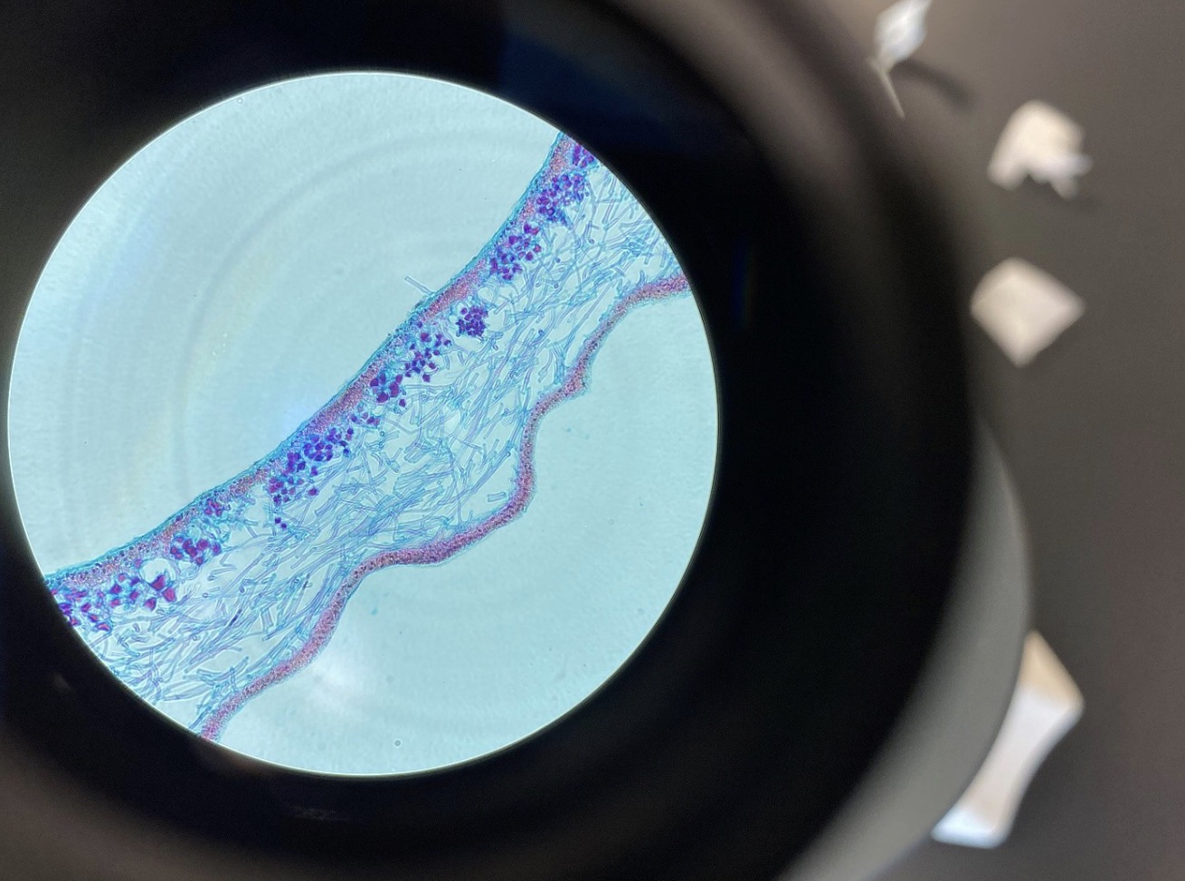
Rhizopus Fungi sporangia 400x (Division Zygomycota). This belongs to the Kingdom Fungi. The filaments are called
hyphae, and the reproductive structure is called a sporangium. Fungus with sporangium are placed in the Division Zygomycota
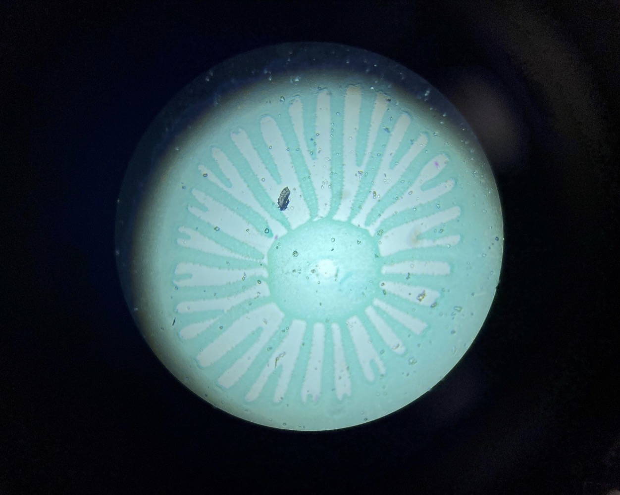
Fungi Basidiocarp 40x (Division Basidiomycota). This is a mushroom cap. The stalk can be seen in the center with gills
radiating out toward the peripheral portion of the cap. The gills contain basidiospores. Fungi with a basidiocarp are placed in
the Division Basidiomycota
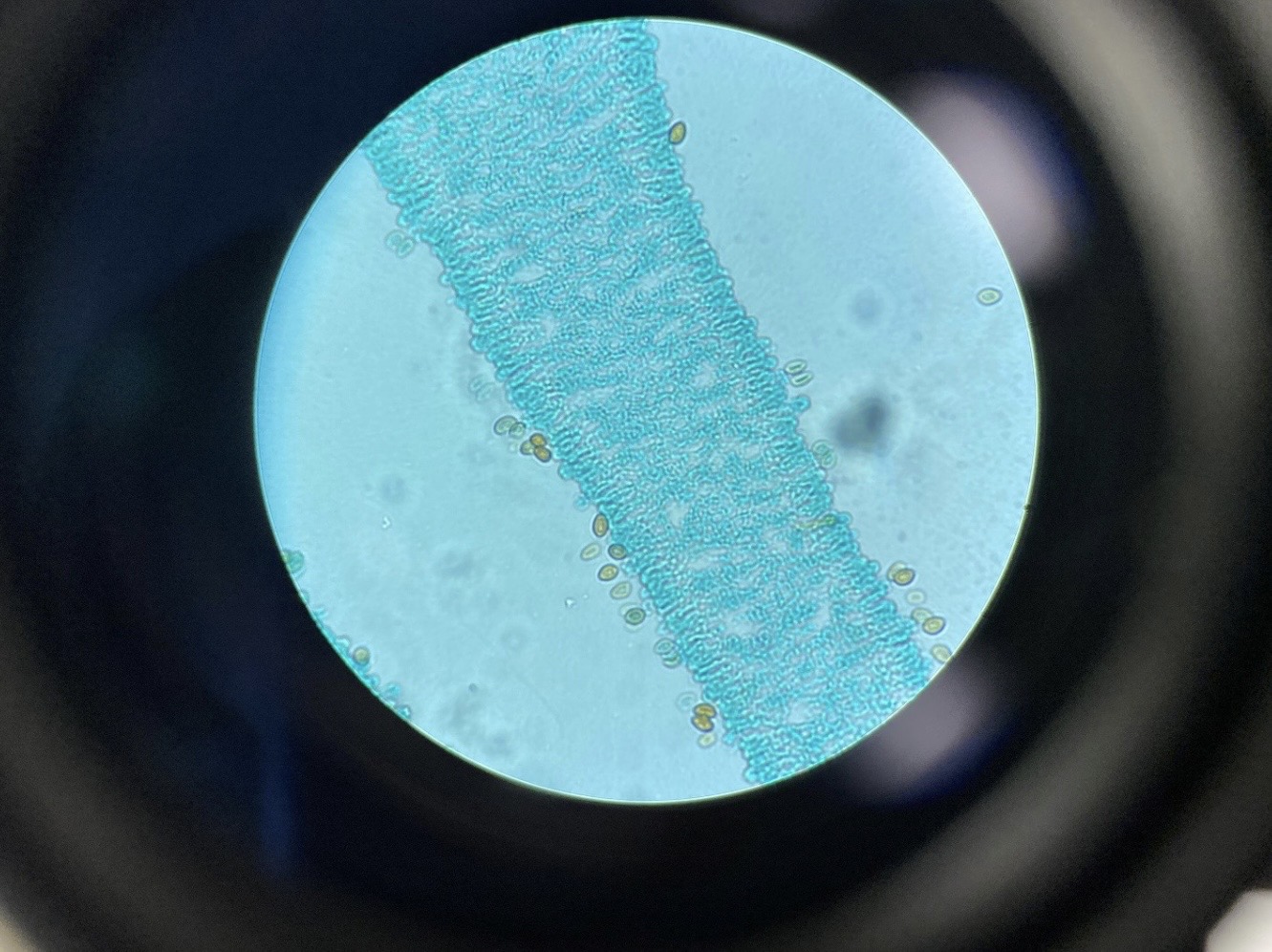
Fungi Basidiocarp with spores 400x (Division Basidiomycota). A gill with basidiospores can be seen in this photograph
Puccinia Wheat Rust 400x, Division Basidiomycota (This is the Wheat Rust bursting out of the surface of the Wheat
leaf.
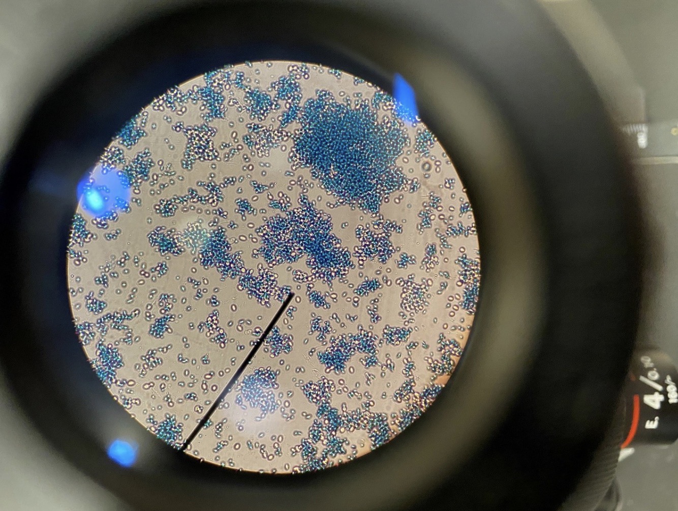
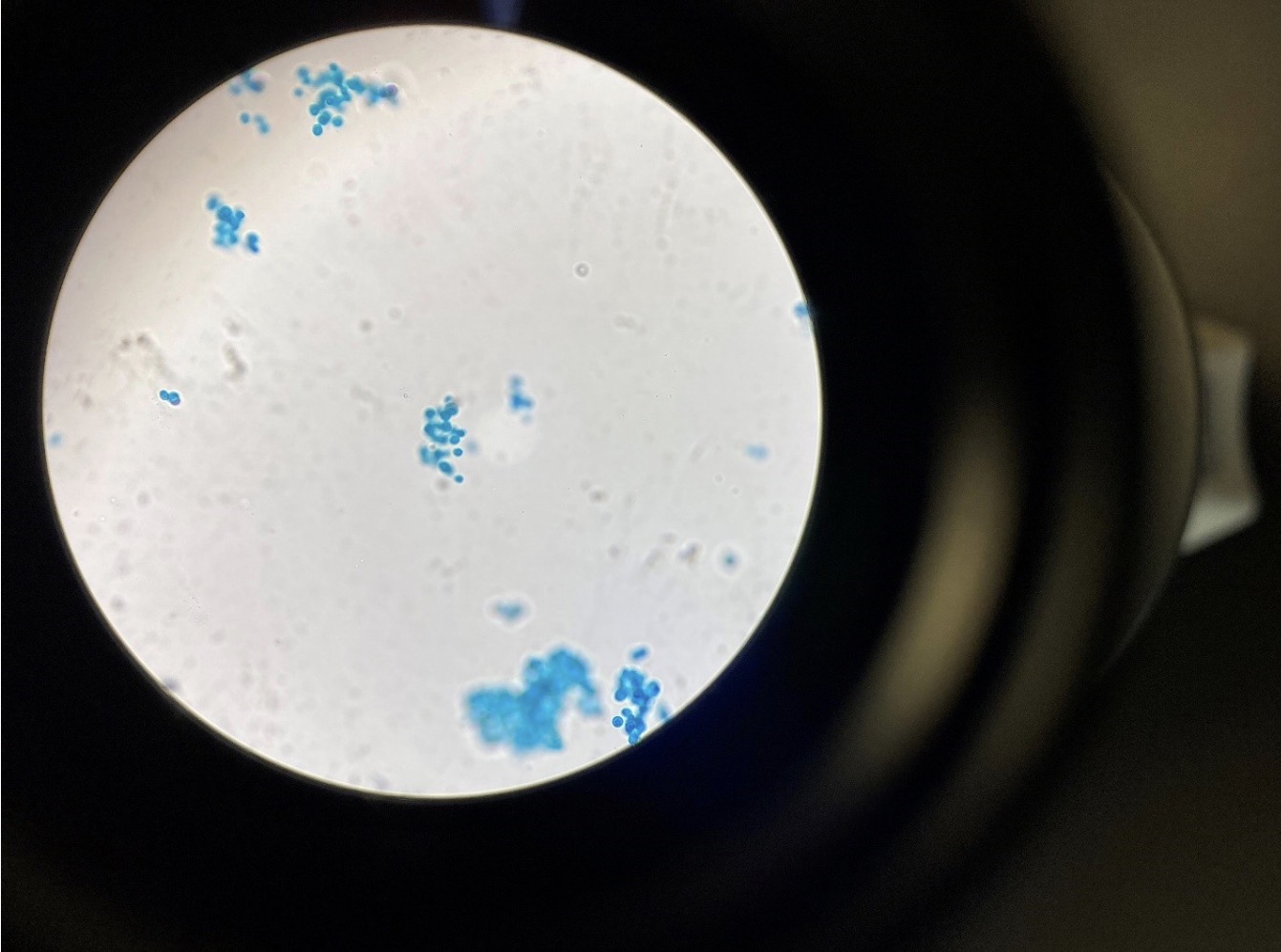
Yeast, Saccharomyces cerevisiae 400x (Kingdom Fungi, Division Ascomycota) (Baking or Brewing YeasT
Lichen 100x, A Lichen is a symbiotic relationship between an algae and a fungus. Algal cells can be seen below the
upper cortex while fungal hyphae can be seen below them
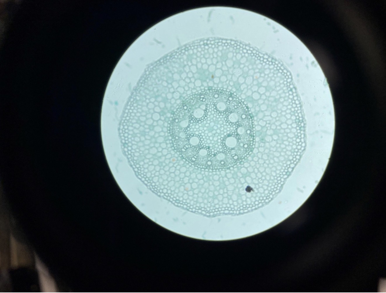
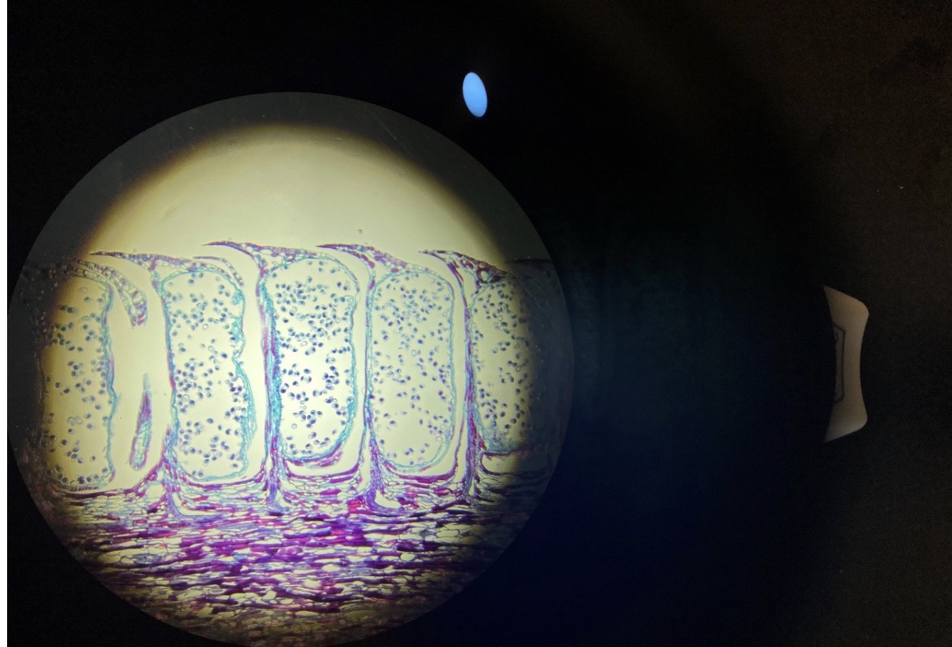
Monocot Plant Root 40x. A variety of cells can be viewed from the periphery inward; epidermal cells, cortex cells,
endodermal cells, xylem cells, phloem cells, and parenchyma pith cells
Dicot Plant Root 40x. A variety of cells can be viewed from the periphery inward; epidermal cells, cortex cells,
endodermal cells, phloem cells and xylem cells
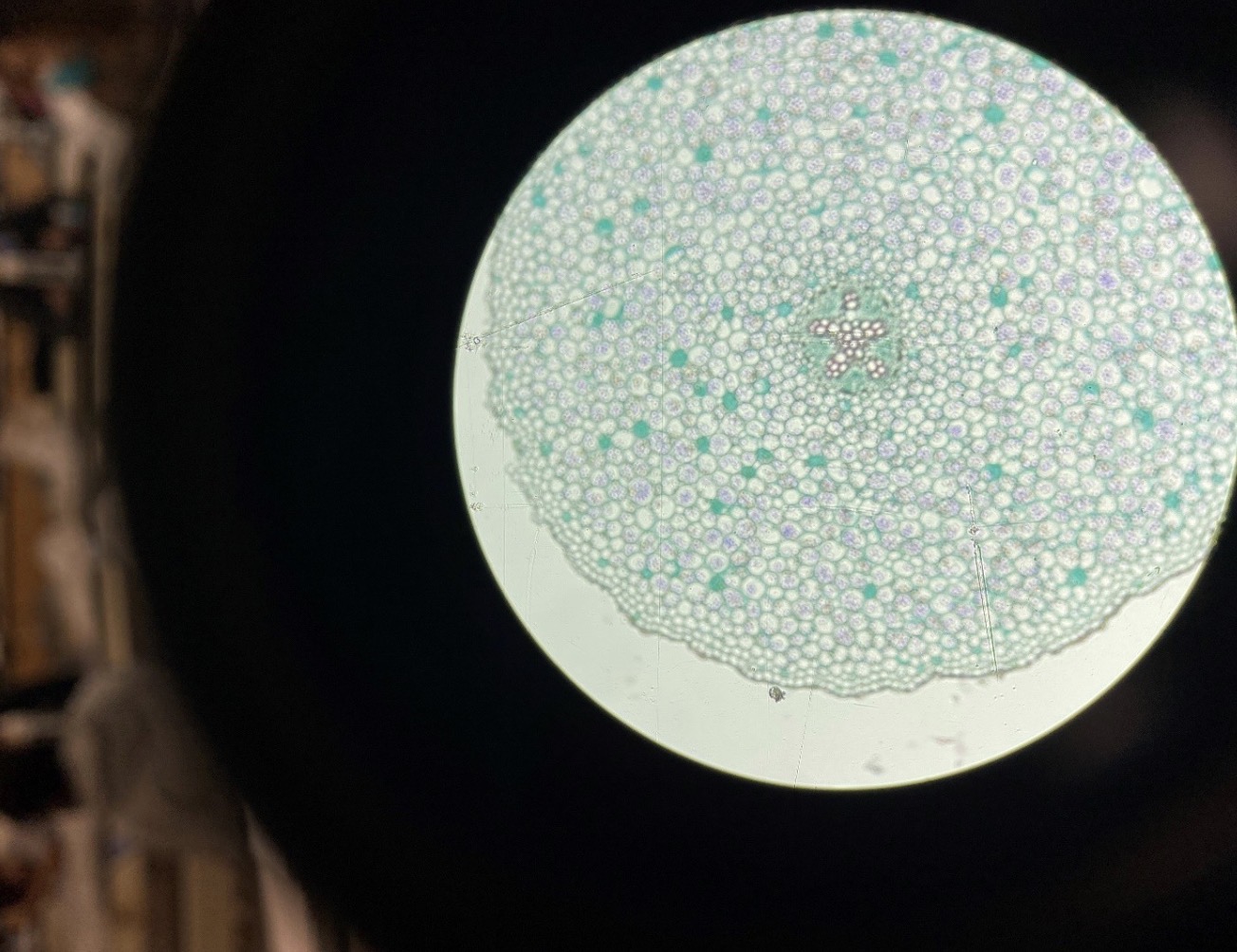
Monocot Plant Stem 40x. A variety of cells can be viewed from the periphery inward; epidermal cells, sclerenchyma
cells, phloem cells and xylem cells in vascular bundles, and parenchyma cells
Monocot Plant Stem 40x. A variety of cells can be viewed from the periphery inward; epidermal cells, sclerenchyma
cells, phloem cells and xylem cells in vascular bundles, and parenchyma cells
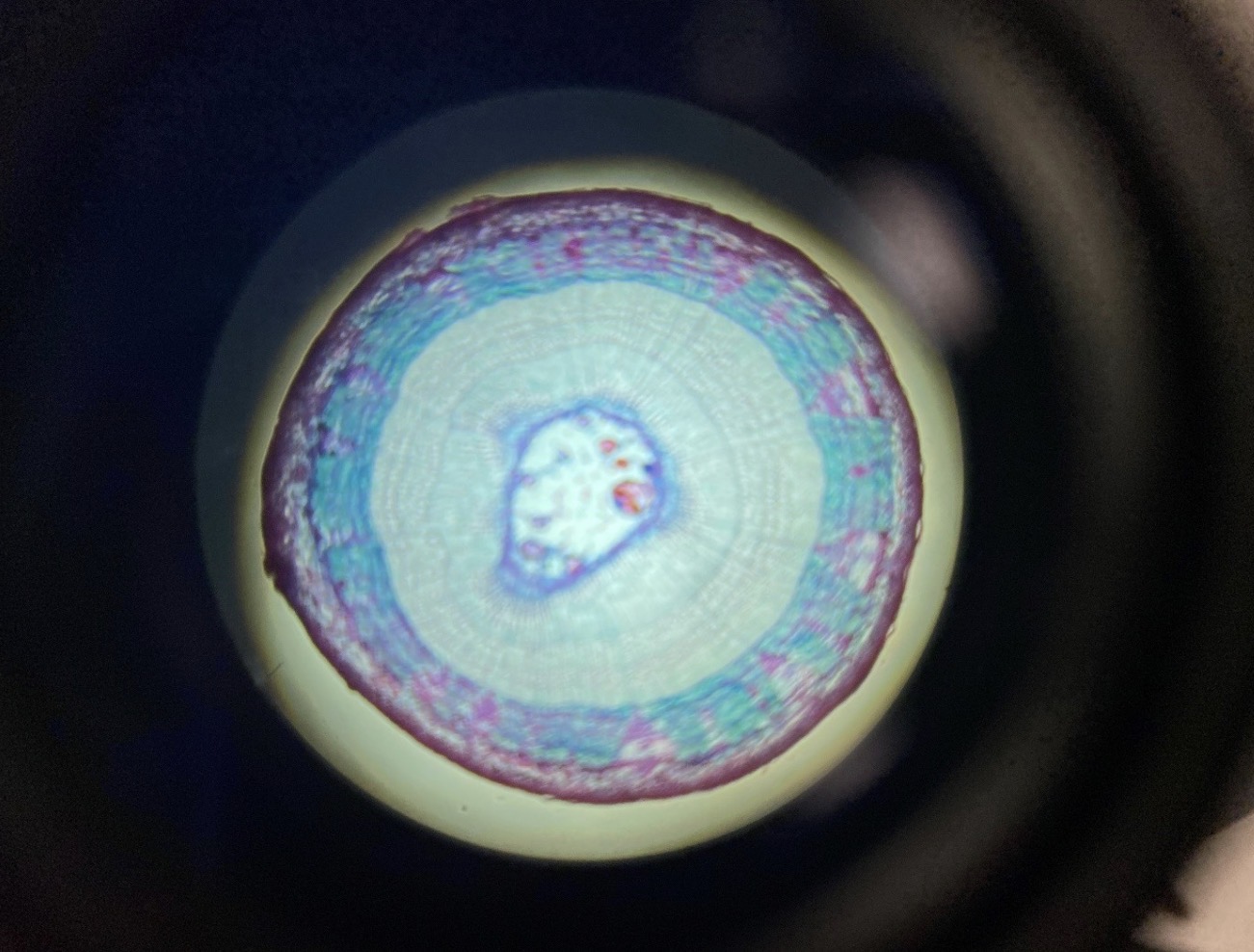
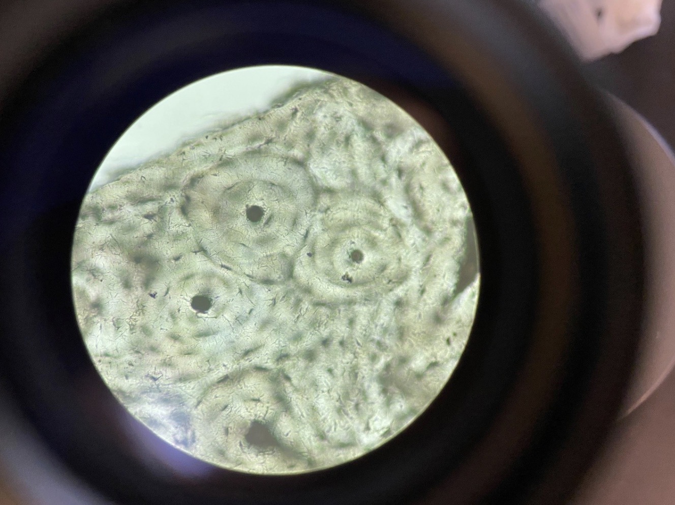
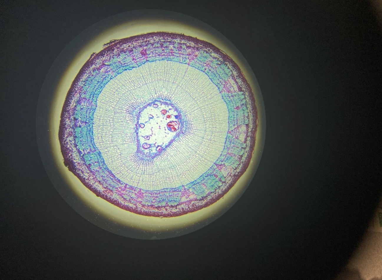
Woody Dicot Plant Stem 40x. From the center outward are cells of the pith, endodermis, xylem, vascular cambium,
phloem, and periderm (bark
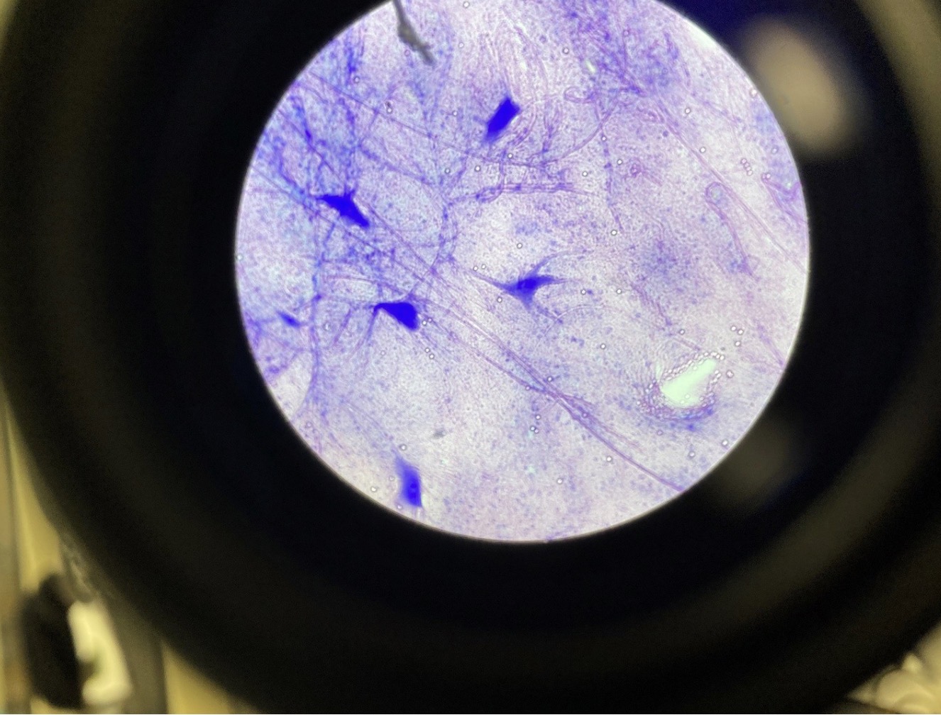
Giant Multi Polar Neurons 40x. Neurons have dendrites that receive signals and an axon that sends a signal
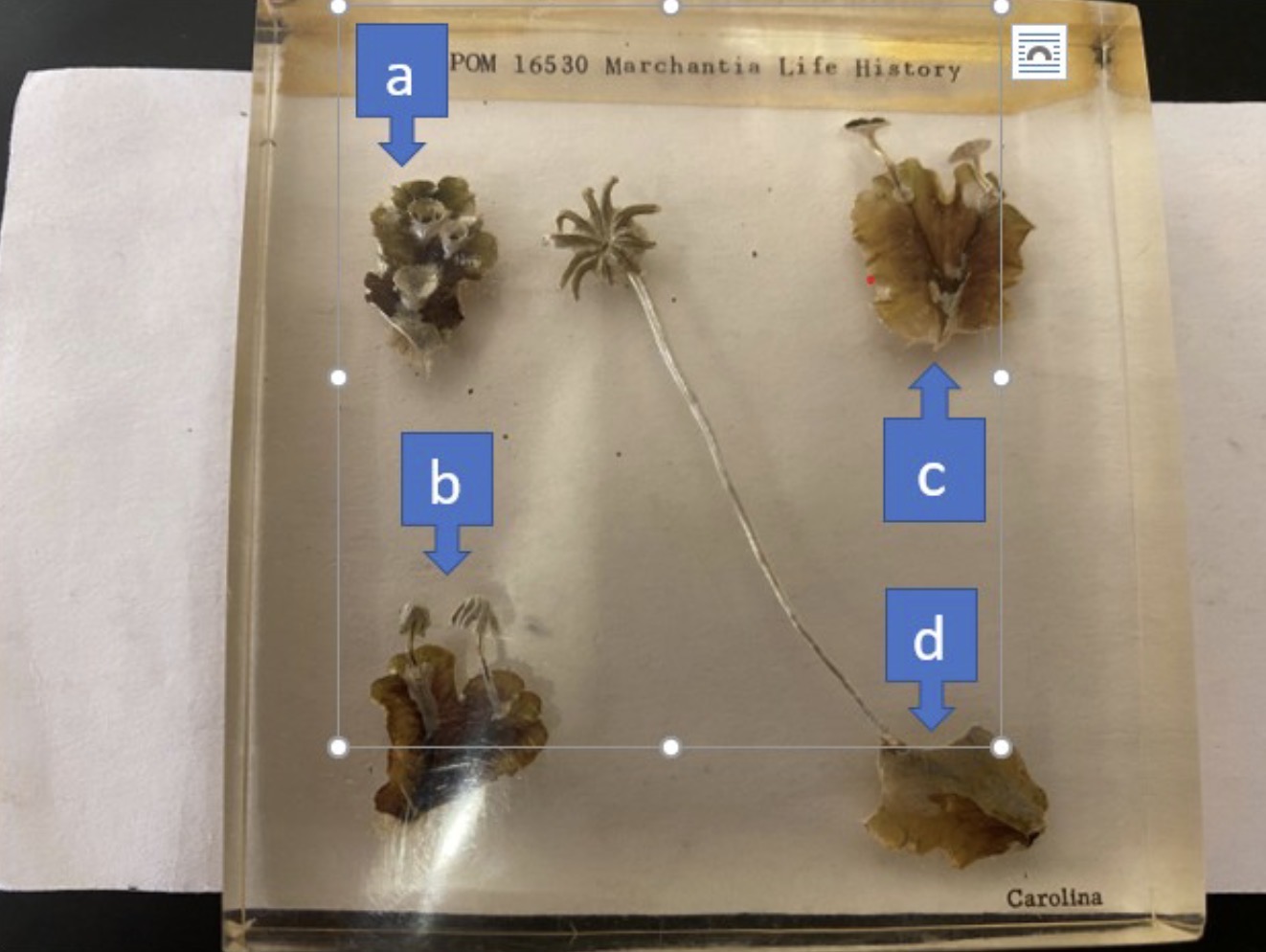
Liverwor Marchantia, a) thallus with gemma cups, b) thallus with antheridiophores, c) thanllys with archegoniophores,
d) thallus with archegonniphore bearing sporophytes
Liverwort Marchantia, gemma cup with gemmae 40x
Liverwort Marchantia, gemma cup with gemmae 40x
Liverwort Marchantia, Antheridiophore 40x
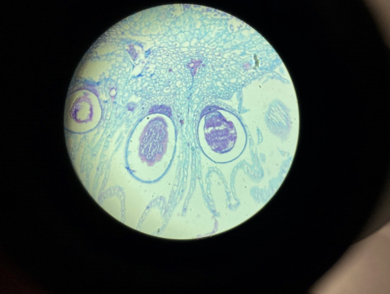
Moss Marchantia, Antheridia containing SPerm 400x
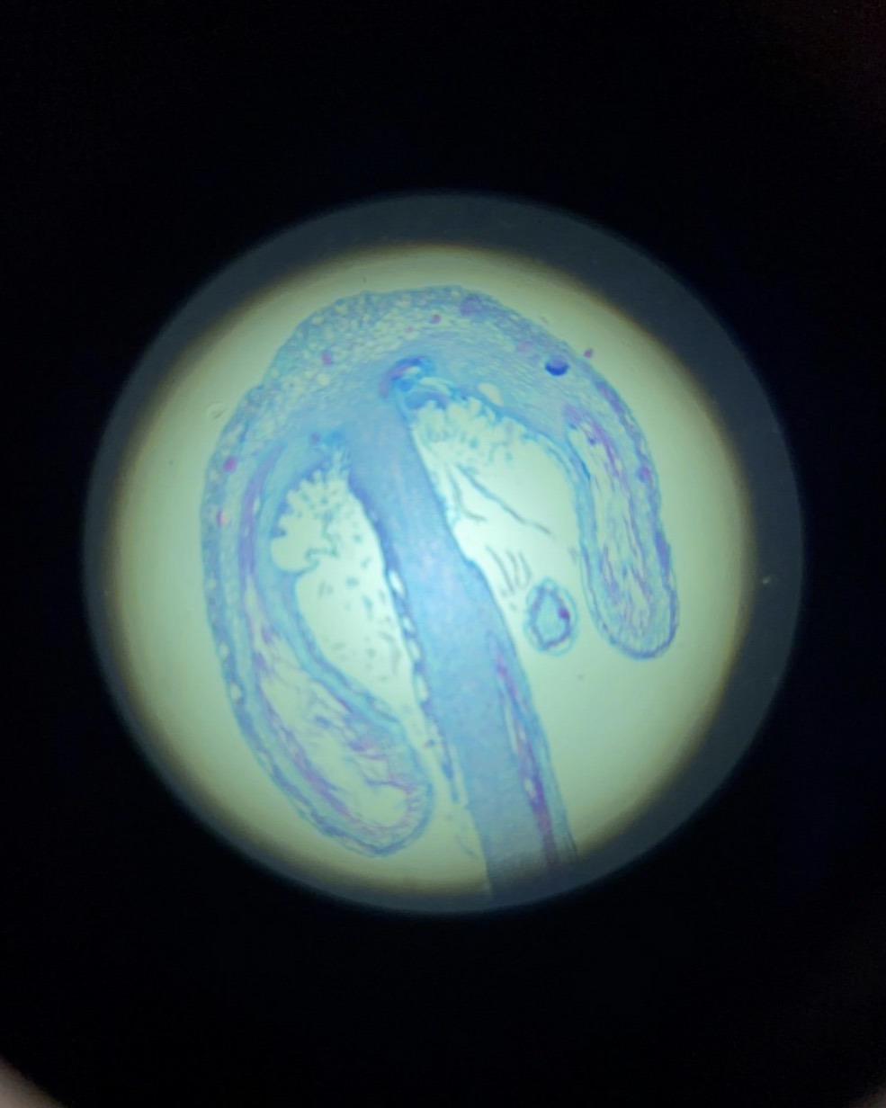
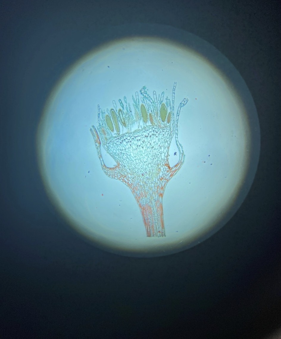
Moss Marchantia Archegoniophore 40x
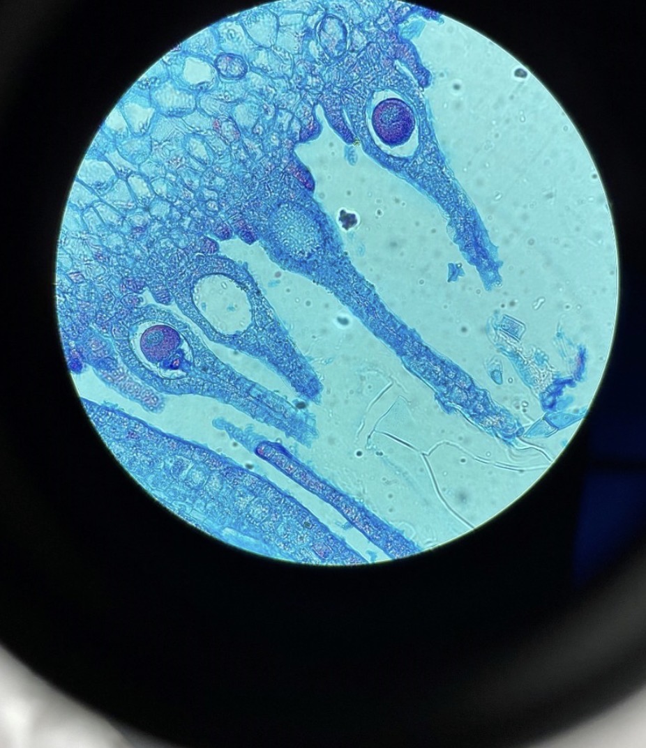
Moss Marchantia Female Archegoniophore with Archegonia containing eggs 400x
Moss Marchantia, Archegoniophore with Sporophytes 400x
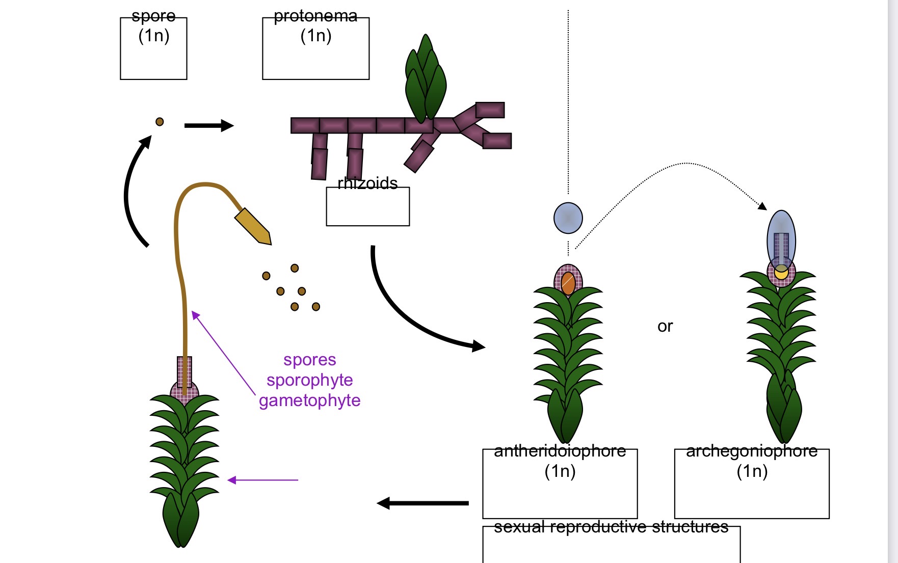
Life cycle of moss
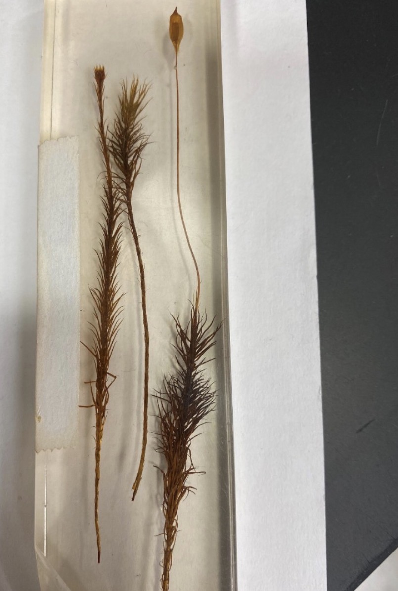
Moss Polytrichum, a) antheridiophore, b) archegoniophore, c) archegoniophore with sporophyte
Moss Polytrichum protonema 400x
Moss Polytrichum, antheridiophore with antheridia 40x
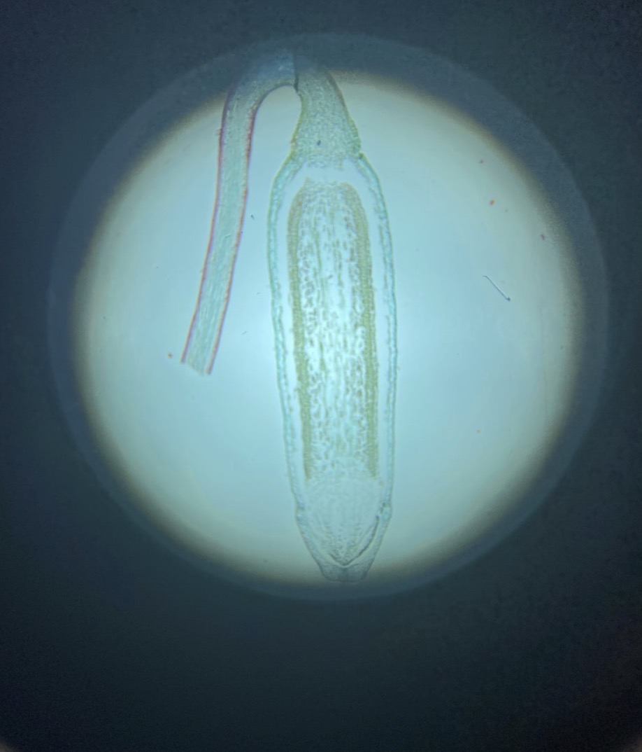
Moss Polytrichum, calyptra 40x
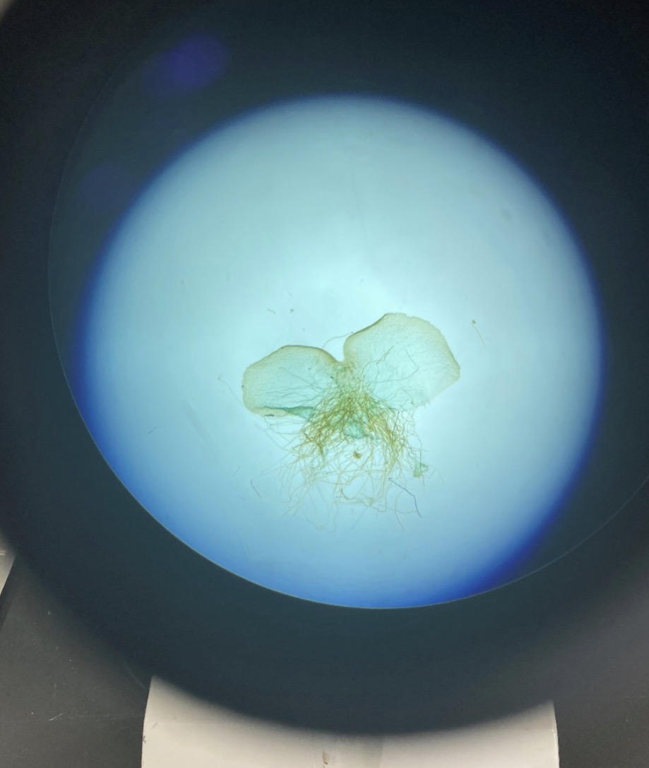
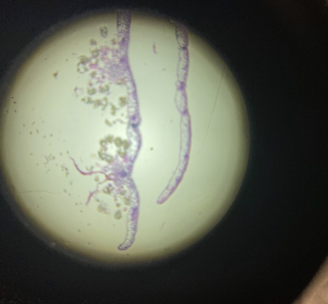
Haploid Fern Prothallium with rhiziods
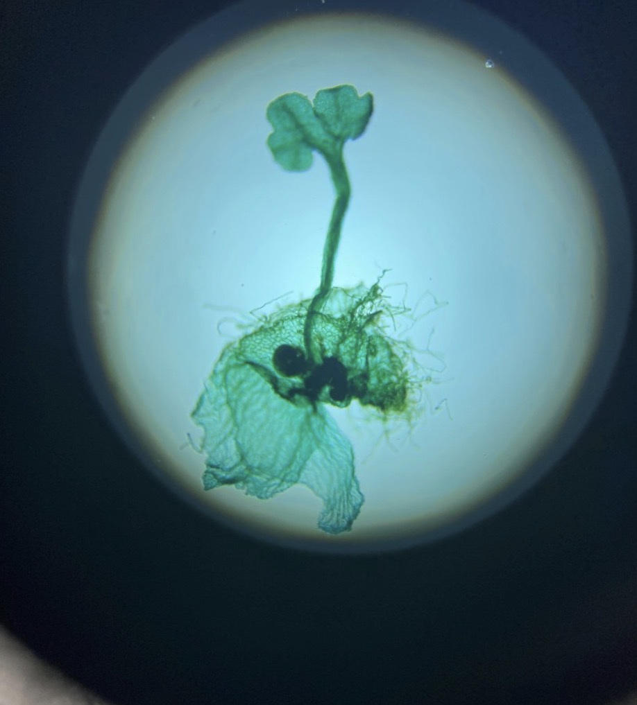
Haploid Fern Protallium with Diploid Primary Leaf
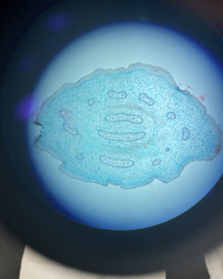
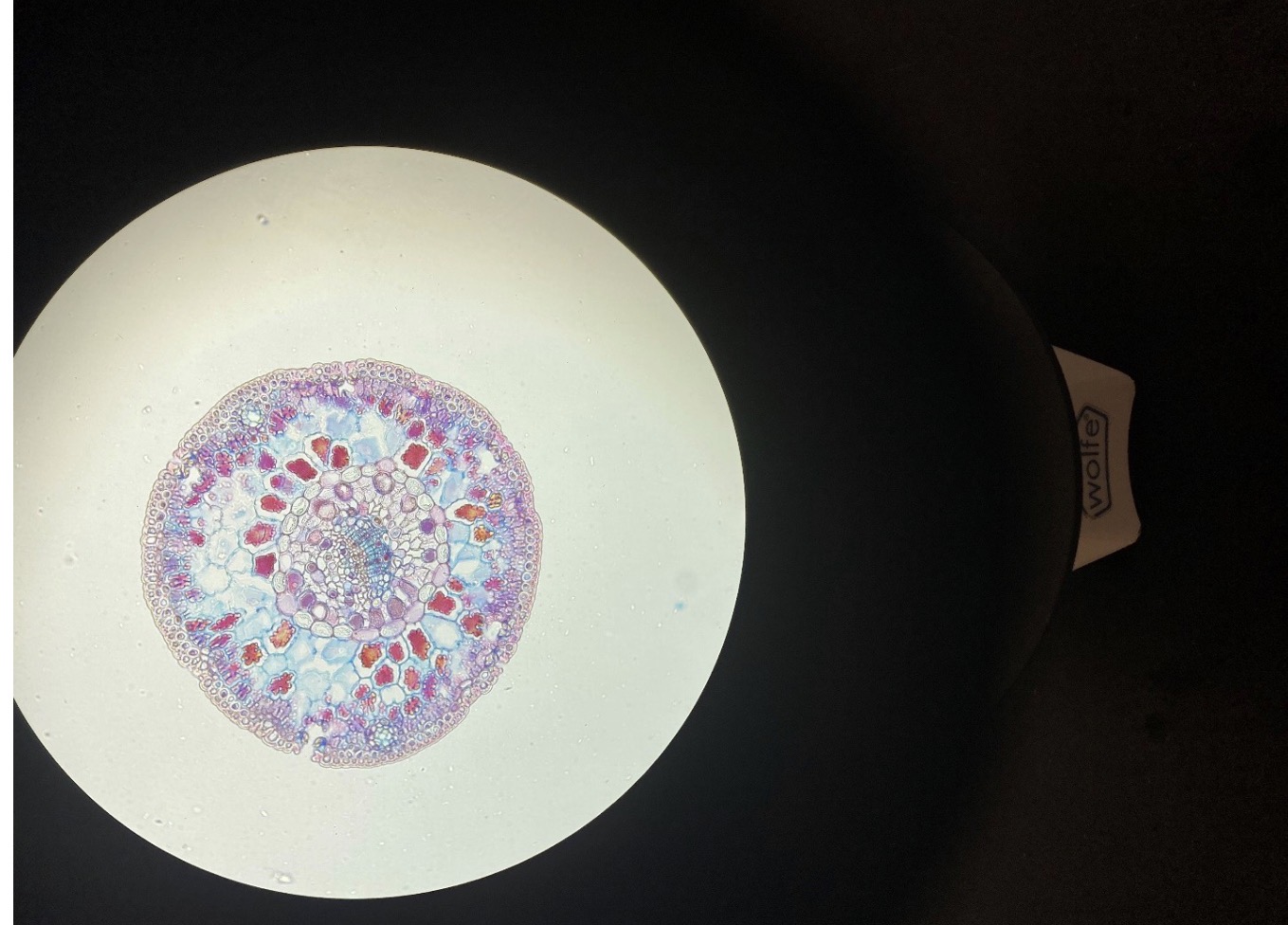
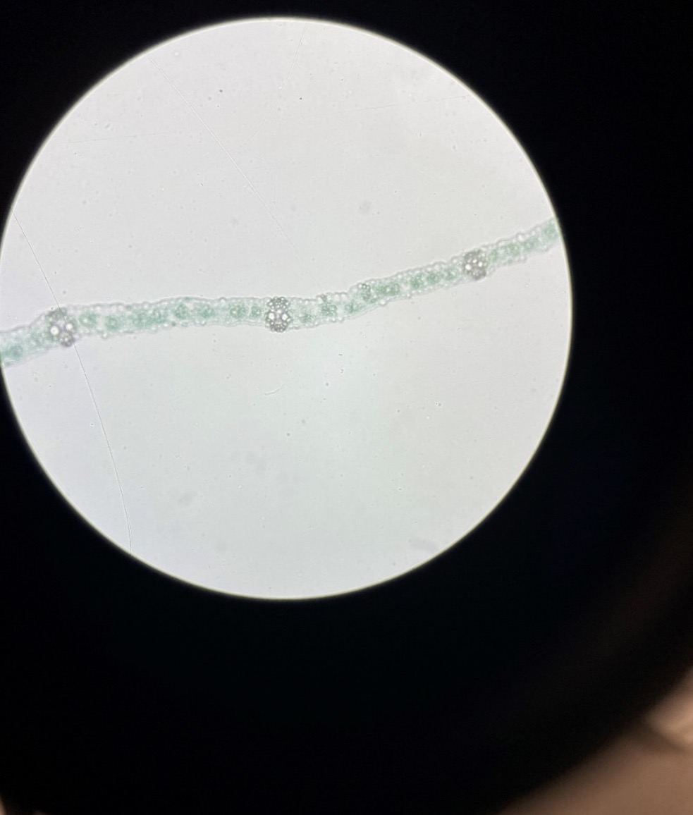
Fern horizontal root with vascular tissue
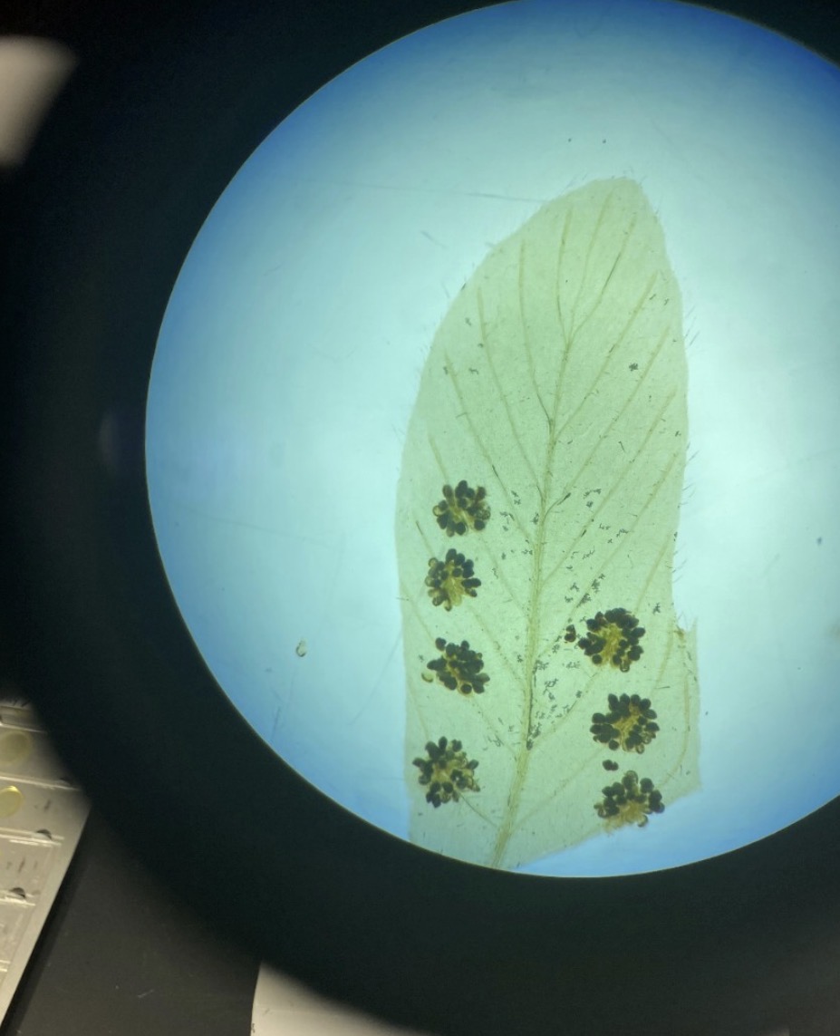
Fern Leaf with Sori containing Sporangia 40x
Fern Sorus with Sporangia 400x
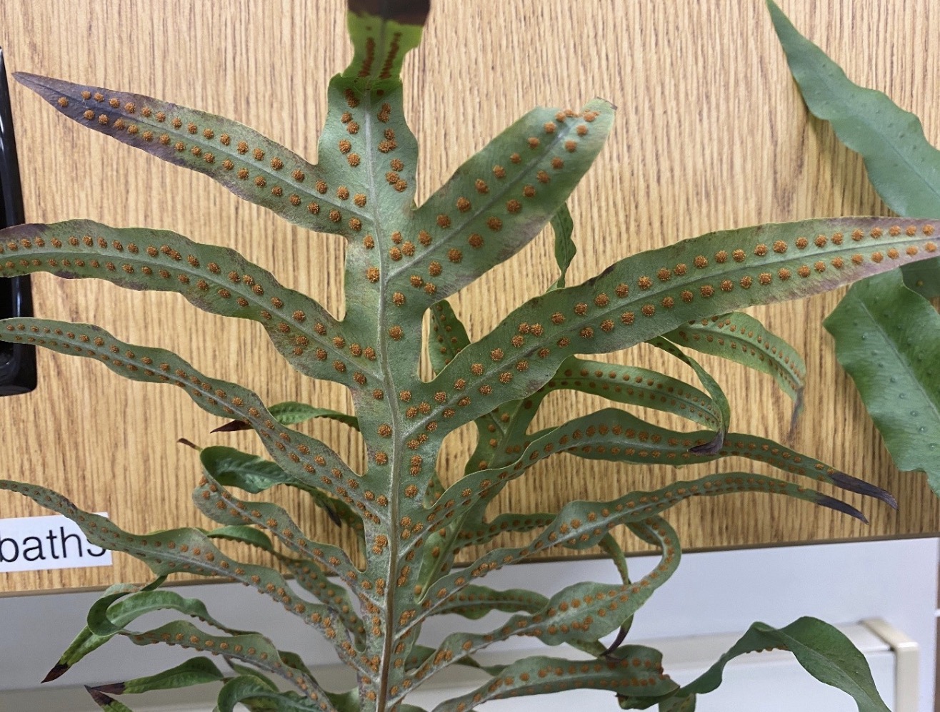
Sori under a Fern Leaf
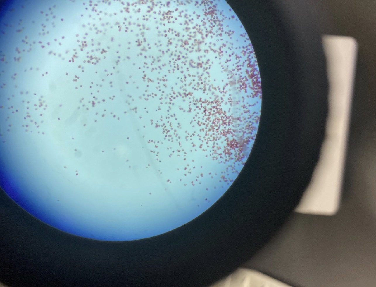
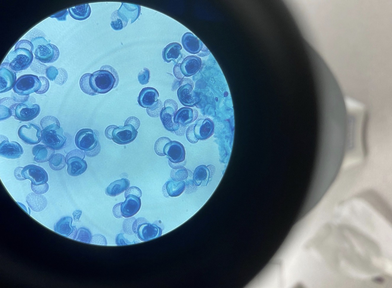
Fern Spores 400x
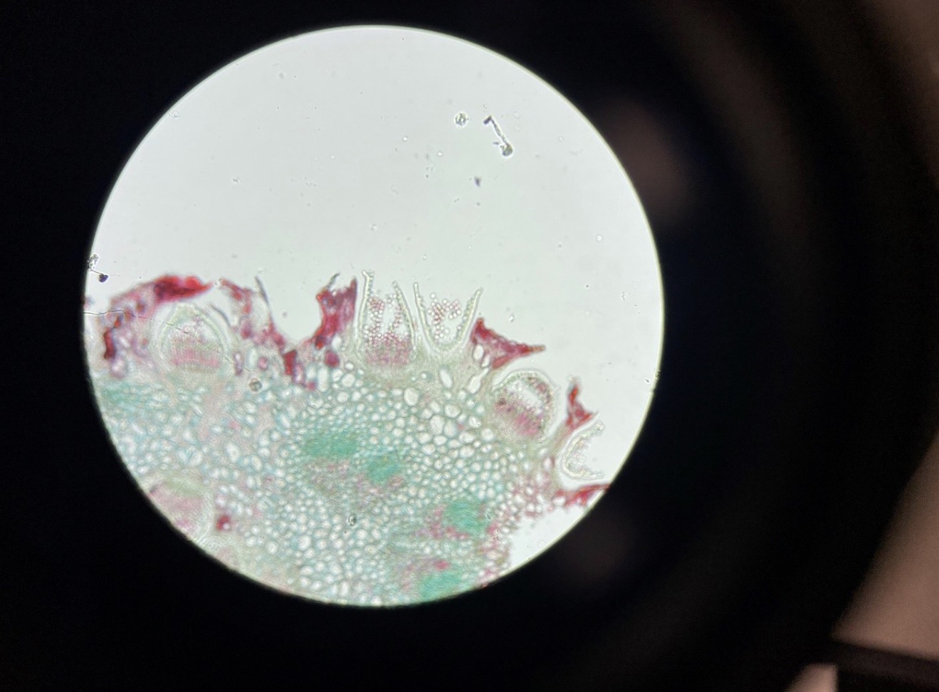
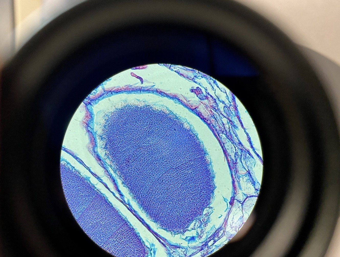
Pine Needle 100x
Female Ovulate Pine Cone 40x
Ovulate Pine Cone with Ovules 400x
Male Pine Cone 40x
Confer Pollen Grains 400x
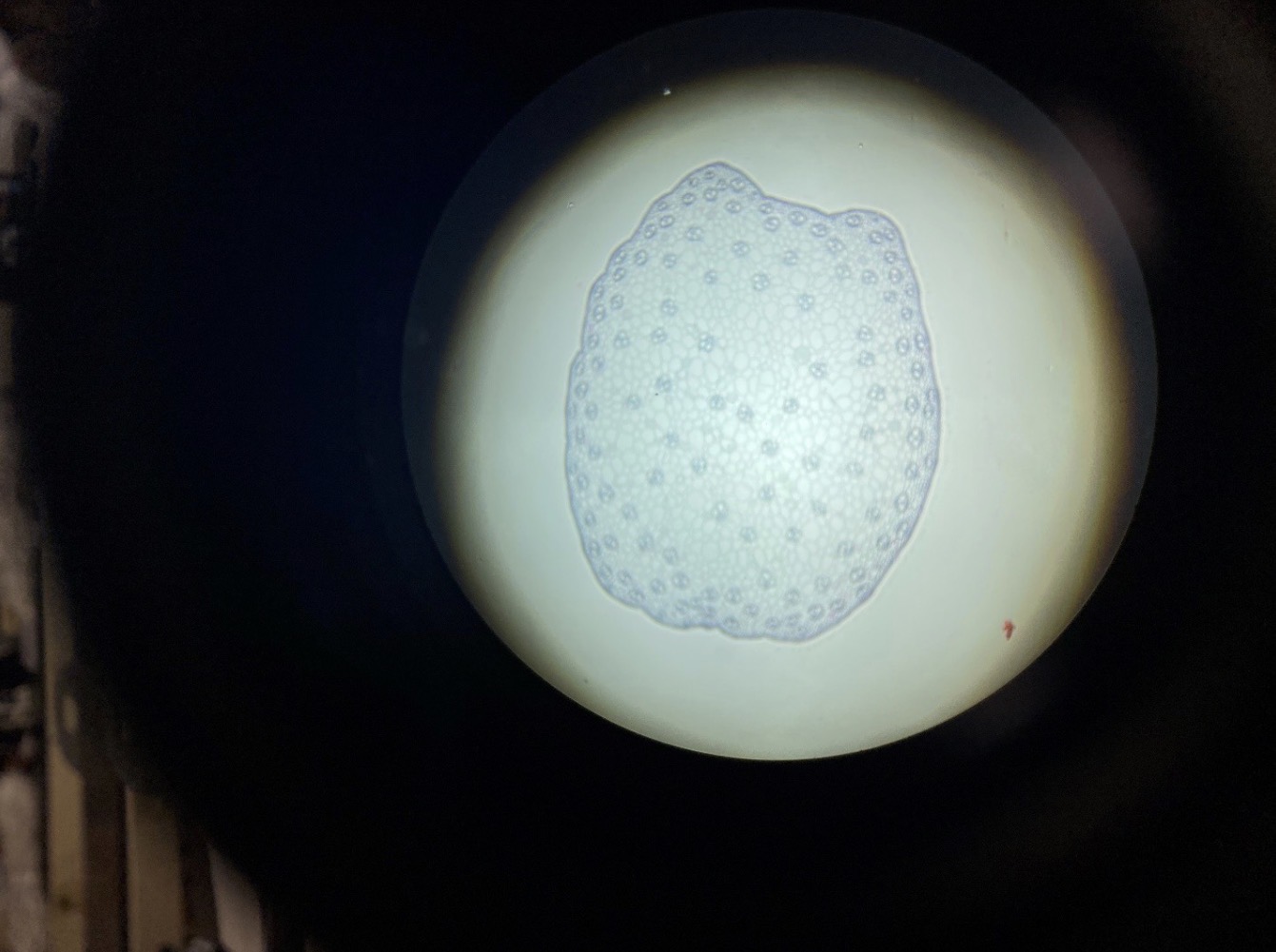
Dicot Root 40x
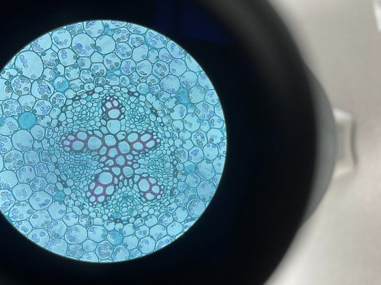
Vascular Cylinder in a Dicot Root 400x
Dicot Stem 40x
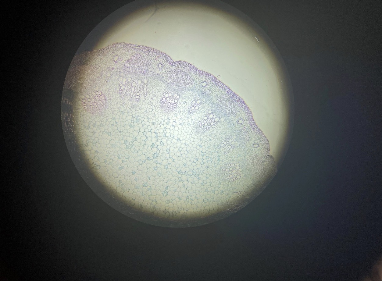
Dicot Stem 100x
Dicot Woody Stem 40x
Perennial Woody Dicot Stem 40x
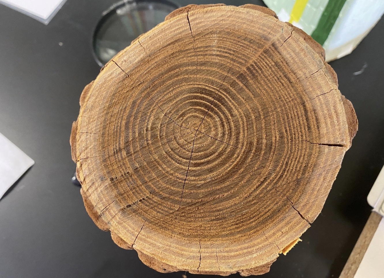
Woody Dicot Stem with Tree Rings
Monocot Leaf showing Parallel Leaf Venation 40x
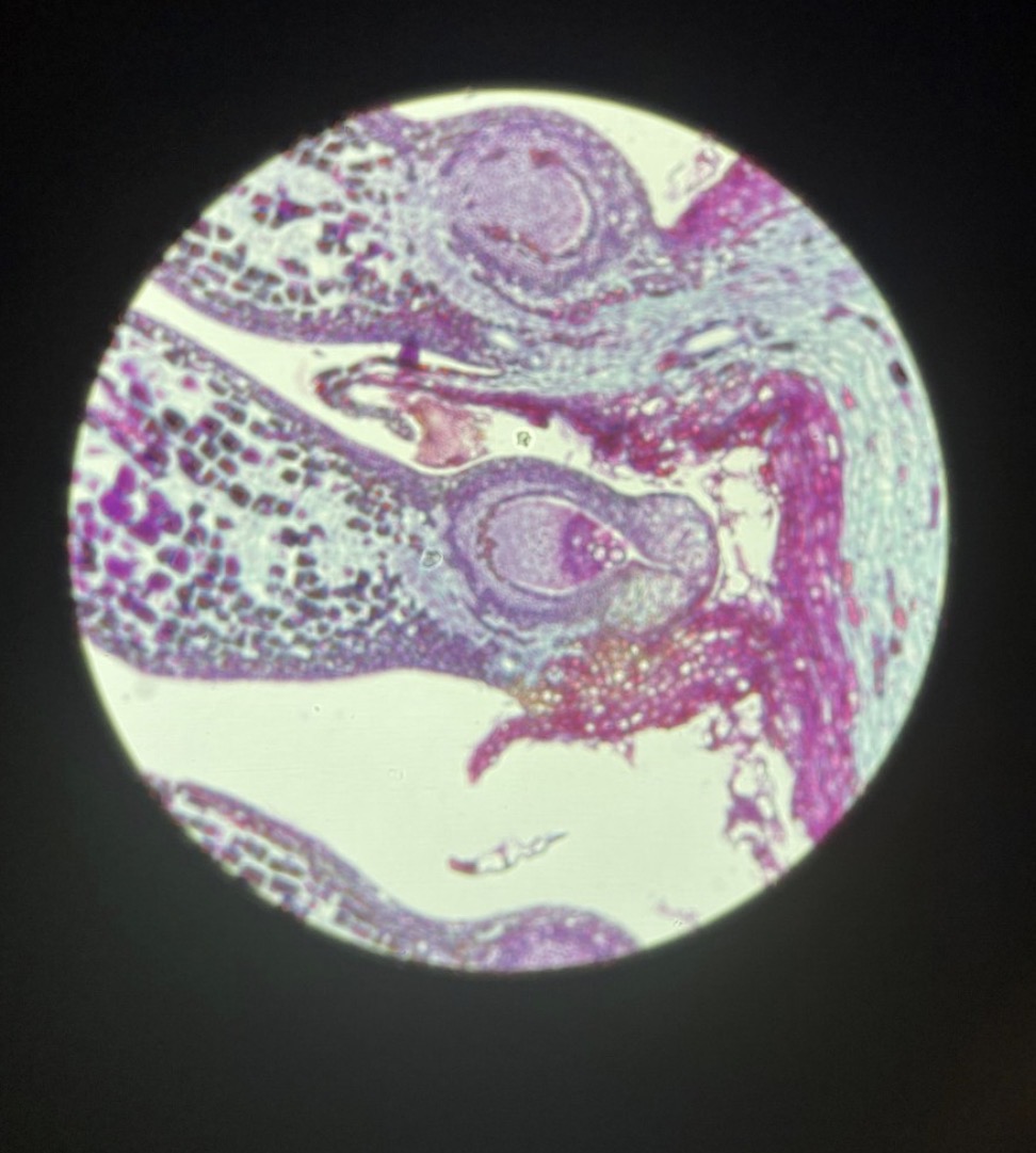
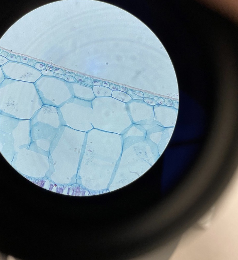
Dicot Leaf with Stomata
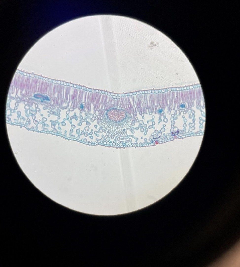
Leaf Vein in a Dicot Leaf 400
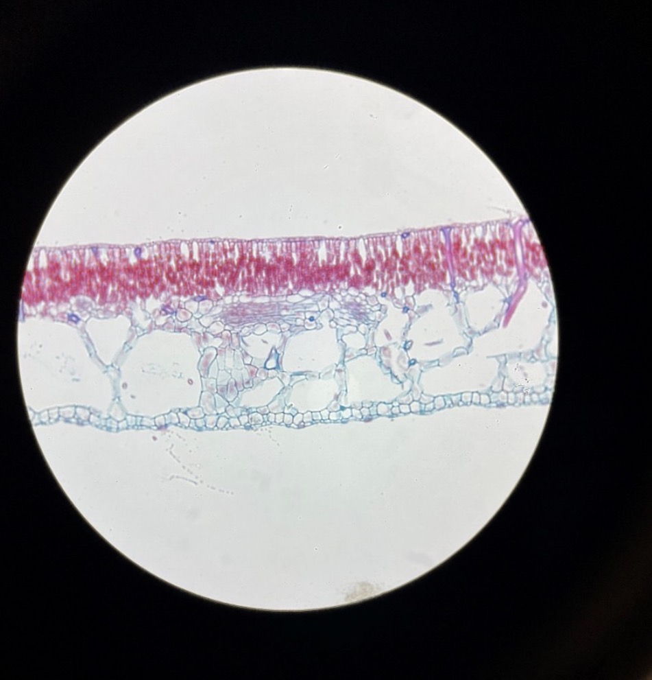
Hydrophyte Leaf without a waxy Cuticle because it Lives in Water 400x
Xerophyte Leaf with Thick Waxy Cuticle because it Lives in a Dry Environment 400x