Histology Unit 3 Review
1/72
There's no tags or description
Looks like no tags are added yet.
Name | Mastery | Learn | Test | Matching | Spaced | Call with Kai |
|---|
No analytics yet
Send a link to your students to track their progress
73 Terms
What organs make up the CNS?
Brain and spinal cord
What organs make up the PNS?
Cranial and spinal nerves with their associated ganglia
What is the Soma?
cell body; contains nucleus and organelles
What are dendrites?
receive input from other neurons and from external environment and carry it to cell body
what are Axons?
carry information from cell body to another neuron or effector cell
What are Synapses?
cell-to-cell junctions utilizing chemical messengers (neurotransmitters)
Describe Multipolar Neurons
most common type; has numerous branched Dendrites and 1x Axon
describe Bipolar Neurons
1x Dendrite + 1x Axon
describe Pseudo-unipolar Neurons
Appears to have 1x process from Soma—Dendrite + Axon fused @ Soma (false appearance of 1 process instead of 2)
describe Neuroglia
supporting but non-conducting cells that are in apposition to neurons
what type of Glia does CNS have?
Oligodendrocytes, Astrocytes, Microglia, and Ependymal cells
what type of Glia does PNS have?
Schwann cells and Satellite cells (capsule cells)
describe Astrocytes
main aid in bld brain barrier; surrounds Somas and processes of neurons
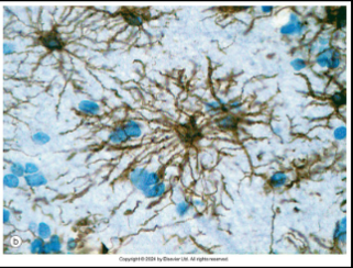
what’s significant of foot processes of Astrocytes on bld-brain barrier?
restricts passage of substances b/w bld and cerebral spinal fluid (CSF): limited diffusion and active transport
describe Oligodendrocytes
1 cell creates Myelin sheath for multiple axons or axon segments
describe Ependymal cells
columnar cells—similar functions to epithelial cells; lines central canal of spinal cord & ventricles of brain
Stem cell for other neuroglia:
Ependymal cells
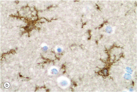
describe Microglia
smallest neuroglia; macrophages resident to CNS (Phagocytic); only Nuclei are visible unless special stain is used
describe Schwann cells
produces lipid-rich myelin sheath the surrounds axons; 1 cell insulates 1 segment of 1 axon; surrounds unmyelinated fibers, too; clears debris & guide regrowth of PNS axons
describe Myelin sheath
speeds up rate of conduction of Action potential; not all Neurons are surrounded by it; appears as white ring around axons unless stained
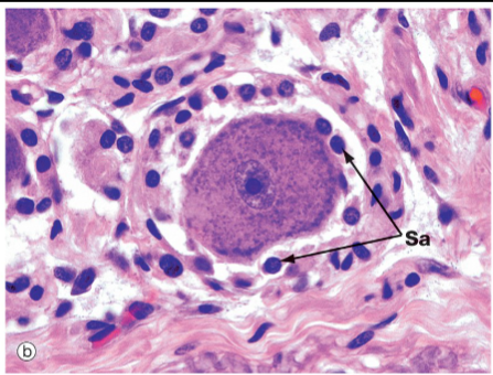
describe Satellite (Sc) & Capsule cells (Cc)
small, flat cells that surround neurons of (PNS) ganglia; Soma structural support; insulate neurons; reg. exchange of metabolic substances
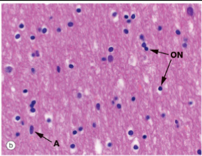
in the Brain, Gray matter is located where?
the outer covering

in the Brain, the White matter is located where?
the inner core

in the Spinal cord, where is the White matter located?
in the outer covering
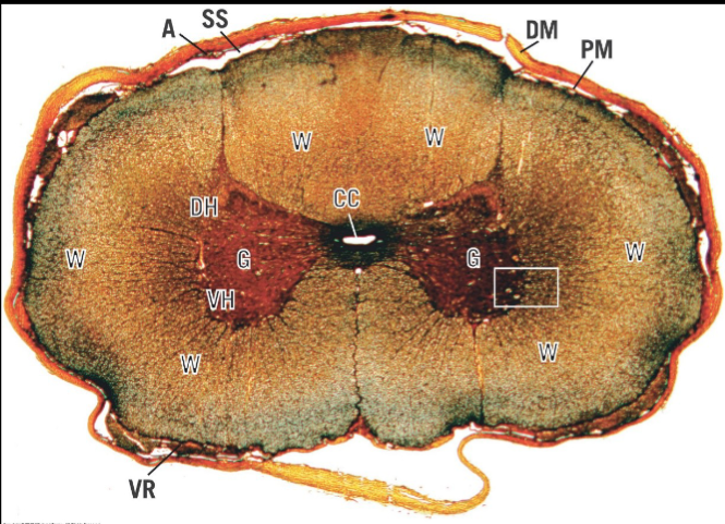
in the Spinal cord, where is Grey matter located?
in the inner core
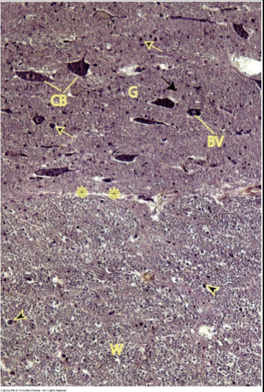
in the Spinal cord, what does its Grey matter contain?
somas of multipolar neurons & neuroglia nuclei

in the Spinal cord, what does its White matter contain?
collection of myenlinated axons w/ neuroglia nuclei
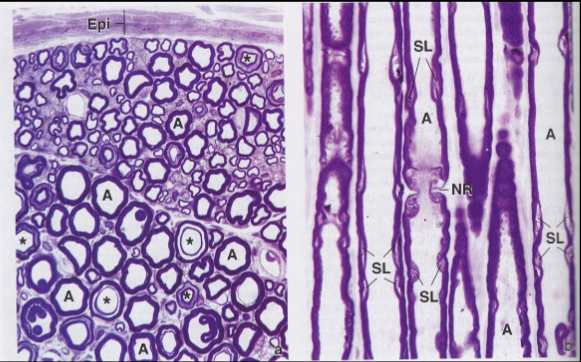
describe Peripheral neve of PNS
Many axons (nerve fibers) that carry sensory and motor information between organs/tissues and brain/spinal cord; Surrounded by connective tissue
sheaths—like muscle
describe Ganglia of PNS
small accumulations of neuron cell bodies & glia surrounded by CT capsule
describe Endoneurium
loose CT + reticular fibers surrounding each indv nerve fiber (axon) and Schwann cells
describe Perineurium
specialized CT + epithelial-like cells and tight junctions surrounding each nerve fascicle (diffusion barrier)d
describe Epineurium
dense irregular CT that surrounds peripheral nerve (blood vessels; adipose)
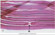
what are Nodes of Ranvier?
space b/w adjacent Schwann cells
what type of neurons are Dorsal root ganglion (snesory)
pseudo-unipolar neurons
what type of neuron is Sympathetic ganglion (motor)
multipolar neutronsw
what type of neurons is Parasympathetic ganglion?
multipolar neurons
describe Tunica intima
Endothelium, Basement mbrane, & thin layer Loose CT
describe Tunica media
layers of Smooth muscle cells + Elastic fibers—thicker layer in Arteries
describe Tunica adventitia
longitudinal Collagenous tissue that merges w/ Loose CT around Vessels—thinker layer in Veins
describe Large/ elastic Arteries fucntion
recoil of numerous Elastic fibers allows for BP created by Heart to propel bld through vessels
which type of arteries make the majority of arteries in body?
Medium/ muscular (distributing) Arteries
describe Small arteries & arterioles function
control bld flow by altering diameter of Lumen
describe Small Arteries & Arterioles
only 1-2 layers of smooth muscle cells in tunica media for arterioles; Gap junct found bw endothelium and smooth muscle; Tunica adventitia thin & blends w surrounding CT
which bld vessels have the smallest diameter?
Capillaries
describe Capillaries
1x layer of endothelial cells + basement mbrane; fluid exchange network(s)
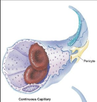
describe Continuous capillaries
most common; 8-10 um diameter
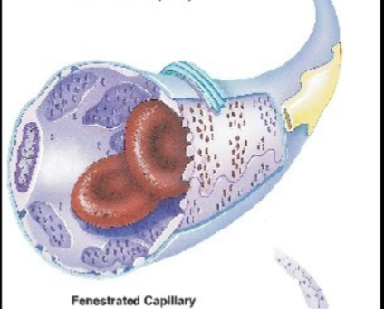
describe Fenestrated capillaries
occurrence of filtration/ absorption; 30-40 um diameter
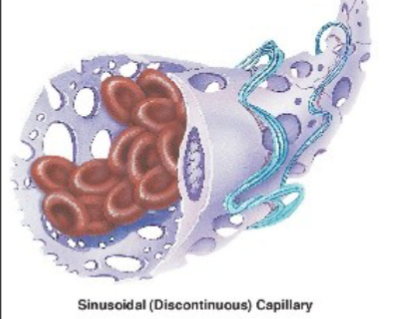
describe Discontinuous capillaries (sinusoids)
cells can pass through vessel wall; 60-80 um diameter
where are Cont. capillaries found in the body?
Muscle lung, CT, and CNS
where are Fenestrated capillaries found in the body?
Endocrine glands, Gall bladder, Kidney, and Intestinal tract
where are Discont. capillaries found in the body?
Liver, spleen, bone marrow
describe Veins
thinner wall & larger Lumen diameter vs. arteries; Valves present to prevent backflow in larger veins
describe Venules & small veins
Postcapillary venules small diameter and limited Tunica media with very few Smooth muscle cells
describe Medium veins
up to 10mm diameter; Valves appear to prevent backflow
describe Large veins
>10mm diameter; thin Tunic media but thick Adventitia (collagen/ elastic fibers; long. smooth muscle cells)”
“Vessels of the Vessels” is referring to what?
Vasa vasorum
describe Vasa vasorum
small bld Vessels that provide nutrients to Tunic media and adventitia of larger Arteries and veins whom have thick walls
Lymphatic caps are sinusoids, describe this:
More permeable than bld Capillaries; Unidirectional flow of fluid from tissues → veins; Collect excess protein-rich fluid from interstitial fluid called Lymph
what does Lymph vessels NOT carry?
bld, presence of bld means internal issues occurring
describe Aneurysm
damage to bld vessels which weakens its wall, bld pools in defected area and is no longer confined to its vessel(s); if left untreated, rupture of vessels causes internal bleeding and potential death
Valves in the heart separate chambers for what purpose?
it’s to prevent backflow
describe Heart wall layer: Endocardium
endothelium lining Artia & Ventricles
describe Heart wall layer: Myocardium
thick layer of cardiac muscle fibersdescribe Heart wall layer:
describe Heart wall layer: Epicardium
Mesothelium (simple squamous) + CT
describe Purkinje fiber
non-contractile: send electrical signal to contractile cardiac muscle fibers; clear cytoplasm; no Intercalated disks w/ less myofibrils but larger in diameter
where are Purkinje fibers found?
in the Endocardium of the Heart
what kind of tissue is found in the Epidermis?
Epithelial tissue
what kind of tissue is found in the Dermis & Hypodermis?
Connective tissue
what kind of muscle tissue is found near hair follicles?
Smooth muscle
describe Epidermis and its derivatives
keratinized stratified squamous epithelium; nonvascular
describe Dermis and its derivatives
dense irregular CT, provides mechanical support, strength, and thickness; vascularized
describe Hypodermis and its derivatives
deep to dermis (subcutaneous/subcutis), composed of variable amounts of adipose tissue lobules and dense irregular CT