GRISTO Module 4 Anatomy: Visual and Auditory Pathways
1/67
There's no tags or description
Looks like no tags are added yet.
Name | Mastery | Learn | Test | Matching | Spaced | Call with Kai |
|---|
No analytics yet
Send a link to your students to track their progress
68 Terms
VISUAL PATHWAY
VISUAL PATHWAY
primary visual cortex in the occipital lobe
Vision is initially processed in the
retina
Light passes through the cornea, aqueous humor, pupil, lens, and vitreous humor to reach the
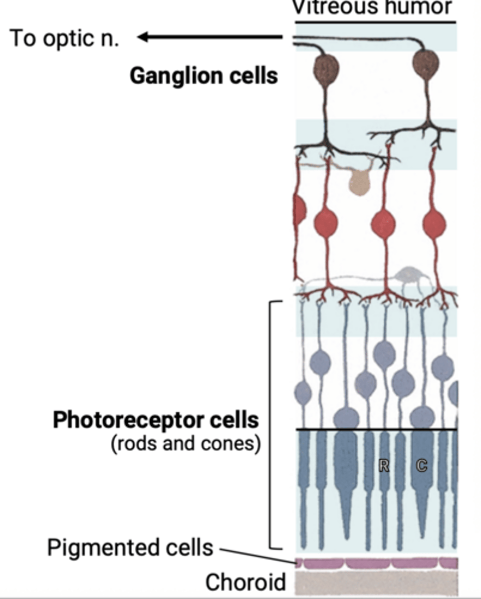
pass through all cell layers in the retina
To reach photoreceptor cells, light must
optic nerve
Photoreceptors send signals back through layers of the retina to reach the
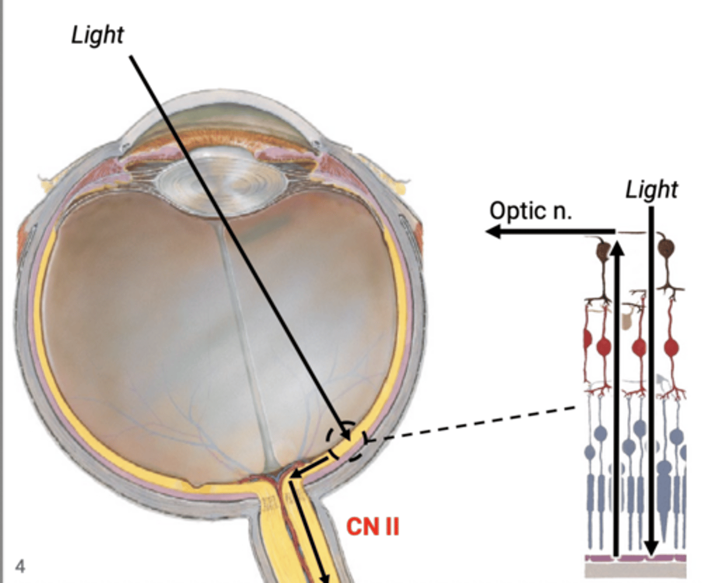
2nd order ganglion cells that run the length of the retina and send central axons into the optic n.
Instead of one large ganglion, bipolar cells relay to
initial receptors that detect the light
Photoreceptor cells are the
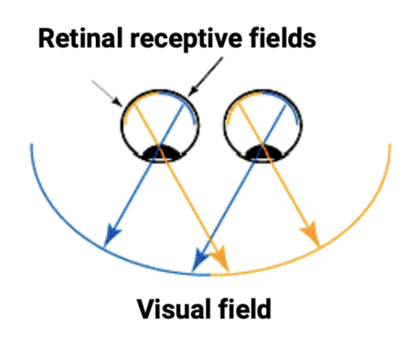
area in which objects are visible at the same moment during steady fixation of gase
The visual field is the
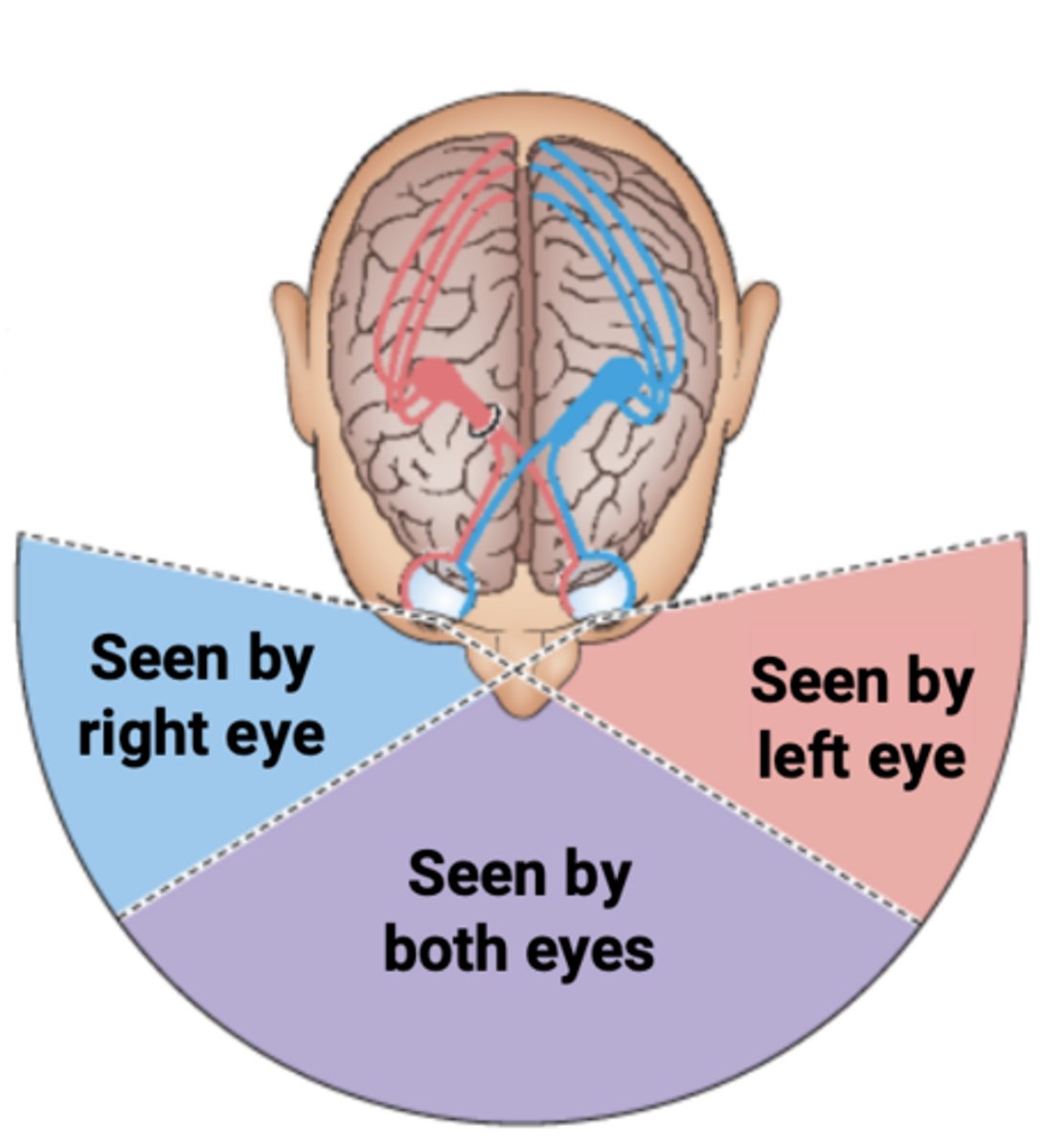
right-left reversed and inverted
Images in the visual field are what on the retina
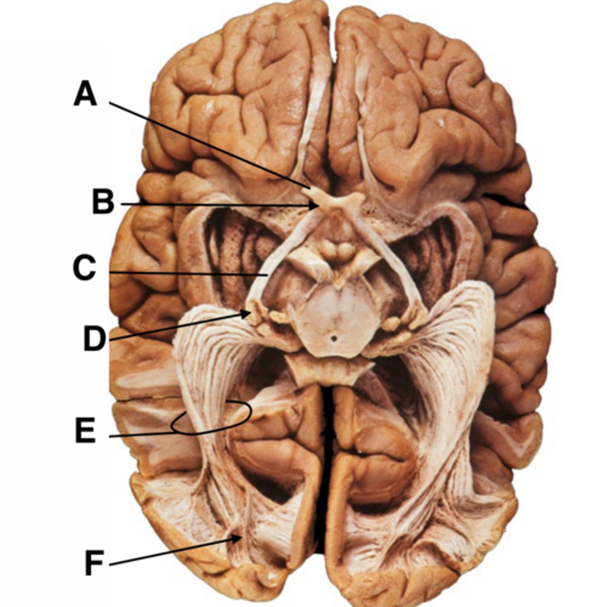
A. Optic nerve
B. Optic chiasm
C. Optic tract
D. Lateral geniculate nucleus (LGN) of thalamus
E. Optic radiation
F. Primary visual cortex
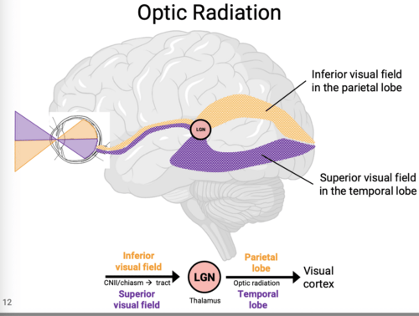
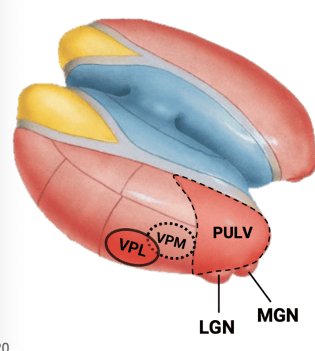
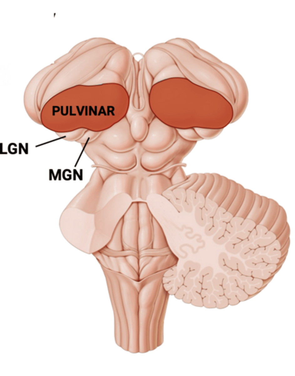
posterior part of the thalamus: the pulvinar
Lateral Geniculate Nucleus (LGN) hangs off the
optic radiation, which relay to the primary visual cortex
The LGN give off the
predictable locales
Just like the nasal and temporal regions of vision the superiro and inferior regions run in
the temporal lobe a
Fibers from the superior visual field run through
parietal lobe
Fibers from the inferior field run through the
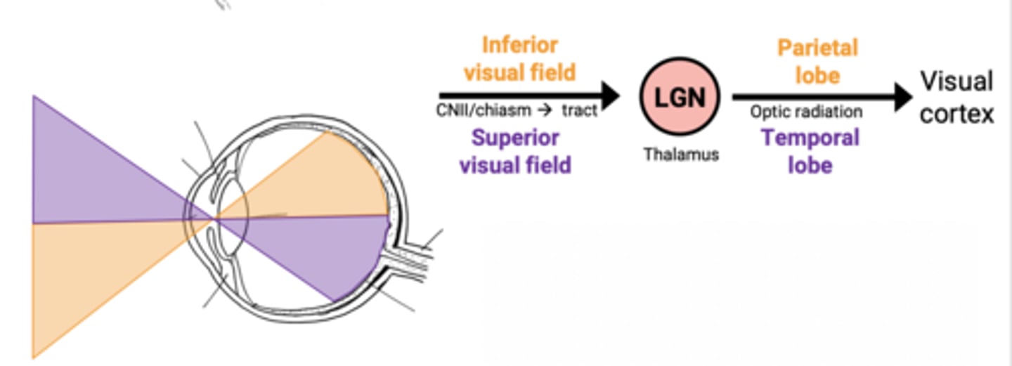
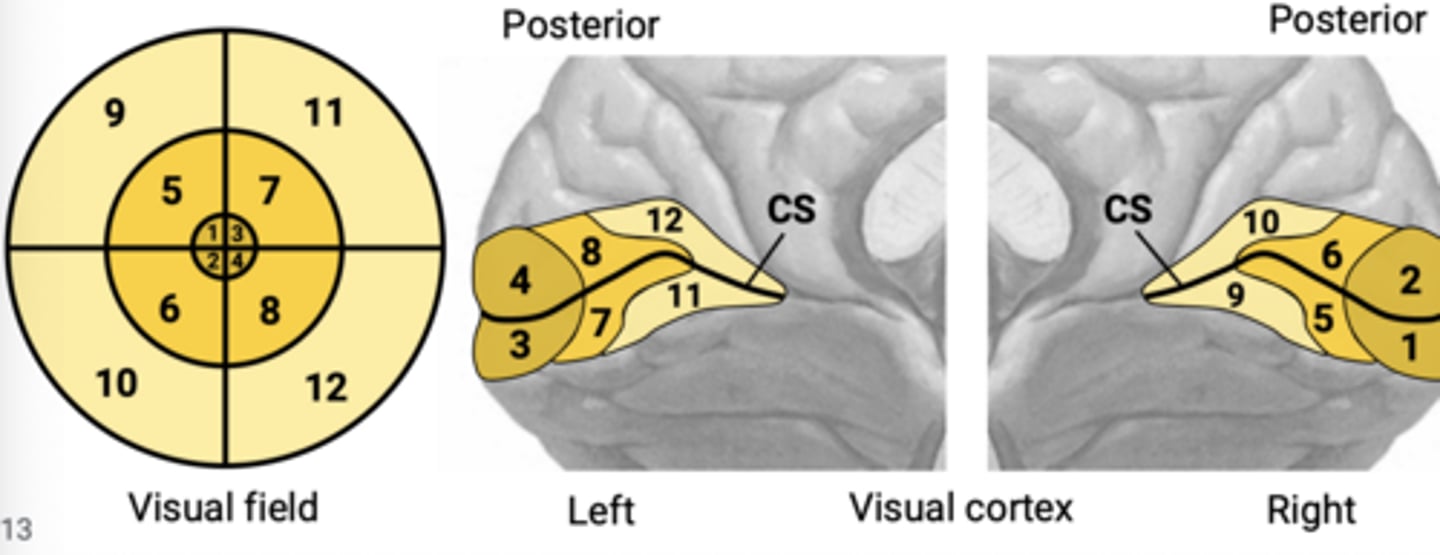
calcarine sulcus (CS)
The primary visual cortex is above and below the
Reversed: Left visual field maps to the right cortex (and vice versa)
Upside down: The inferior visual field is on the superior half of visual cortex- meaning in the brain the image is inverted (and vice versa)
Inside-out: The center of visual field is posterior (outer) and peripheral vision is anterior (inner)
Relative to the visual field the visual cortex is
one sided blindness
Visual field Dysfunction:
Optic nerve
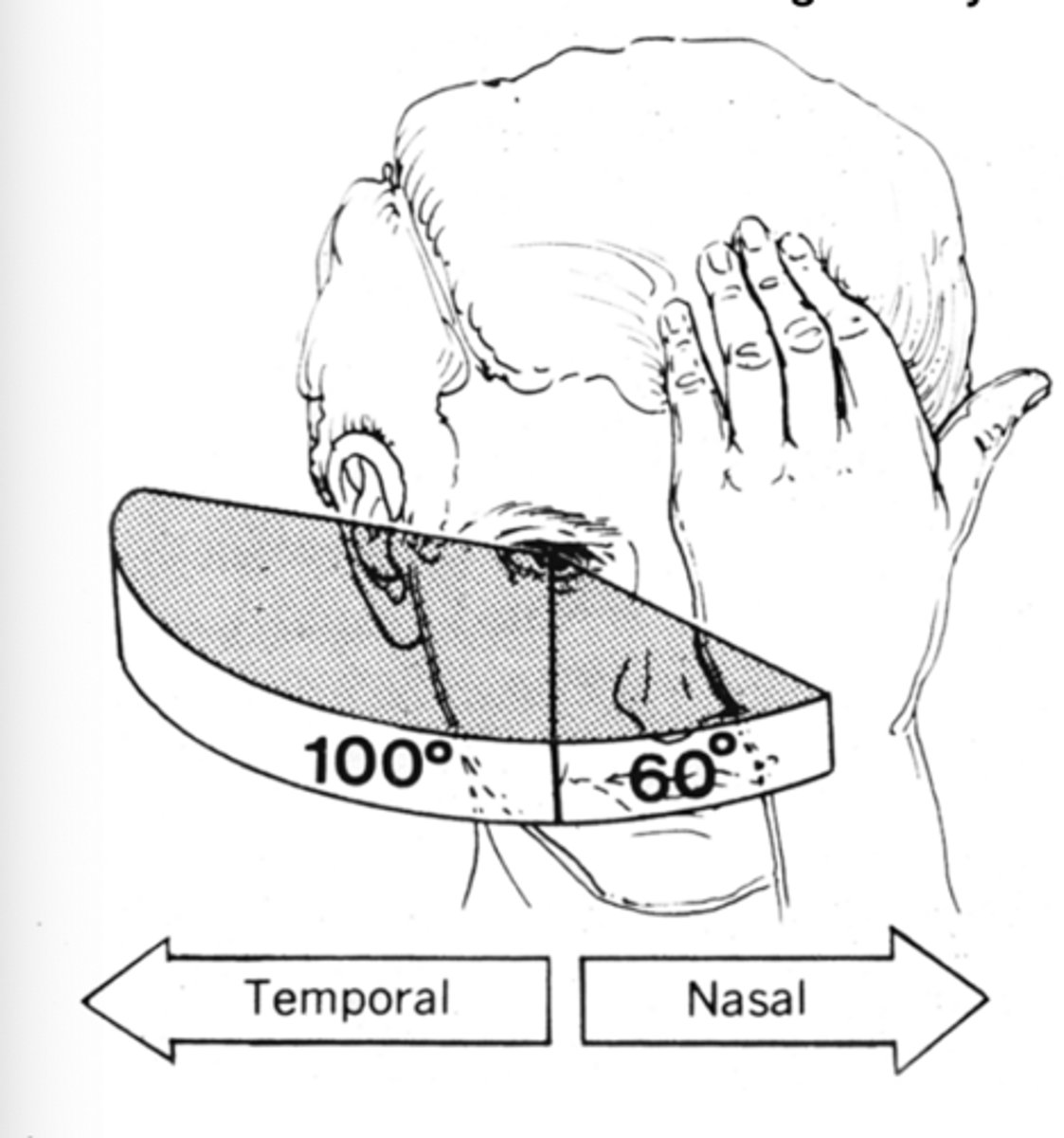
loss of peripheral fields (temporal fields cross)
Visual field Dysfunction:
Optic chiasm
Loss of an entire visual field (right or left)
Visual field Dysfunction:
Optic tract
suggests temporal lobe damage
Visual field Dysfunction:
Homonymous superior quadrantanopia (pie in the sky)
suggests parietal lobe damage
Visual field Dysfunction:
Homonymous inferior quadrantanopia (pie on the floor)
Optic nerve damaged

Optic chiasm damaged

Optic tract damaged

Homonymous superior quadrantanopia

Homonymous inferior quadrantanopia

YES it does
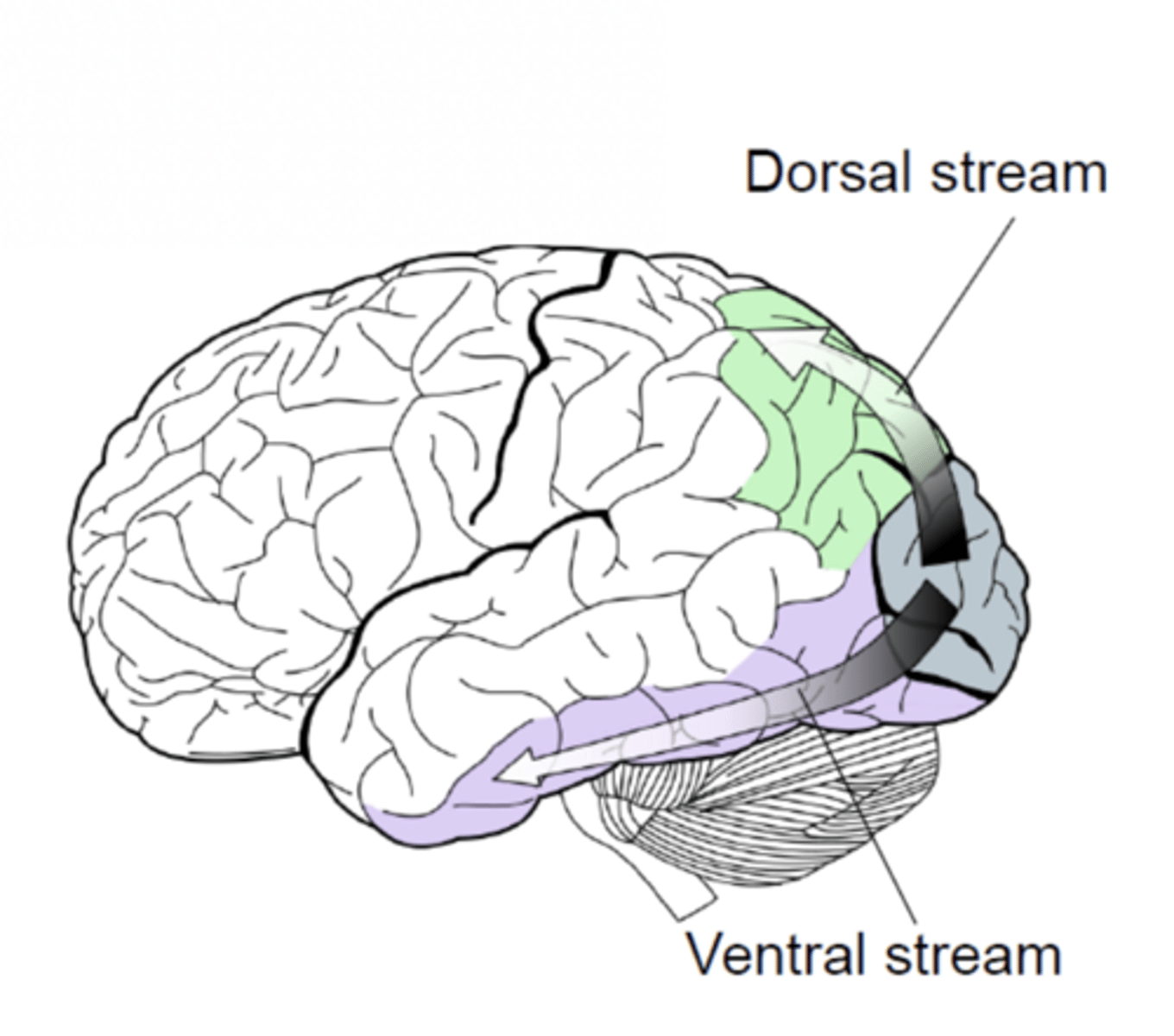
The primary visual cortex relays to other regions of the brain to further process what the visual cortex sees
the where and how stream
Occipital --> parietal
- For processing of spatial and contextual relationships, movement
Dorsal stream
The what stream
Occipital --> temporal
- for processing of face and scene recognition
Ventral stream
AUDITORY PATHWAYS
AUDITORY PATHWAYS
Is the big important guy for this stuff
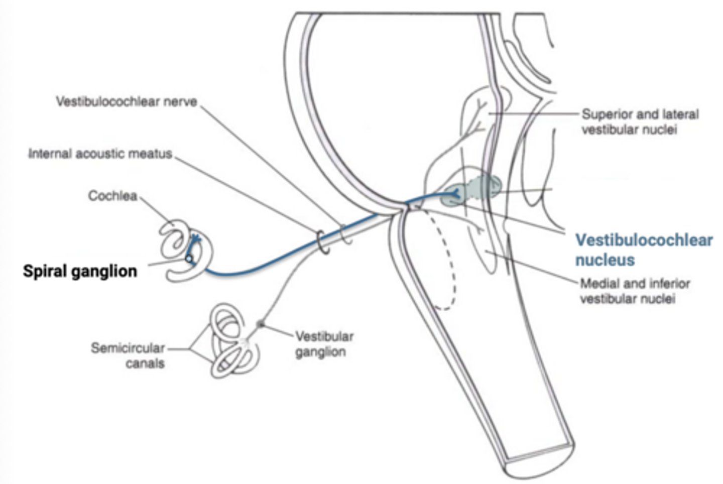
Vestibulocochlear nerve
Primary auditory cortex
The MGN projects to the
inferior quadrigeminal brachium from the inferior colliculus to the MGN
Auditory sensations then follow the
auditory reflexes
The inferior colliculus also relays to regions that participate in
ipsilaterally and contralaterally to the inferior colliculus in the midbrain
CN VIII synapses on neurons in the CN VIII nucleus. From there fibers ascend
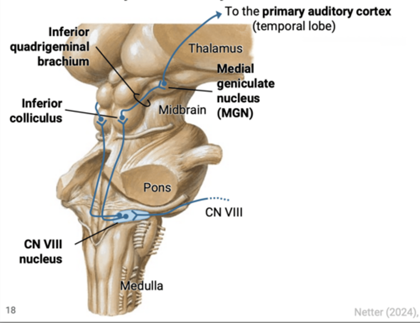
not all of those bitches
Most fibers emanating from the CN VIII nucleus decussate but
unilateral central lesions rarely produce more than subtle changes in hearing, generally contralateral to the lesion
Functionally, this means
pathology in peripheral auditory structures (middle and inner ear, cochlear nerve)
Most disturbances in auditory functions are associated with
bottom of the pulvinar on the posterior thalamus
The medial geniculate nucleus (MGN; auditory pathway) and the Lateral geniculate nucleus (LGN; vision pathway) hang off the
transverse temporal gyrus (Heschl's gyrus)
Primary Auditory cortex is located in the
may lead to loss of awareness of sound, but reflexes involving sound remain intact
Lesions to the primary auditory cortex
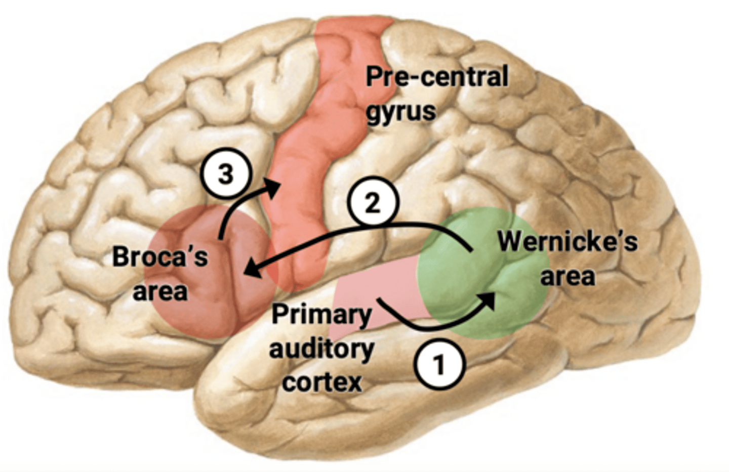
1. Primary auditory cortex --> Wernicke's area (language comprehension)
2. Wernicke's area --> Broca's area (motor speech)
3. Broca's area --> Pre-central gyrus (primary motor cortex)
When a spoken word is heard by the primary auditory cortex:
- CN V: Motor nucleus of V (pons)
- CN VII: Motor nucleus of VII (pons)
- CN IX, X: Nucleus ambiguus (medulla)
- CN XII: Motor nucleus of XII (medulla)
Corticobulbar tract to cranial nerve nuclei involved in speech:
QUIZ
QUIZ
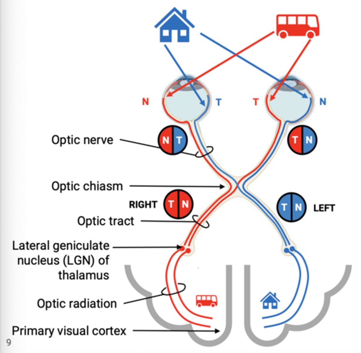
right, left
Images in the left visual field are sseen by the _____ retina and images in the right visual field are seen by the _____ retina
BDAEC
Put in order the pathway of vision from the retina to the primary visual cortex.
Retina
A. Optic tract
B. Optic nerve
C. Optic radiation
D. Optic chiasm
E. Lateral geniculate ganglion
Primary visual cortex
The nasal visual field of the left eye
The temporal visual field of the left eye
The left optic nerve contain fibers from
the temporal visual field of the right eye
the nasal visual field of the right eye
the nasal visual field of the left eye
the temporal visual field of the left eye
The left-sided nasal and temporal visual fields
The right optic tract contains
The inferior nasal and temporal visual fields from the right eye
The left optic radiation in the parietal lobe carries
Parietal, Where and how, Spatial and contextual relationships, movement
The dorsal visual association stream enters the _____ lobe and relays _____ visual information for processing ______
Temporal, What, Face and scene recognition
The ventral visual association stream enters the _____ lobe and relays ______ visual information for processing ______
Wernicke's area
From the primary auditory cortex, the first place a spoken word is relayed is