Assessing the Peripheral Vascular and Regional Lymphatic Systems
 Call Kai
 Learn
Learn Practice Test
 Spaced Repetition
 Match
Match Flashcards
 Knowt Play
1/44

Earn XP
Description and Tags
Name | Mastery | Learn | Test | Matching | Spaced | Call with Kai | Chat |
|---|
No analytics yet
Send a link to your students to track their progress
45 Terms
Lymphatic system structure
Spleen, thymus, bone marrow, tonsils and adenoids, appendix, parts of the liver and lungs, lymph nodes, peyer’s patches
Right lymphatic duct
Receives lymph from the right upper part of the body
Thoracic duct
Receives lymph from the rest of the body
Diagnostics
Computer tomography (CT) scan, duplex ultrasound, magnetic resonance angiography (MRA), angiogram, angioplasty, lymph node biopsy
Unilateral edema
Local or peripheral cause
Lymphedema
Swelling due to an abnormal accumulation of lymph fluid within the tissues
Rubor
Redness
Elevational pallor
Pale skin when raising up the affected extremity
Raynaud’s phenomenon
Periodic ischemic attacks affecting extremities of the body, especially fingers, toes, ears, and nose, becoming cyanotic and very painful; attacks are brought on by arterial constriction due to extreme cold or emotional stress
Atherosclerosis
Condition where fatty deposits called plaque build up on the inner walls of the arteries
Intermittent claudication
Pain in leg muscles that occurs during exercise and is relieved by rest
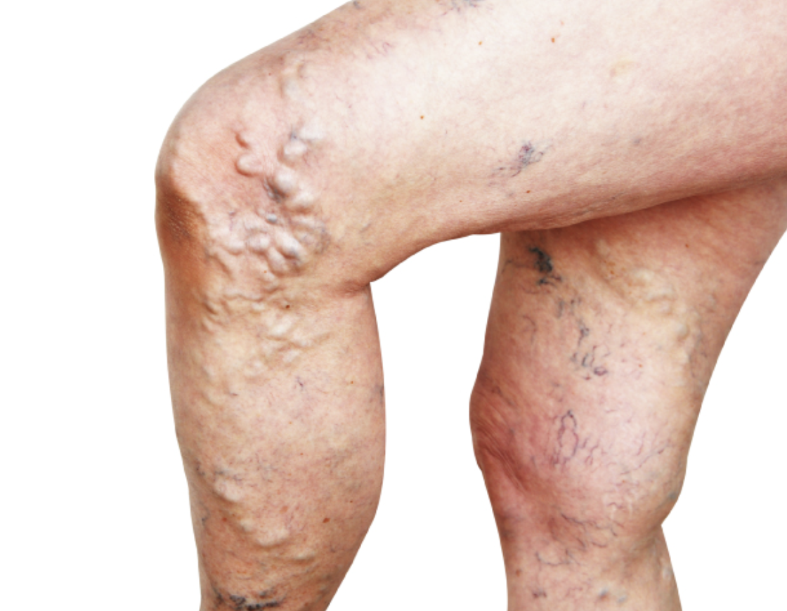
Varicose veins
Abnormally swollen, twisted veins with defective valves; most often seen in the legs
Diabetes
Body is unable to produce enough insulin, the hormone required for the metabolism of sugar
Peripheral arterial disease
Blockage of arteries carrying blood to the legs, arms, kidneys and other organs
Lymph node sites
Pre and postauricular, suboccipital, tonsilar, submandibular, superficial and posterior cervical, superclavicular
Assessing lymph nodes normal findings
No nodes are palpated; if palpated, nodes are <1 cm; discrete, moveable, nontender
Assessing lymph nodes abnormal findings
Enlarged, >1cm, matted, hard, tender
Inspecting and palpating the carotid arteries normal findings
Visible symmetrical pulsations, pulse rate between 60-100 bpm, rhythm is regular, amp +2
Inspecting and palpating the carotid arteries abnormal findings
No pulsations or asymmetrical pulsatile bulge, pulse rate <60 or >100 bpm, rhythm irregular, amplitude absent, weak, or bounding
Auscultating carotid arteries normal findings
No bruit heard
Auscultating carotid arteries abnormal findings
Bruit heard
Bruit
Blowing, swooshing sound heard through a stethoscope when an artery is partially occluded
Inspecting for jugular venous distention normal findings
No visible pulsation or jugular vein distention
Inspecting for jugular venous distention abnormal findings
Visible distention
Inspecting/palpating upper extremities normal findings
Arms symmetrical, color uniform, no edema or ulcerations, fingernail beds pink, venous pattern normal, fingernails have even thickness, fingernails 160 angle of attachment, epitrochlear nodes not felt, temperature warm to touch, +2 radial and brachial pulse
Inspecting/palpating upper extremities abnormal findings
Signs of altered peripheral circulation, discoloration, change in skin texture, cool extremities, unilateral edema, enlarged epitrochlear nodes, cellulitis, edema, ulcerations
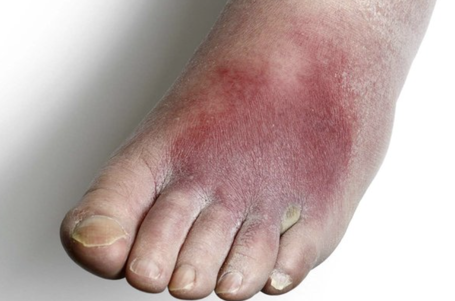
Cellulitis
Inflammation of cellular or connective tissue
Ulcerations
Blisters or open-sores
Assessing capillary refill normal findings
Pink color returns in <2 sec
Assessing capillary refill abnormal findings
Pink color returns in >2 sec
Inspecting/palpating lower extremities normal findings
Legs symmetrical, uniform color, no edema or ulceration, toenails pink with even thickness, temp warm to touch, even hair distribution, inguinal lymph nodes palpable, +2 symmetrical pulses, nondistended veins
Inspecting/palpating lower extremities abnormal findings
Discoloration, decreased texture, loss of hair, cool skin temp, thick toenails, arterial or venous insufficiency, presence of bilateral or unilateral edema, lymphedema
Pitting edema
Retains an imprint when touched
Nonpitting edema
Does not retain an imprint when touched
Edema grading
0-4+
0 edema grading
No pitting edema
1+ edema grading
Mild pitting edema, 2mm depression that disappears rapidly
2+ edema grading
Moderately pitting edema, 4mm depression that disappears in 10-15 seconds
3+ edema grading
Moderately severe pitting edema, 6mm depression that may last more than 1 minute
4+ edema grading
Severe pitting edema, 8mm depression that can last longer than 2 minutes
Deep vein thrombosis
Blood clot forms in a large vein, usually in a lower limb
Peripheral vascular disease
Disease of veins with inadequate blood flow from extremities
Peripheral arterial disease signs and symptoms
Intermittent claudication, pain, no pulse or weak pulse at affected site, black eschar, cyanosis
Peripheral vascular disease
Dull achy pain, lower leg edema, erythema, drainage, varicose veins
Risk factors for deep vein thrombosis
Birth control, surgery, smoking, sitting for long periods of time