NPB101 MT1: homeostasis/muscles/cardio syst
1/86
There's no tags or description
Looks like no tags are added yet.
Name | Mastery | Learn | Test | Matching | Spaced | Call with Kai |
|---|
No analytics yet
Send a link to your students to track their progress
87 Terms
mitochondria
creates ATP (energy) in the body
nucleus
cell control center
cytoplasm
consistent pH, intracellular transport
ribosome
protein synthesis
lysosome
breaks down certain proteins
endoplasmic reticulum
synthesize exterior cell + proteins/lipids
golgi complex
enclosed sacs that modify from ER, secrete vesicles, sort and direct
4 primary types of tissues
muscle, nervous, connective, epithelial
organ
structure composed of several types of tissues
homeostasis
maintenance of a dynamic steady state in the internal environment
what 3 fluids are exchanged in homeostasis?
intracellular fluid, intersitial fluid, blood (plasma)
homeostatic control system
sensors (detect change), control center (integrates info), effectors (makes adjustments)
negative feedback
counteracts deviations from the set point in the OPPOSITE direction of intial change
positive feedback
amplifies initial change AWAY form the set point
what are the two forms of direct intercellular communication?
gap junctions and transient direct linkup of cell’s surface markers
what are the two forms of indirect intercellular communication via chemical messengers?
paracrine secretion and NT secretion
function of muscles
turn chemical potential energy into mechanincal energy (smooth, cardiac, and skeletal)
smooth muscles
no striations, involuntary and found in stomach, airways, and blood vessels
cardiac muscles
striated, involuntary, one nucleus per cell, intercalated disks
skeletal muscles
long, multinucleated, striated, voluntary
sarcomere
smallest unit of muscle cells containing all of the elements for contraction in striated muscles. composed of myosin and actin
thick filament
myosin composed of long interwoven protein with globular head
thin filament
actin, troponin, and tropomyosin in a double helical structure
what happens to parts of sarcomere during contraction?
the sarcomere shortens, H zone shortens, I band shortens, all other parts stay same length
cross bridge cycle/activity
ACh produced in MN, Na+ ch open, AP flows thru sarcolemma and t-tubules, Ca2+ release, Ca2+ binds to troponin C, I, and T, then moves tropomyosin out of blocking position for binding, crossbridge binds to actin, Pi released then ADP released, powerstroke and filaments slide, sarcomere gets smaller and new ATP binds to head, ATP is hydrolyzed and cycle resets
excitation contration coupling
muscular contraction when the thick and thin filaments in sarcomere slide past one another by a power stroke
motor unit
motor neuron and all the fibers it innervates (1 MN has many fibers, each fiber has 1 MN)
muscle tension
tension that depends on the number of motor untis recruited based on frequency, length, extent, and thickness
twitch summation
increase in tension by repetitive stimulation of a muscle fiber (muscle can never fully relax)
tetanus
smooth, sustained contraction of maximal strength so rapid cannot relax between stimuli
muscle fatigue
inability of muscle to maintain tension
smooth muscle contraction
muscle contraction by sliding filament mechanism (no sarcomere): excited, Ca2+ messenger activated calmodulin, myosin light chain kinase, cross bridge cycle
multi-unit smooth muscle
activated by neuronal input for an independent response
single-unit smooth muscle
capable of pacemaker activity by gap junctions
circulatory system
heart, blood vessels, and blood. function: to supply O2 and nutrients, remove waste, regulate temp, distribute hormones, and immunovigalance
right atrium
receives O2 poor blood from systemic venous circulation
right ventricle
receives O2 poor blood from right atrium and pumps blood through pulmonary (semilunar) valve to pulmonary artery
left atrium
receives O2 rich blood from pulmonary circulation from left and right pulmonary veins
left ventricle
receives O2 rich blood from left atrium and pumps this blood through aortic (semilunar) valve into aorta

what does #1 correspond to on this image?
vena cava

what does #2 correspond to on this image?
right atrium

what does #3 correspond to on this image?
tricuspid (right AV) valve

what does #4 correspond to on this image?
right ventricle

what does #5 correspond to on this image?
left ventricle

what does #6 correspond to on this image?
pulmonary/semilunar valve

what does #7 correspond to on this image?
mitral/left AV valve
what does #8 correspond to on this image?
aortic/semilunar valve
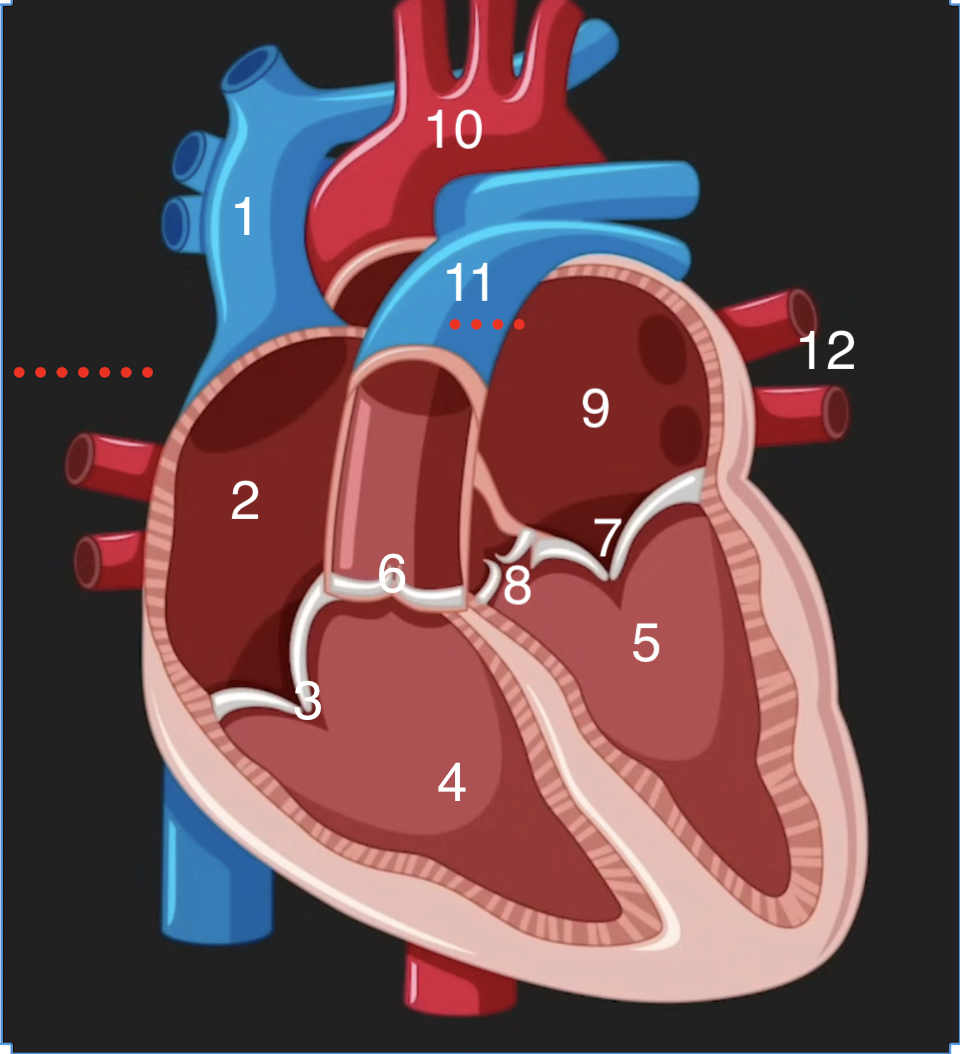
what does #9 correspond to on this image?
left atrium

what does #10 correspond to on this image?
aorta

what does #11 correspond to on this image?
pulmonary artery

what does #12 correspond to on this image?
pulmonary veins
why are heart valves important and which is strongest/weakest valve?
important for one-way blood flow. semilunar is the strongest, tricuspid is the weakest
what are the 3 layers of the heart wall?
endocardium, myocardium, epicardium
contractile cells
99% of cardiac muscle cells, do mechanical work of pumping
autorhythmic cells
1% of cardiac cells, conduct APs for contraction, pacemaker
nodes
pacemaker cells grouped together for control rate and coordination
SA node
pacemaker node in right atrium. acts as a sensor, fastest and most dominant node (70 APs/min)
AV node
pacemaker node at base of right atrium (50 APs/min)
Bundle of His
starts at AV node and divides into left and right ventricles (40-60 APs/min)
Purkinje Fibers
fibers of pacemaker cells in bundle of his to ventricular myocardium (30 APs/min)
interatrial pathway
conduct pacemaker activity from right atrium to left atrium
internodal pathway
conduct pacemaker activity from SA node to AV node
electric flow in heart
SA node —> AV node —> Bundle of His —> L/R bundle branches —> Purkinje Fibers
vein
blood vessel that carries blood TOWARDS the heart
artery
blood vessel that carries blood AWAY from the heart
flow of deoxygenated blood
blood from the body —> vena cava —> right atrium —> tricuspid valve —> right ventricle —> pulmonary valve —> pulmonary artery —> lungs
flow of oxygenated blood
oxygenated blood from the lungs —> pulmonary veins —> left atrium —> mitral valve —> left ventricle —> aortic valve —> aorta —> body
systole
contraction and emptying
diastole
relaxation and filling
isometric ventricular contraction
valves are CLOSED and pressure increases
isometric ventricular relaxation
valves closed and pressure decreases
stroke volume
amount of blood pumped out of the chamber with each contraction
action potential in contractile muscle cells
rapid rising phase due to Na+ entry, repolarization due to K+ ch open and Na+ inactivated, plateau due to slow Ca2+ entry, falling phase with K+ efflux and K+ ordinary ch opening, resting potential maintained by leaky K+ ch
excitation contraction coupling in cardiac cells
Ca2+ entry —> dihydropine receptors —> power stroke —> cross bridge with graded response to regulate heart beat
when does the heart make a sound
when a valve CLOSES
first heart sound
“lub”, closure of AV valve
second heart sound
“dub”, closure of semilunar valve
what is an electrocardiogram (ECG)?
electrical currents generated by APs of heart muscle

p wave of ECG
depolarization of atria, pacemakers go from SA node to AV node
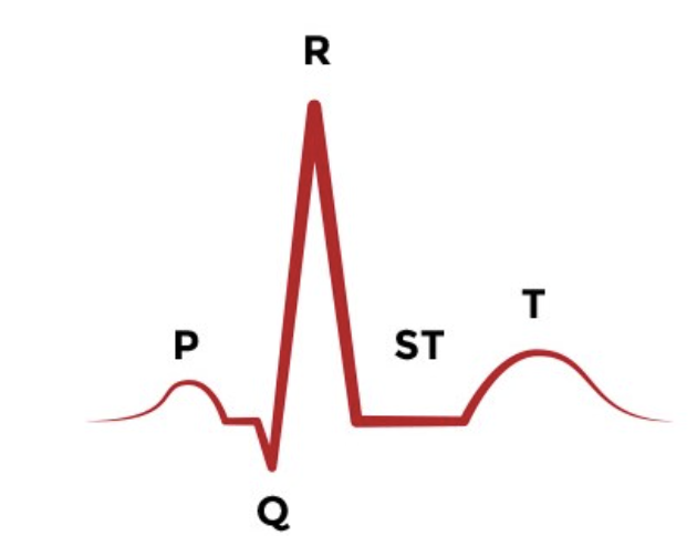
PR segment of ECG
AV nodal delay

QRS segment of ECG
depolarization of the ventricles

ST segment of ECG
ventricles contracting and emptying

t wave of ECG
repolarization/relaxation of ventricles
regulation of cardiac output (C.O.)
volume of blood pumped in each ventricle per minute determine by heart rate and stroke volume: C.O. = H.R. x S.V.
heart murmur
abnormal heart sounds due to malfunctioning valves
stenotic valve
valve does not OPEN completely: whistling sound
insufficient valve
valve does not CLOSE properly: swishing sound