The seeing brain
1/29
There's no tags or description
Looks like no tags are added yet.
Name | Mastery | Learn | Test | Matching | Spaced | Call with Kai |
|---|
No analytics yet
Send a link to your students to track their progress
30 Terms
The vision problem
vision is computationally complex
Sensory input is ambiguous (more than one meaning)
Different perceptions can arise from the same input
brain processing, not just sensory input
The inverse problem
→ challenge of converting 3D image of the world into a 2D retinal image
Ambiguity = many different configurations of a surface could lead to a single retinal input
-requires context, prior knowledge and assumptions
Gestalt e.g. Necker cube = sudden involuntary perceptual change where the brain flips from one interpretation of an ambiguous image to another without the image itself changing
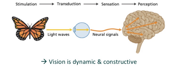
Sensation vs perception
Sensation = the raw input
when a stimulus (light, sound etc.) activates our sensory organs
Perception = the brains interpretation
using past knowledge to organise and make sense of that sensory input
The retina
→the light sensitive inner surface of the eye, made up of multiple layers of:
Photoreceptors = convert light to neutral signals
Signal flow within the retina = photoreceptors → intermediary neurons → ganglion cells
Types pf photoreceptors
rod cells - specialised for low light intensity
Cone cells - specialised for high light intensity
Fovea - highest conc of cones and visual acuity
Receptive field
Region of visual space a neuron responds to
Early feature detection
Early visual neurons respond to:
light vs dark regions
Edges and orientation
Motion direction
→ early visual areas extract simple features that form building. Locks for later more complex visual processing
Lateral inhibition - early detection
neighbouring neurons inhibit each other
Enhances contrast
Sharpens edge detection
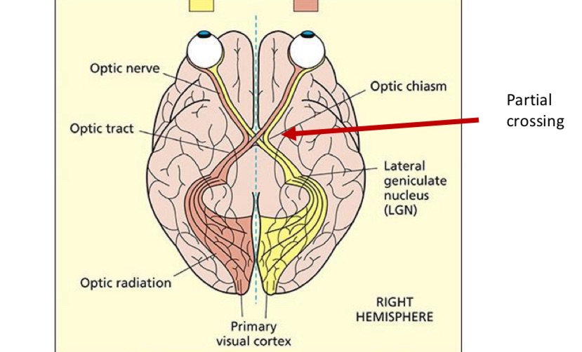
From the eye to the brain
Info leaves each eye via Optic nerve
then branches to multiple brain targets
Main and best studied pathway (about 90% of fibres) = optic nerve → lateral geniculate nucleus (LGN) → primary visual cortex
What is V1
primary visual cortex
Retinotopic organisation
Basic visual features processed here
Early visual processing
V1 sends info feed forward → higher order visual areas
These include V2, V3, V4 and motion sensitive area MT
Examples of specialised visual regions:
fusiform face area (FFA) = face perception
Lateral occipital cortex (LOC) = object recognition
Parahippocampal place are (PPA) = scene and place perception
Dorsal vs ventral stream
Dorsal = ’where/how’, spatial awareness and motion
Ventral = ‘what’, object recognition

The role of feedback between high and low
for visual awareness to emerge, it needs feedforward and feedback between higher order and lower order cortical areas
Feedforward is automatic and leads to abstract and categorical representations in higher cortical eras
Feedback is needed to analyse visual information in a more detailed manner
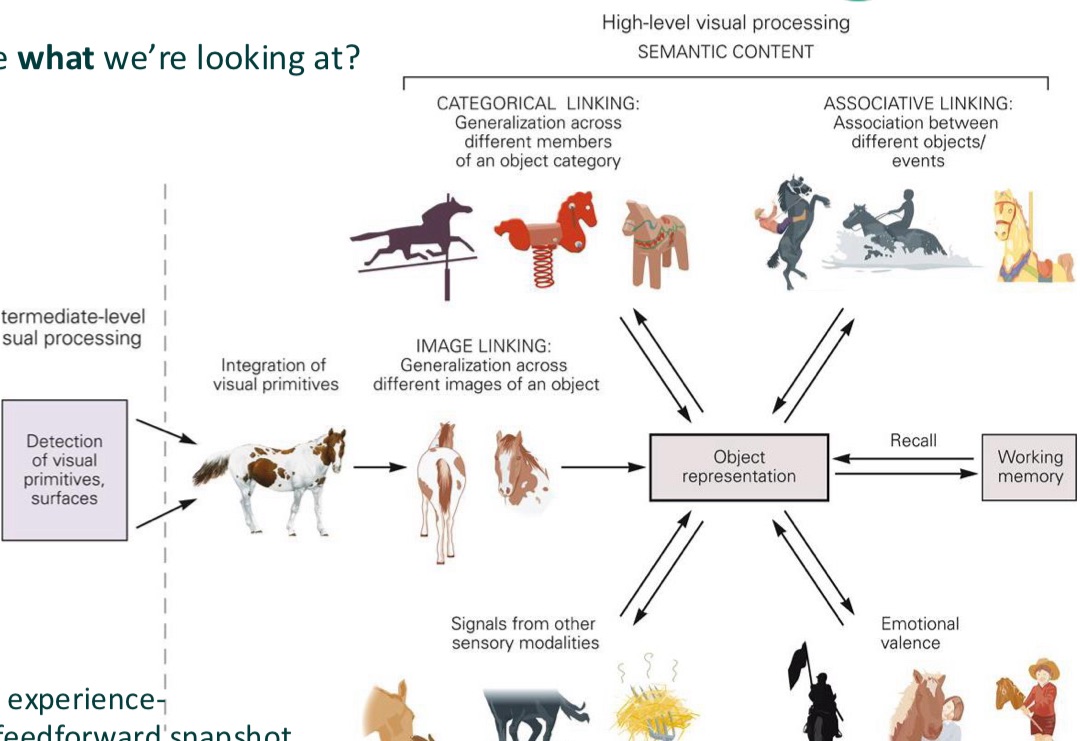
What does object recognition depend on
combining simple features into complex shapes
Matching input to stored memories
Feedback signals that refine and disambiguate perception
Experience shaping and tuning visual representations
→Object recognition is an active, experience-dependant process
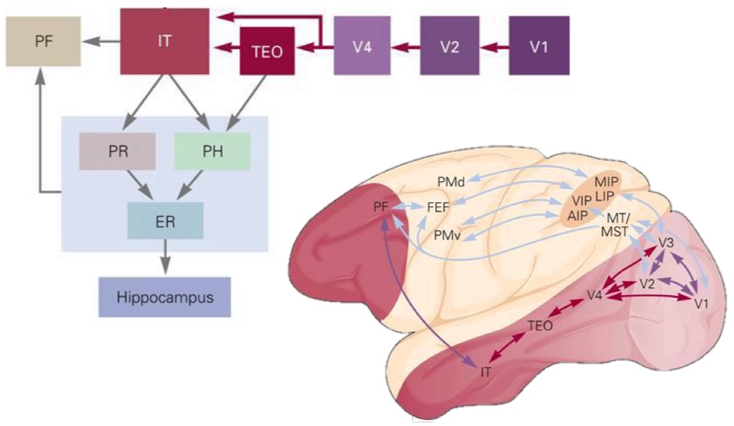
Where is object recognition
Relies on an interconnected network:
Inferior temporal cortex (IT) = recognising complex objects/shapes
Perirhinal cortex (PR) = links visual features to memory and object identity
Parahippocampal cortex (PH) = recognising scenes/places
Prefrontal cortex (PF) = decision making, categorisation, task goals
Factors that influence object recognition
environment = perceptual constancy - the visual system has to identify the same object under different environmental conditions
Intrinsic variability = categorical perception - objects within same category show some level of variability
Previous experience = associative recall - creation of object templates to compare new objects to
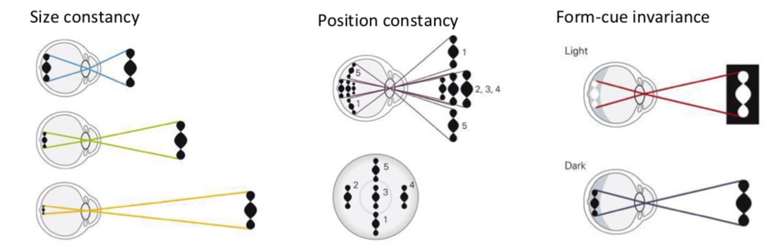
What is perceptual constancy
→ ability of our brains to see objects as stable and unchanging despite dramatic shifts in sensory info hitting our eyes
When is an object perceived to be the same
SIZE - The size of its image on the retina becomes larger or smaller due to the viewing distance
POSITION - The location in the retinal image changes
FORM-CUE - There are changes in reflectance e.g. changes in properties that define it (colour/texture)
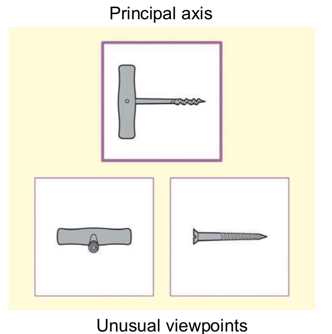
What is a viewpoint
structural descriptions: 3D memory representations of objects
Principal axis: usual viewpoint representation of an object
Right parietal lobe: sensitive to viewpoint
Left IT: insensitive to viewpoint
Two parallel routes for perceptual constancy
Categorical perception
ability to perceive different objects as the same category, even if they differ e.g. apples in a fruit bowl
Category-specific responses are common in neurons of the lateral prefrontal context, which receives direct info from IT
Perceptual learning (experience)
object recognition is modified by experience
Two forms of learning:
Implicit = improve at recognising objects without conscious effort
Explicit = consciously remember and identify new objects
Mediated by response properties of inferior temporal (IT) neurons
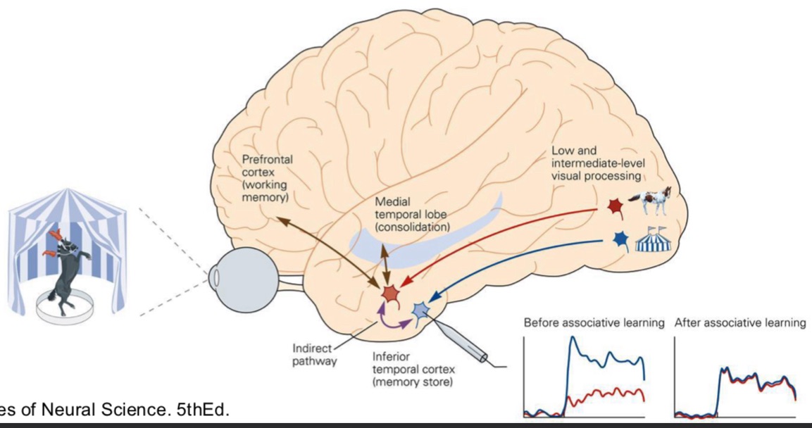
Associative recall
→ cognitive process of retrieving a memory triggered by a related cue or stimulus
contribution of inferior temporal cortex (IT) to object recognition can be modified by pervious experience
Consolidated by inputs from memory structures in the temporal lobe
Activation of working memory from the prefrontal cortex can also mediate this effect
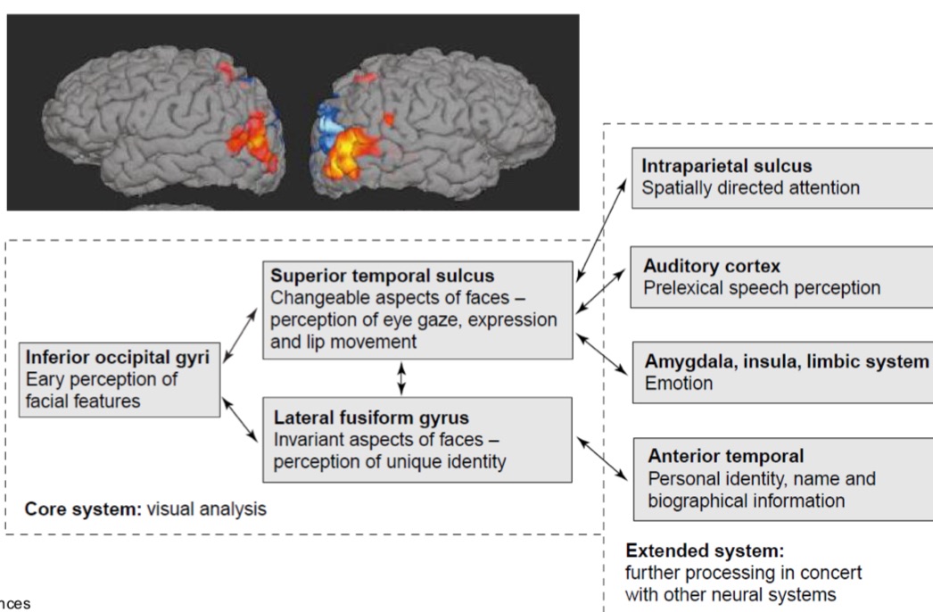
Face recognition
relies on different neural substrates to the recognition of other objects
System has to recognise the identity of a face, in addition to its changeable aspects (expresison)
Mediated by a distributed bilateral neural system located in the occipitotemproal extrastritae visual cortex
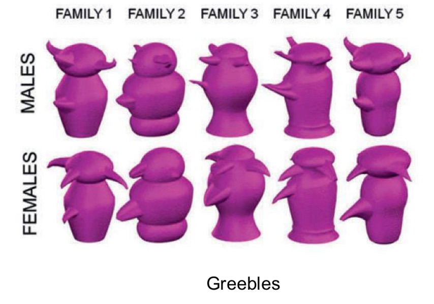
Greebles
face selective neurons also respond to other categories of objects e.g. artificially generated objects called Greebles
Visual awareness
regions involved in object perception become more active when we are aware of that object
Not one single brain area responsible
Activity evoked by a stimulus in a specific brain area is necessary, but NOT sufficient to generate a conscious perception of that stimulus
visual awareness results from distributed interaction between different lower-order (sensory) and higher-level associative regions
Different disorders of vision
Damage to different visual regions produces characteristic patterns of deficits: V1 → cortical blindness, blindsight
Dorsal stream → hemispatial neglect
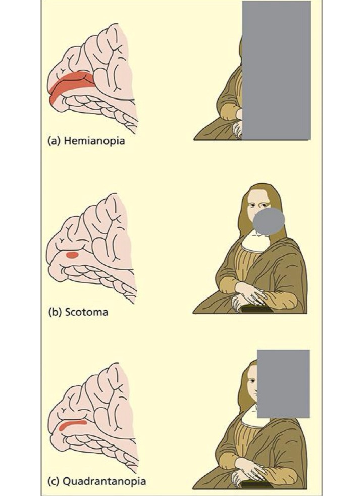
Damage to V1
Complete damage to one side of V1 = cortical blindness for one side of space (hemianopia)
Partial damage can lead to specific areas of blindness:
upper part of V1 = bottom part of space
Lower part of V1 = top part of space
→ quadantanopia
Small region of damage = scotoma
Disorders of V1
Anton’s syndrome
cortical blindness with anosognosia - complete unawareness of total visual loss in both hemifields
Lack of concern, denial and confabulation - ansodiaphoria
Bilateral lesions to V1 likely involving interruption of V1 from association cortices but no definitive cause
Blindsight
visual perception is preserved, but visual awareness is not
Dorsal stream - hemispatial neglect
visual scenes contain multiple elements that compete for limited attentional resources
Spatial attention is modulated by a dorsal attention network, influencing processing even in early visual areas
Key regions = posterior parietal cortex (PPC), front eye fields(FEF) and cingulate cortex
Damage to this network, particularly the right hemisphere, impairs attention to the contralateral (left) side of space
Neglect is not a visual deficit and can affect multiple sensory modalities
Ventral stream - prospagnosia
inability to recognise familiar faces or learn new ones
Other senses intact
May identify individuals based on voice, posture, smell etc
Able to match faces, distinguish between faces and determine sex, age etc.
Damage to the lateral occipital cortex and IT area