Bones of the axial skeleton
1/41
There's no tags or description
Looks like no tags are added yet.
Name | Mastery | Learn | Test | Matching | Spaced | Call with Kai |
|---|
No analytics yet
Send a link to your students to track their progress
42 Terms
What are the joints between the bones of the skull called? What do they do?
Sutures: provide very strong connections between the bones
What are the two major components that make up the skull?
Cranium: houses the brain
Facial bones
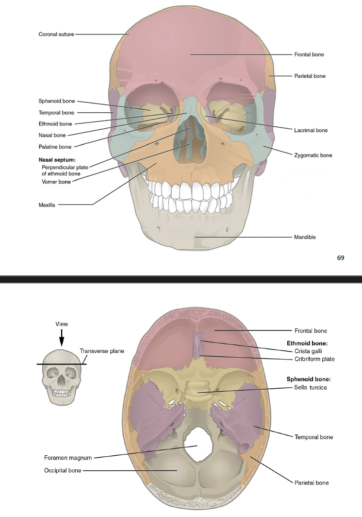
What are the four different sutures of the skull? Describe each one
Sagittal suture: joints the left and right parietal bones
Coronal suture: joins the frontal bone to the left and right parietal bones
Lambdoid suture: joins the occipital bone to the left and right parietal and temporal bones
Squamous suture: joins each temporal bone to the parietal and occipital bones

What are the facial bones? Describe each one?
Nasal bones: support the uppermost part of the nose
lacrimal bones: found at the medial part of the eye socket. They have lacrimal fossa to all tears to drain
Zygomatic bones: the posterior portion of the cheek
Palatine bones: the posterior portion of the bony palate of the mouth
Maxilla: forms the upper jaw, anterior face, and inferior eye sockets
Mandible: the lower jaw
Vomer: forms the posteiror inferior portion of the nasal septum and the superior, posterior part of the nasal cavity
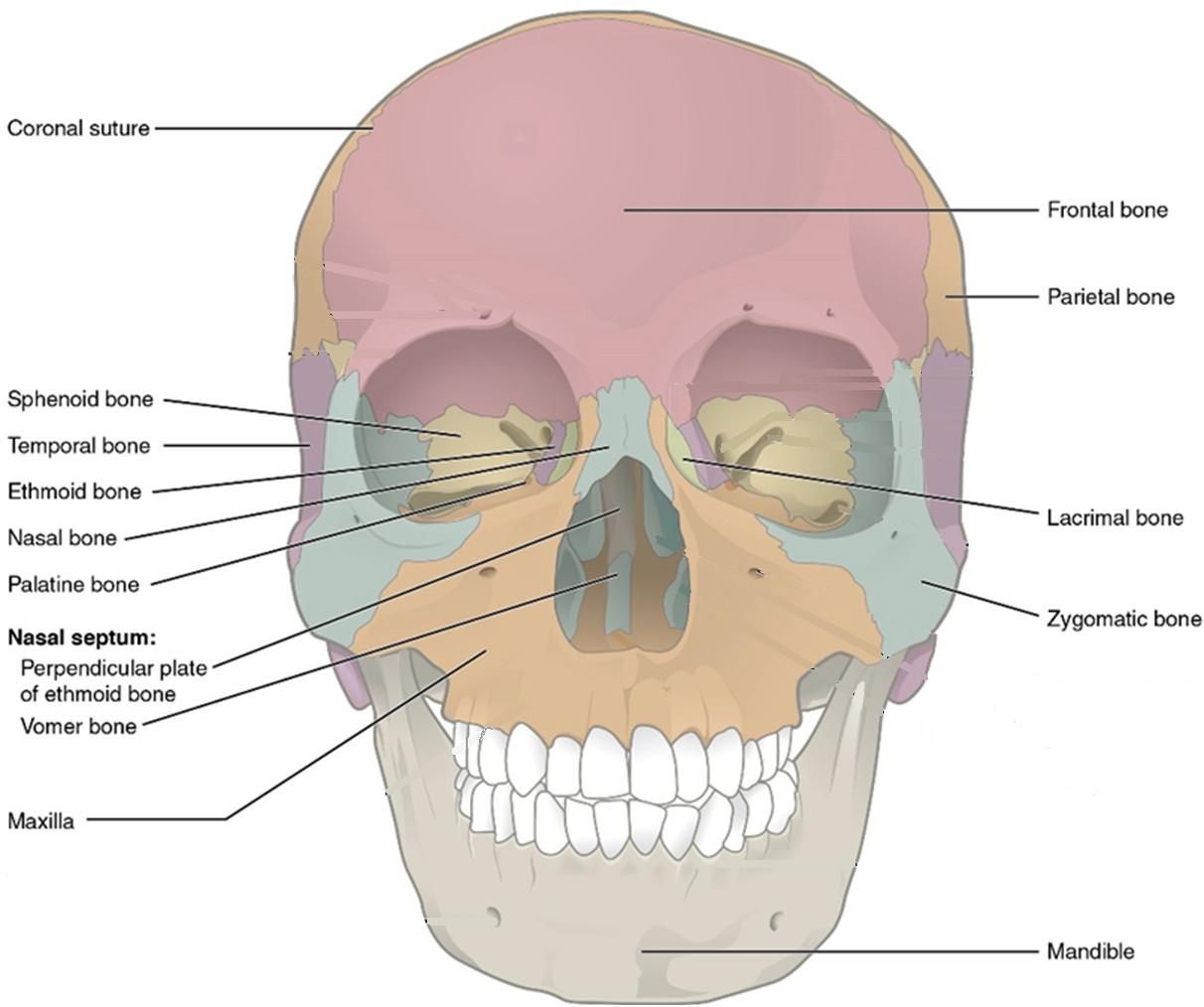
What are the 6 bones of the cranium? Describe them
Frontal bone: the anterior part of the cranium commonly called the forehead
Parietal bones: the two large plates that make up the top of the skull
Temporal bones: make up the sides of the skull above the ears
Occipital bone: makes up the back and the bottom of the skull
Sphenoid bone: a large bone that spans the width of the skull (across from one side of the ear to the other), visible from every direction. It makes up part of the floor of the cranium (brain cavity), the back of the eye sockets, your ‘temples’, and part of the inferior surface of the skull
Ethmoid bone: makes up a small, medial portion of the eye socket, a portion of the cranium anterior to the sphenoid, and the superior portion of the nasal septum

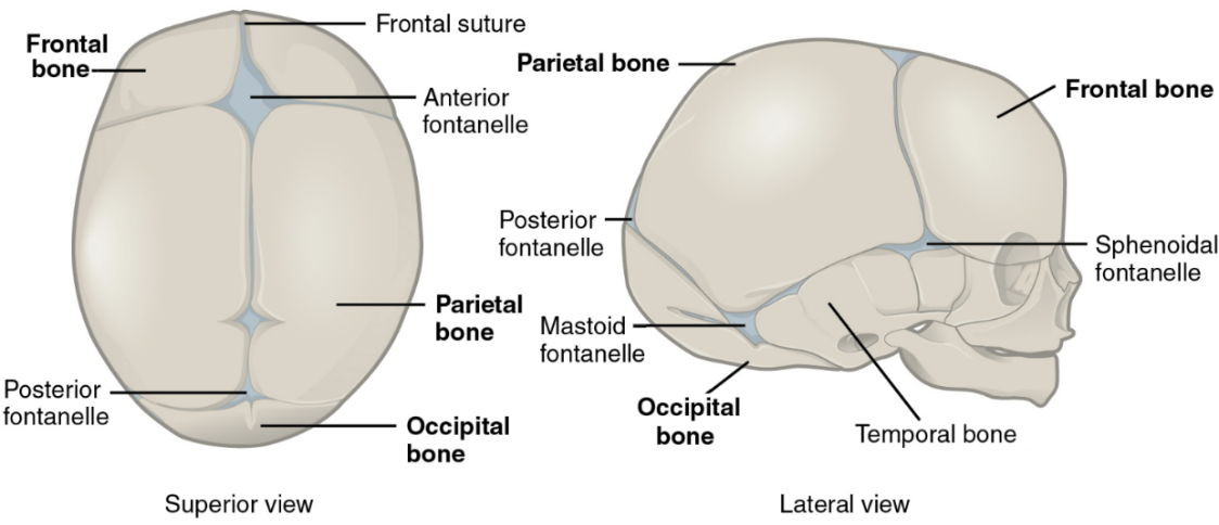
What are the ‘soft spots’ in a fetuses skull that allows for flexibility? What is the largest?
Fontanelles
Anterior fontanelle

In fetuses, the frontal lobe develops as a pair. What is the articulation between these two developing bones called?
Frontal suture
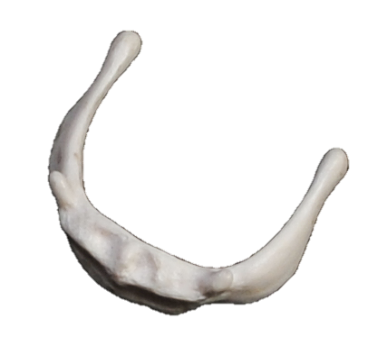
What bone is isolated deep to the mandible and provides structural support for some muscles of the tongue?
The hyoid bone
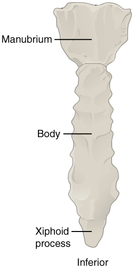
The ____ is a plate of bone at the front of the thorax. What are the three different portions?
Sternum is divided into the Manubrium, body, and the xiphoid process
How many ribs do humans have?
24 (12 pairs)
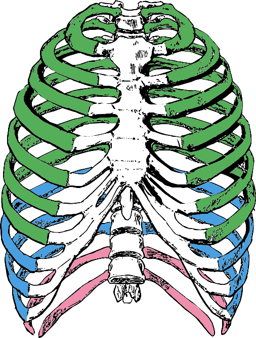
State and describe the three different types of ribs
True ribs: own sternal cartilage - first 7 pairs (hyaline cartilage connects to sternum)
False ribs: share a sternal cartilage - 3 pairs (share common cartilage)
Floating ribs: no sternal cartilage - last 2 pairs
What is the vertebral column compromised of?
24 vertebrae
sacrum
coccyx
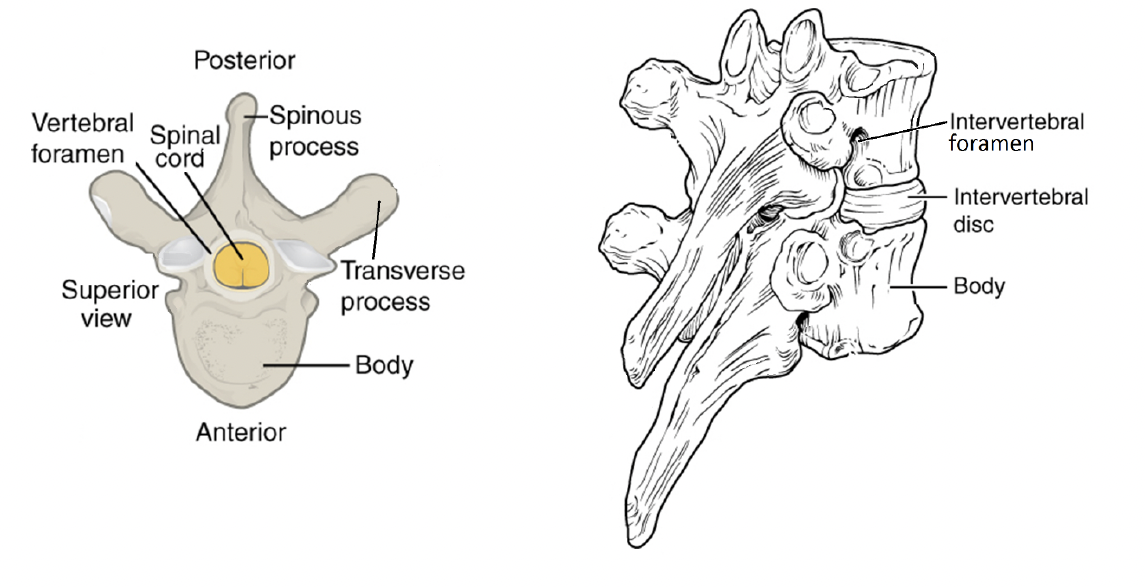
State and describe the common vertebral strucutes
Body: the supporting disc of bone at the anterior side of each vertebra
Vertebral foramen: the central hole through which the spinal cord passes
Transverse process: the pointed extensions to the left and right of the vertebra
Spinous process: the pointed extension toward the posterior. These are the bumps you can see through the skin of the back when you bend forward
Intervertebral foramen: gaps created on the lateral sides of the spinal cord by an upward notch in the posterior part of the bone. Spinal nerves exit here
Intervertebral disc: fibrocartilaginous pad that seperates vertebrae from one another, withstands compression, and provides a slight degree of motion
Transverse foramen: found only on cervical vertebrae, these are passages through the transverse processes for blood vessles
What are the three different types of vertebrae? how many are there?
Cervical: 7
Thoracic:12
Lumbar: 5
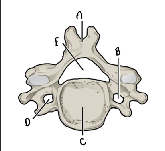
Name and label the bone
Cervical Vertebrae
A: Spinous process (bifid) - divided into two
B: Transverse process
C: Body
D:Transverse process
E: Vertebral foramn
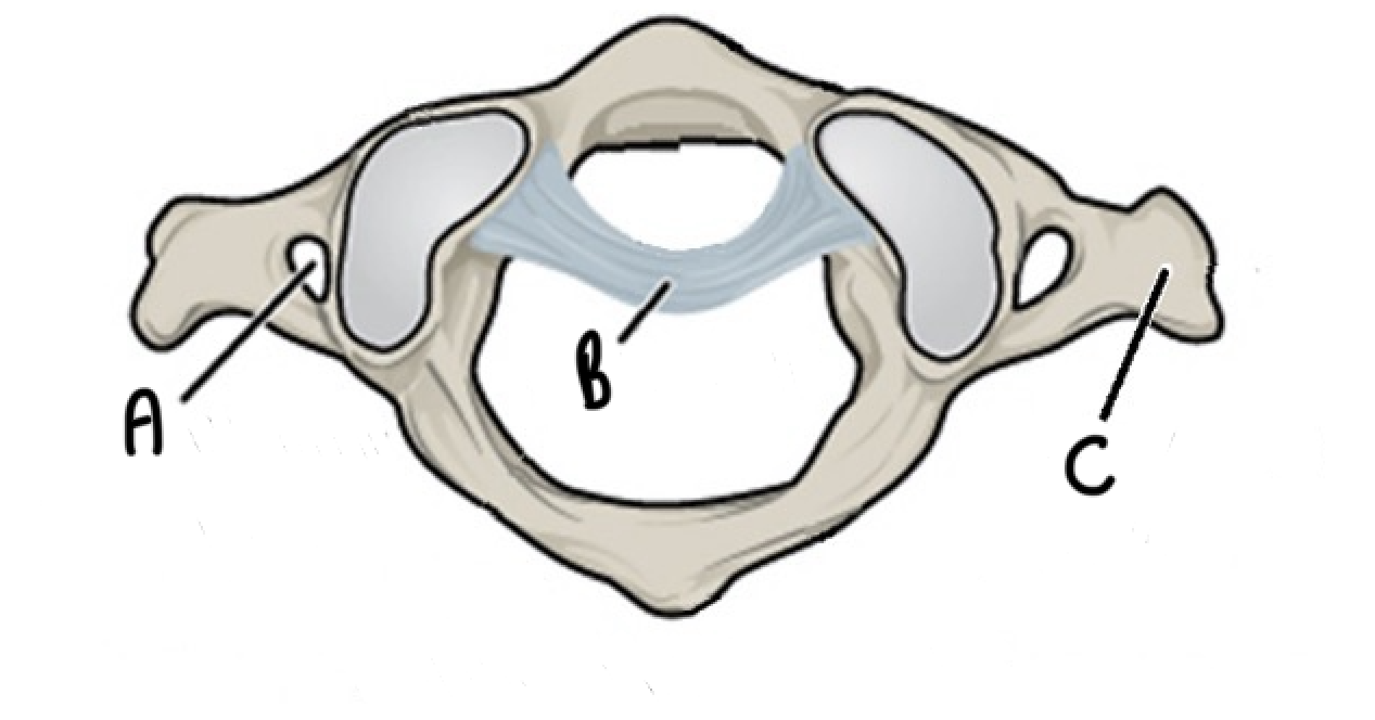
What vertebrae is this and label it
C1: atlas - allows nodding of head “yes”
A: Transverse foramen
B: Ligament
C: transverse process
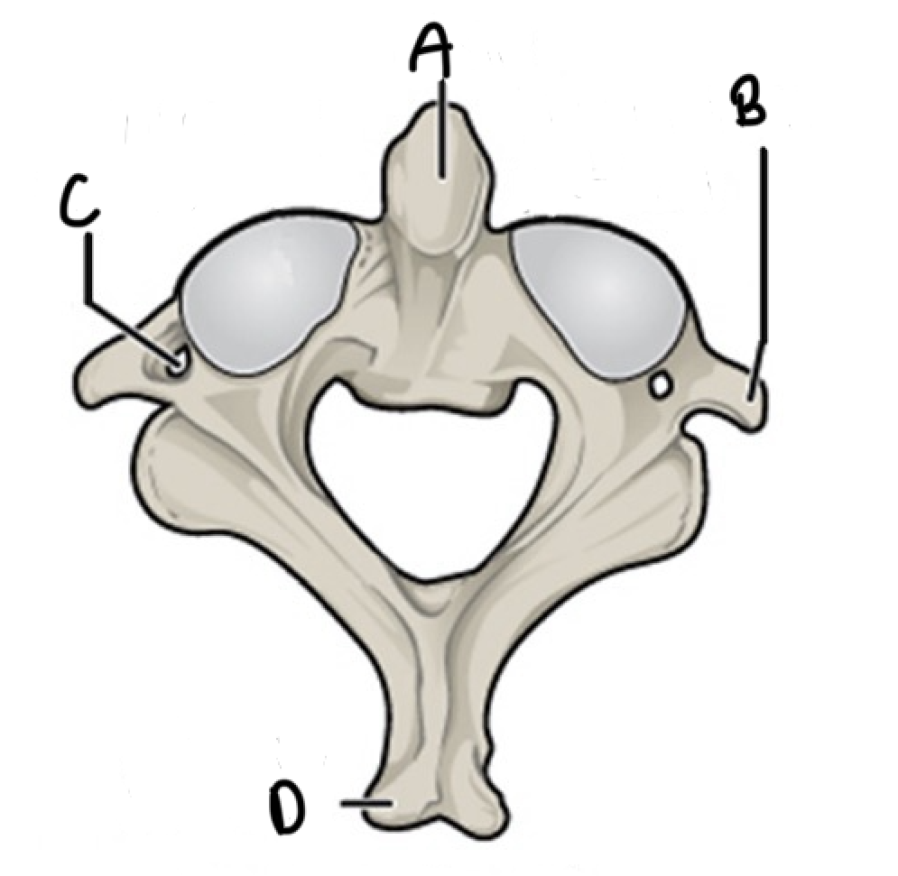
What vertebrae is this and label it
C2: axis - allows nodding of head '“no”
A: Dens (superior projection that allows rotation of the head to say “no”)
B: Transverse process
C: Transverse foramen
D:Spinous process
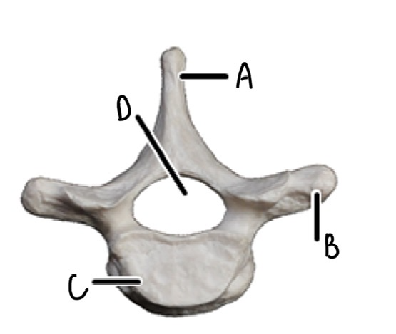
What vertebrae is this and label it
Thoracic vertebrae: each articulate with a pair of your ribs, and make up your thorax (heart shaped, long psine)
A: Spinous process
B: Transverse process
C: Body
D:Vertebral foramen
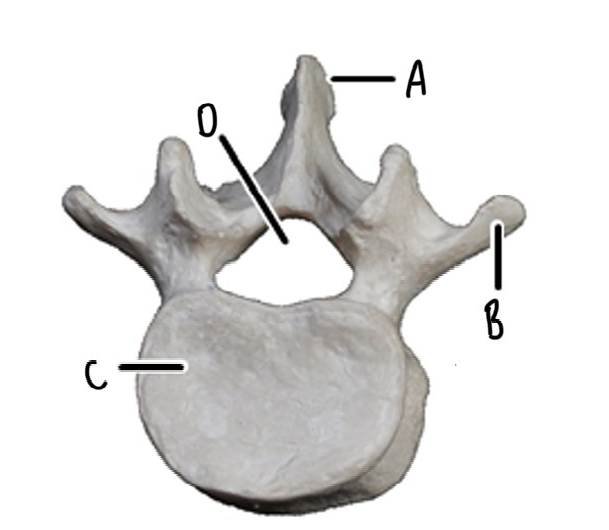
What vertebrae is this and label it
Lumbar Vertebrae: lower back (heave, bean shaped bodies)
A: Spinous process
B: Transverse process
C: Body
D:Vertebral foramen
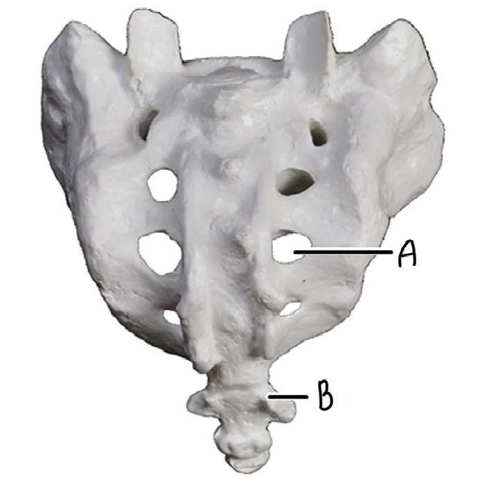
What is this and label it
Sacrum and Coccyx (tail bone)
A: Sacral foramen
B: Coccyx (4 fused, small vertebrae)
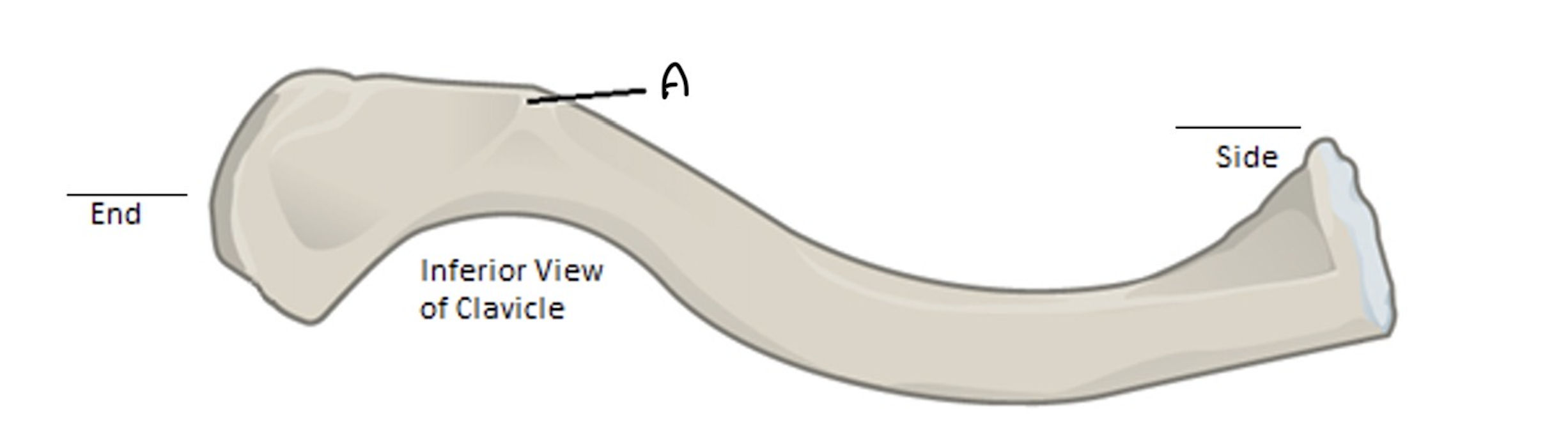
What bone is this? where does it attach too? Label it. What is the cone shaped formation on the inferior surface?
Clavicle: S-shaped bone that curves out from its blocky medial end that articulates with the sternum, then back toward the flattened end that articulates with the scapula.
What type of bone is the scapula?
An irregular bone (many different characteristics_
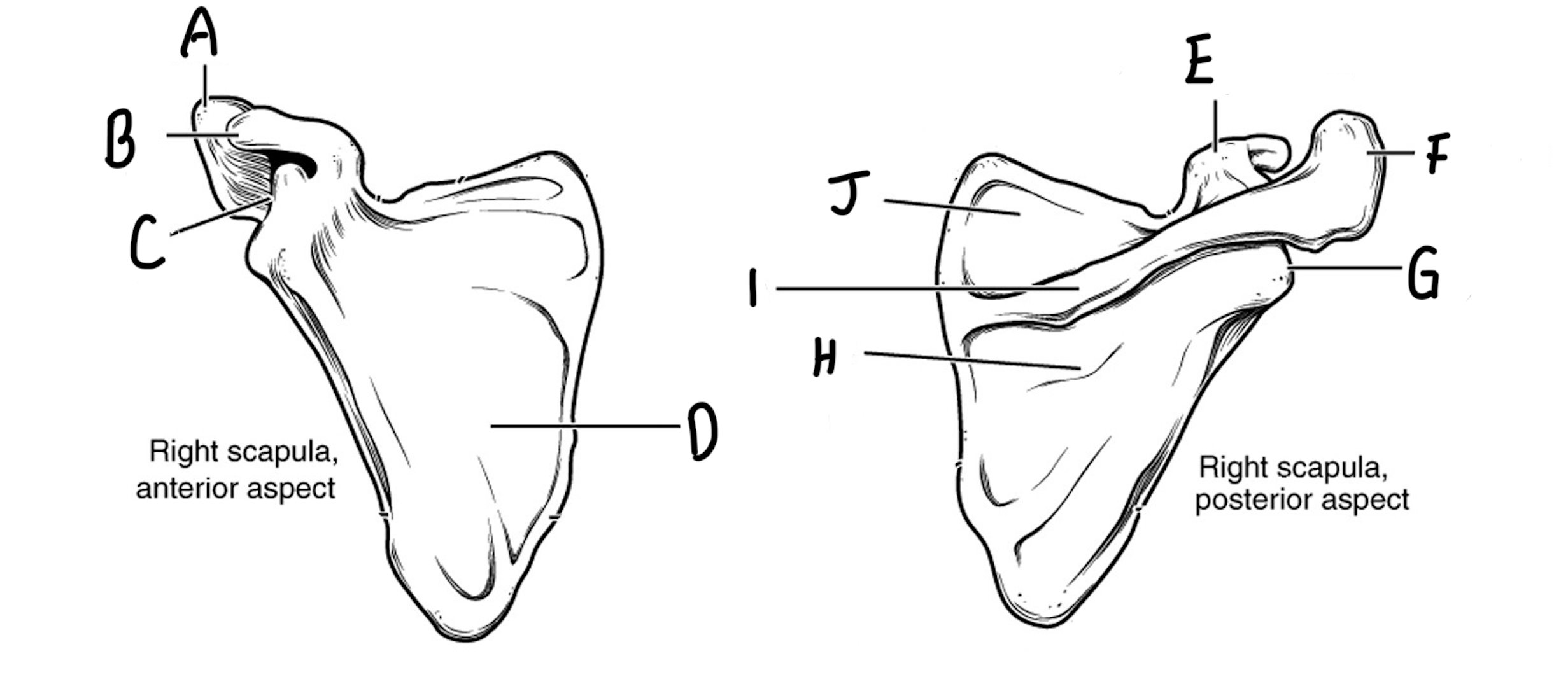
Please label the scapula and describe each bone marking
A: Acromion - larger, superior process that makes up the superior portion of the shoulder joint
B: Coracoid process - smaller, beak-shaped, anterior-facing process anterior & inferior to the acromion
C: Glenoid cavity - lateral-facing, shallow socket for the head of the humerus
D: Subscapular fossa - shallow depression on the anterior surface of the flat part of the scapula
E: Coracoid process
F: Acromion
G: Glenoid cavity
H: Infraspinous fossa - large, open depression inferior to the scapular spine
I: Spine
J: Supraspinous fossa - large depression superior to the scapular spine
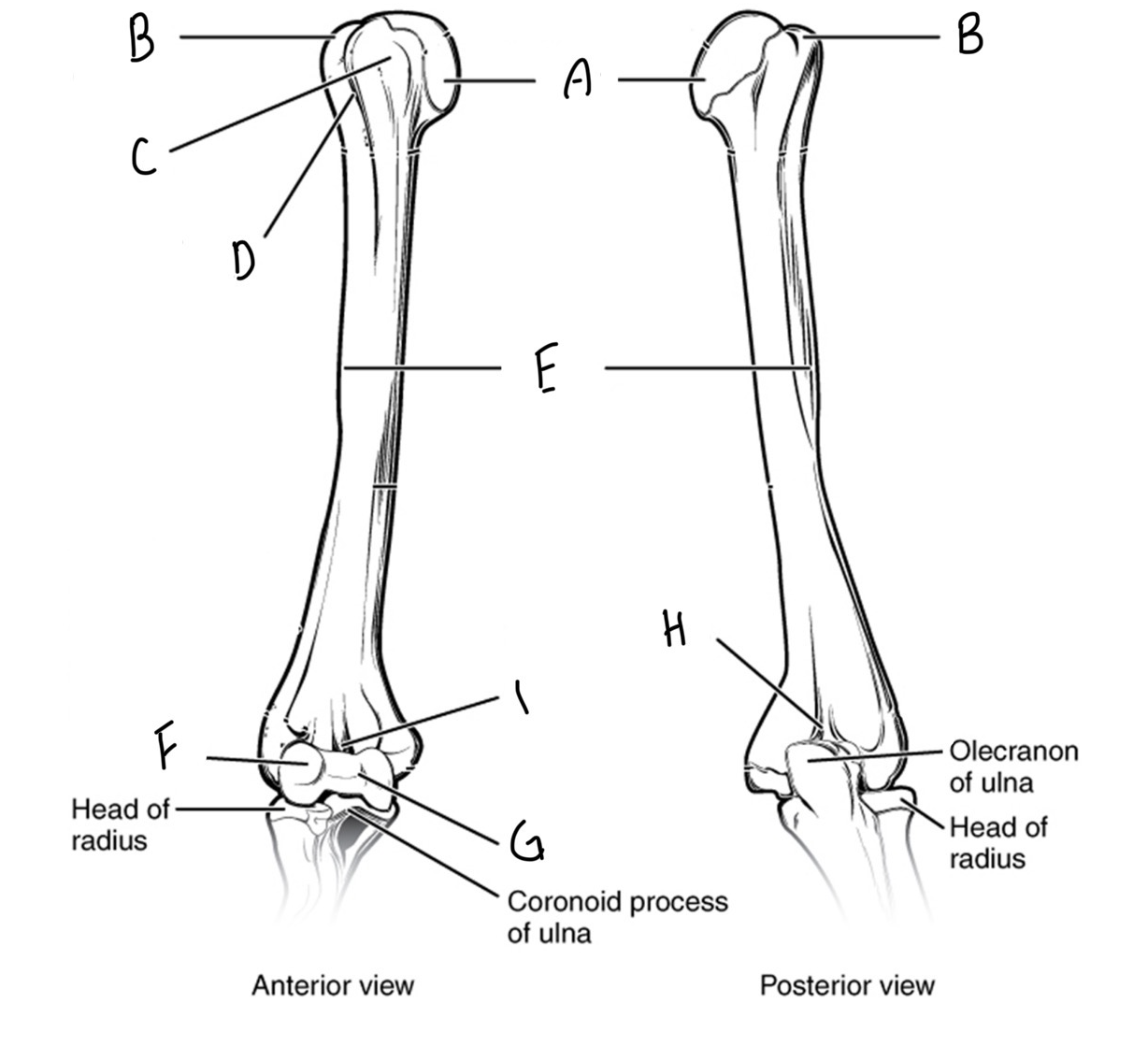
Please label the humerous and describe each bone marking
A: head - the large, smooth ball of the ball - and - socket joint of the shoulder
B: Greater tubercle - the larger, raised, roughened surface at the proximal end of the bone
C: lesser tubercle - the smaller, raised, roughened surface at the proximal end of the bone
D; Intertubercular sulcus - groove between the greater and lesser tubercules. Allows passage of tendon
E: Deltoid tuberosity - roughened area on the lateral mid-shaft. The deltoid muscle attaches here
F: Capitulum - the dome-shaped condyle on the lateral side of the dital end of the humerus. It is the articulation for the radius
G: Trochlea - the grooved condyle of the medial side of the distal end of the humerus. It is the aritculation point for the ulna
H: Olecranon fossa - large indentation on the posterior surface of the humerus, just superior to the trochlea
I: Coronoid fossa - small indentation on the anterior surface of the humerus, just superior to the trochlea
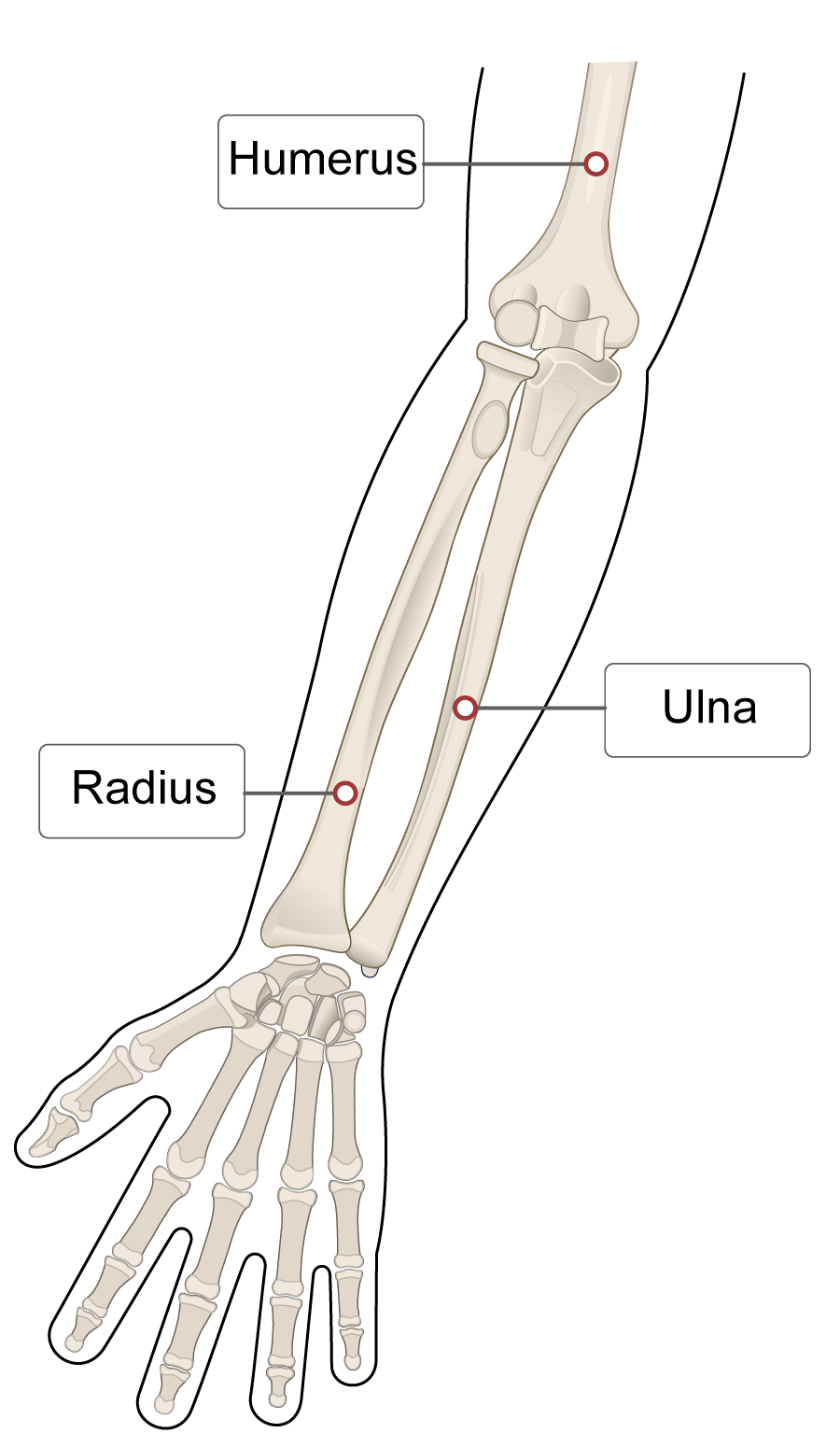
What side is the radius and ulna on?
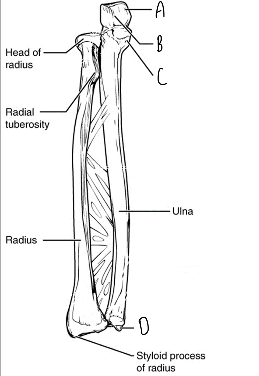
Label the bone markings of the radius
A: Head of Radius - smooth, circular surface on the proximal end of the radius
B: Radial tuberosity - Large, roughened surface near the proximal end of the bone. The biceps muscle attaches here
C: Styloid process - the small, pointed projection at the distal end
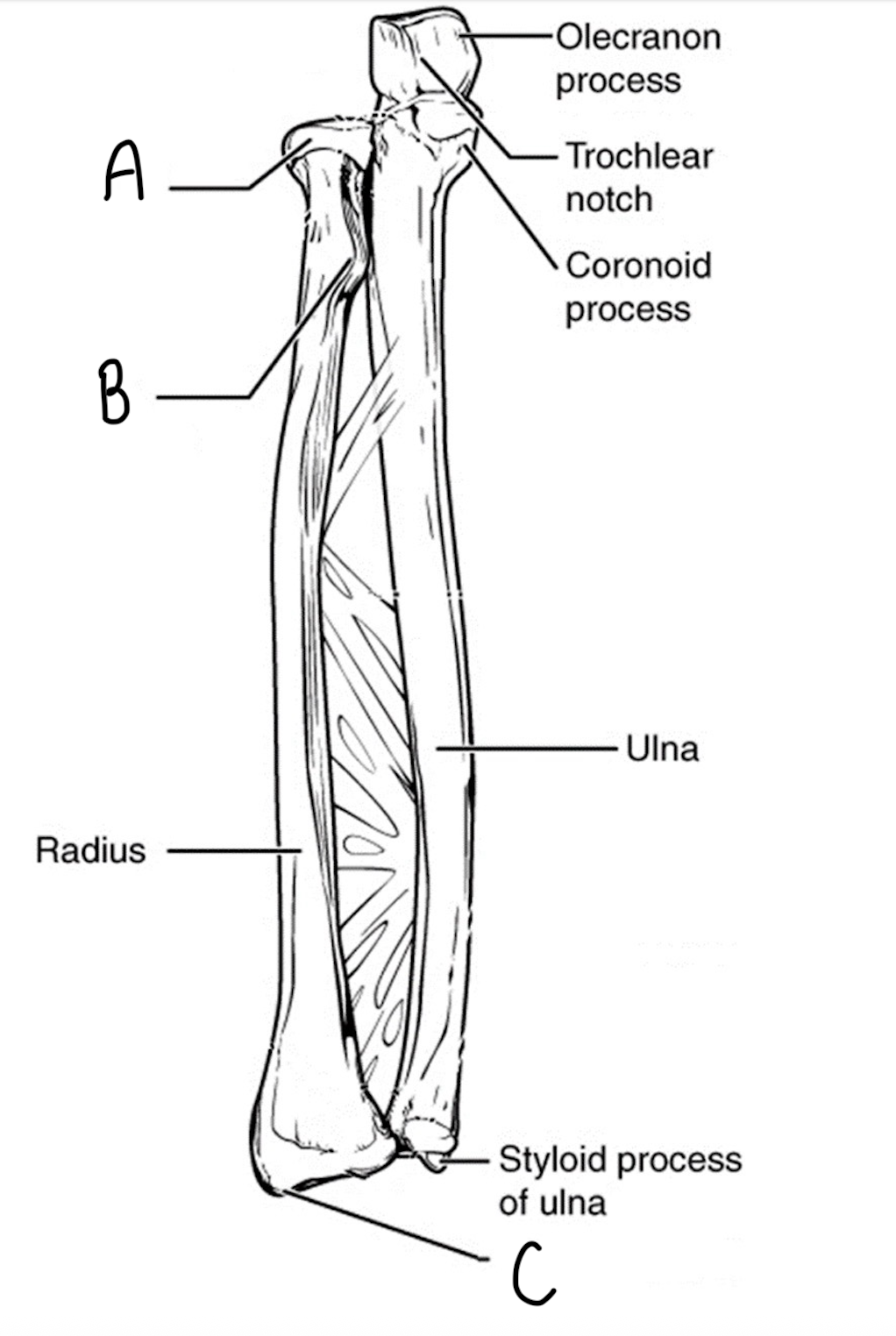
Label the bone markings on the ulna
A: Olecranon process - the large projection on the proximal end of the ulna
B: Trochlear notch - the large ‘U’ shaped depression in the proximal head of the ulna
C: Coronoid process - the smaller projection on the anterior surface of the ulna, just inferior to the trochlear notch
D: Styloid process - the small pointed projection on the distal end of the ulna
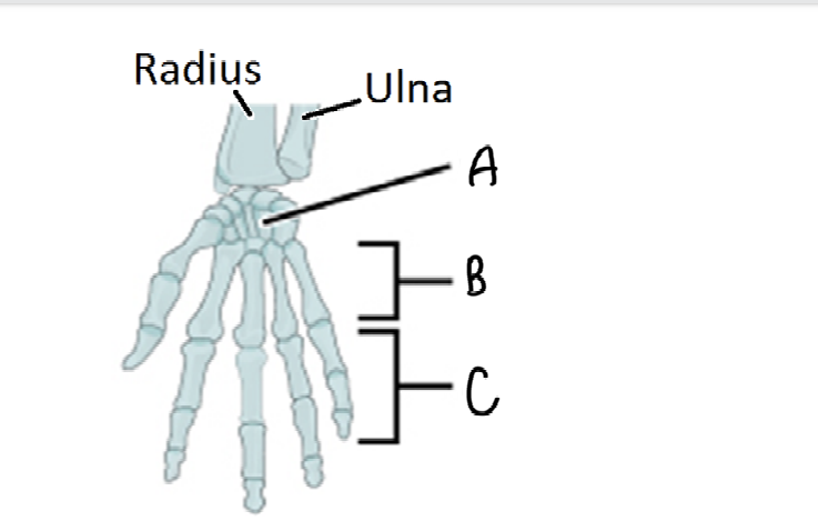
Label the bones of the wrist and hand
A: Carpals (8 bones)
B: Metacarpals
C: Phalanges
What is the pelvic girdle made up of?
The sacrum and the bones of the hip called the os coxae
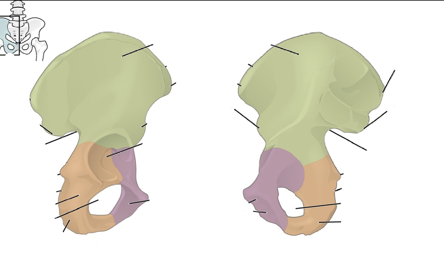
What is the os coxae made up of? Which colour is which?
Three pelvic bones:
ilium (green) - the superior portion of the os coxae. This is the bone most people consider their “hip bones”
ischium (orange) - the posterior, inferior portion of the os coxae
pubis (purple) - the anterior and medial portion of the os coxae
What are the structures that straddle bones in the os coxae? Describe them
Acetabulum - large socket for the articulation of the femoral head
Obturator foramen - large oval hole in the anterior pelvis
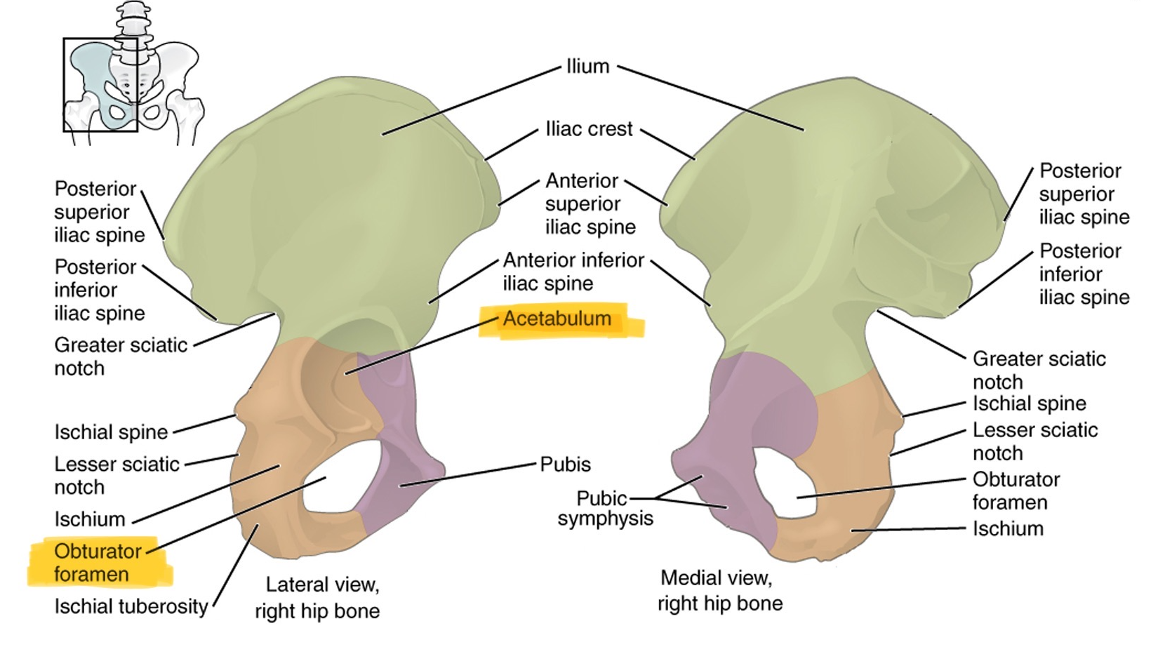
What structure is found on the pubis?
The pubic symphysis - fibrocartilaginous articulation between the pubis of the left and right os coxae

What structures are found on the ilium?
Iliac crest - the superior margin of the ilium
Anterior superior iliac spine - projection at the top, front of the ilium
Anterior inferior iliac spine - projection at the bottom, front of the ilium
Posterior superior iliac spine - projection at the top, back of the ilium
posterior inferior iliac spine - projection at the bottom, back of the ilium
Greater sciatic notch (half heart shaped) - large indentation just inferior to the posterior inferior iliac spine

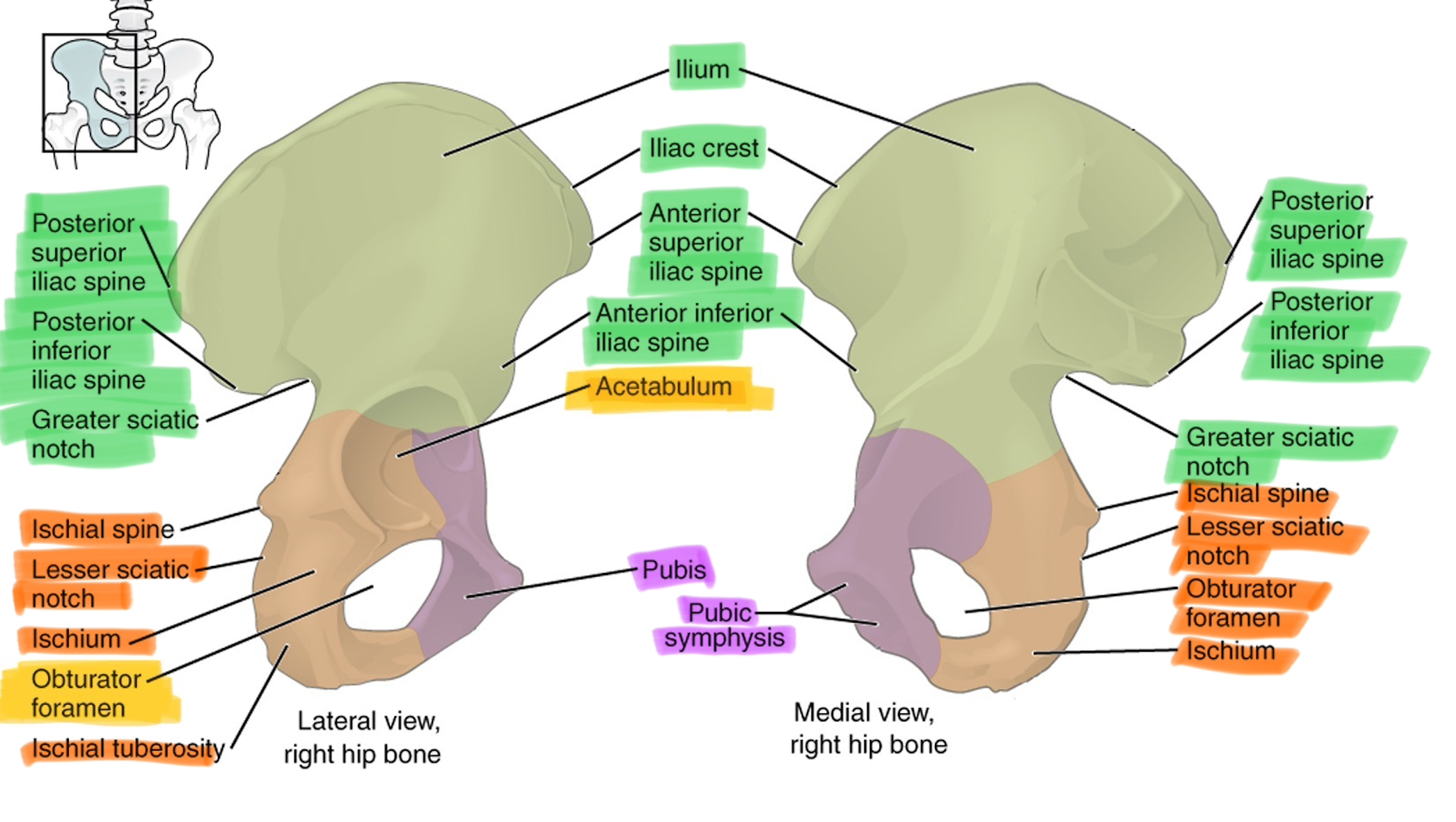
What structures are found on the ischium?
ischial spine - particularly large spine inferior to the greater sciatic notch (small bump under sciatic notch
Lesser sciatic notch - small indentation inferior to the ischial spine
Ischial tuberosity - large roughened area at the most inferior point of the pelvis when sitting
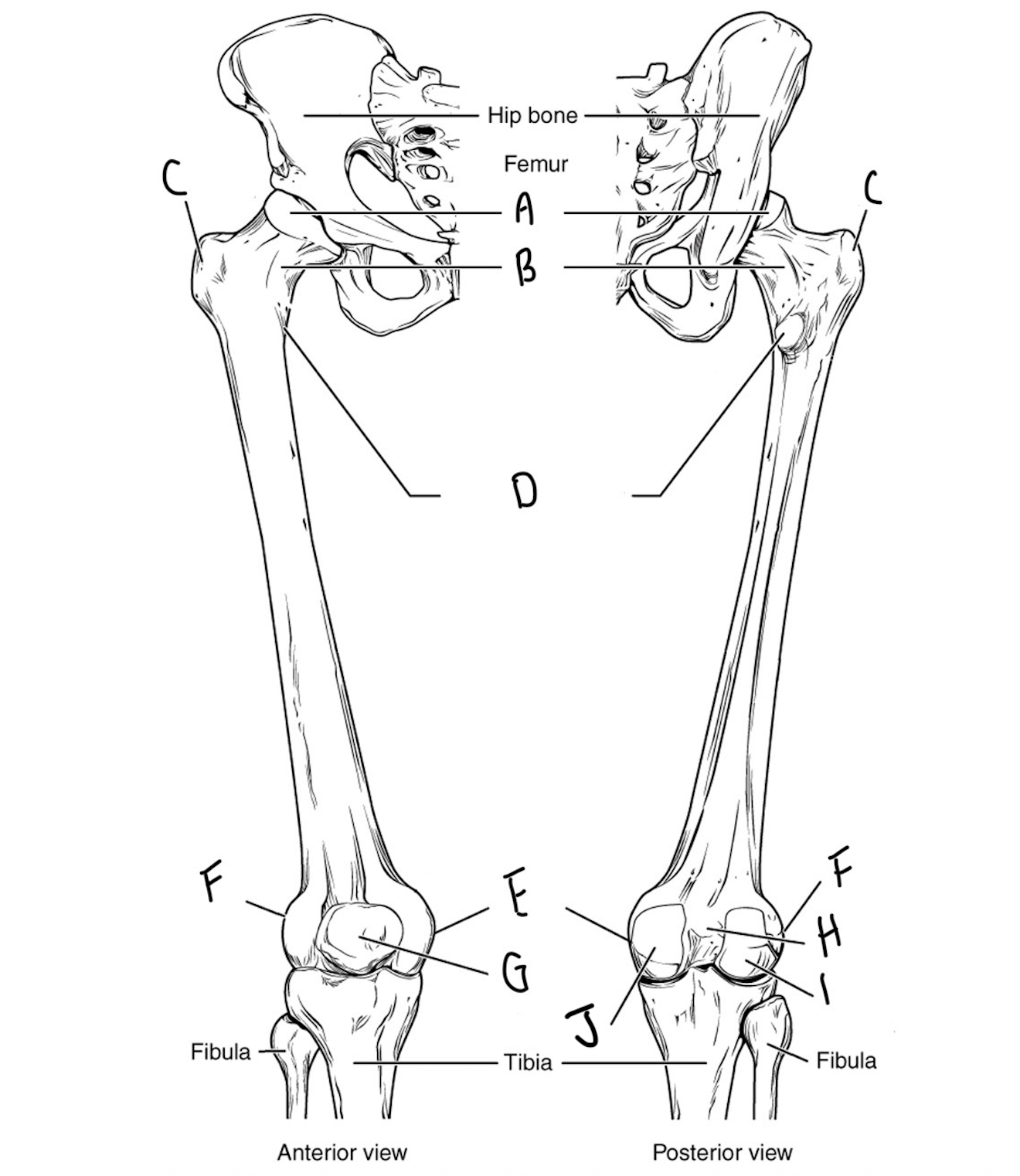
Label the femur and describe each bone marking
A: Head - The large ball-like joint on the proximal end of the bone. Fits into the acetabulum
B: Neck - the narrow connection between the head and the diaphysis
C: Greater trochanter - very large, rough protrusion on the proximal, lateral portion of the bone
D; Lesser trochanter - much smaller and more pointed process on the posterior proximal end of the bone
E: Medial epicondyle - The roughened surface just superior to the medial condyle
F: Lateral epicondyle - The roughened surface just superior to the lateral condyle
G: Patella (knee cap) - largest sesamoid bone with a slight point on the inferior side and a smooth articular surface at the posterior
H: Intercondylar fossa - the indentation between the condyles
I: Lateral condyle - the smooth, distal, rocking surface on the opposite side from the head
J: Medial condyle - the smooth, distal, rocking surface on the same side as the head
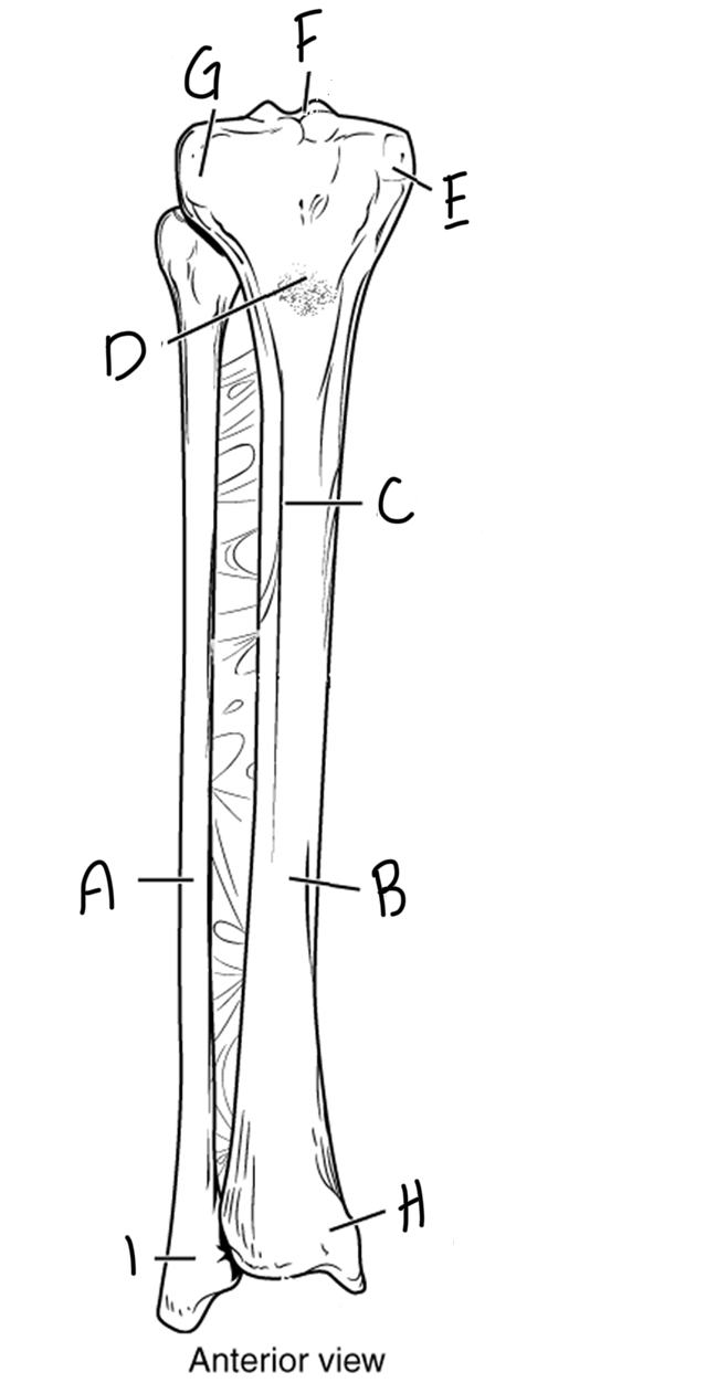
Label and identify the bone markings on the tibia and fibula
A: Fibula - long, slender bone that supports the lateral side of the ankle
B: Tibia - primary support of the lower leg. Has a triangular cross-section that provides resistance to strain from multiple directions
C: Anterior border - the sharp crest down the anterior side of the tibia, also called the tibial crest
D; Tibial tuberosity - a large, very rough, raised area just inferior to the condyles of the tibia. A large portion of the muscles of the thigh attach here
E: Medial condyle - platform-like, smooth articular surface located on the same side of the bone as the medial malleolus
F: Intercondylar eminence - sharply raised, two peaked projection between the condyles. It fits into the intercondylar fossa of the femur
G: Lateral condyle - platform-like, smooth articular surface located on the side opposite of the medial malleolus
H: Medial malleolus - a projection down the medial side of the tibia that creates part of the ankle joint
I: Lateral malleolus
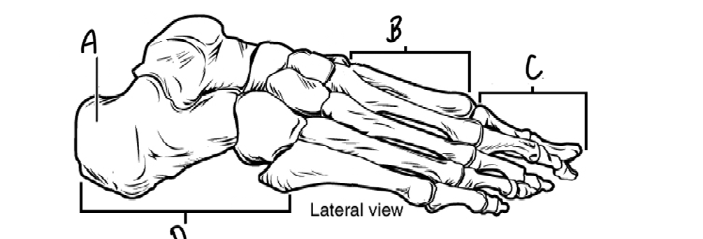
Label the bones of the ankle and foot
A: Calcaneus - the largest of the tarsals
B: Metatarsals
C: Phalanges
D; Tarsals
What bones are found on the temporal bones?
External acoustic meatus: outside opening into the auditory (hearing) canal - E for ear
Styloid process: small, very sharp ‘pen-like’ process on the inferior surface of the skull
Mastoid process: large, rough, cone-shaped process posterior and lateral to the styloid process

What bones are found on the occipital bone?
Foramen magnum: large opening in the bottom of the occipital bone. The spinal cord passes through it
Occipital condyles: small, smooth, oval surfaces that allow the head to rock on the spinal column as in nodding ‘yes’
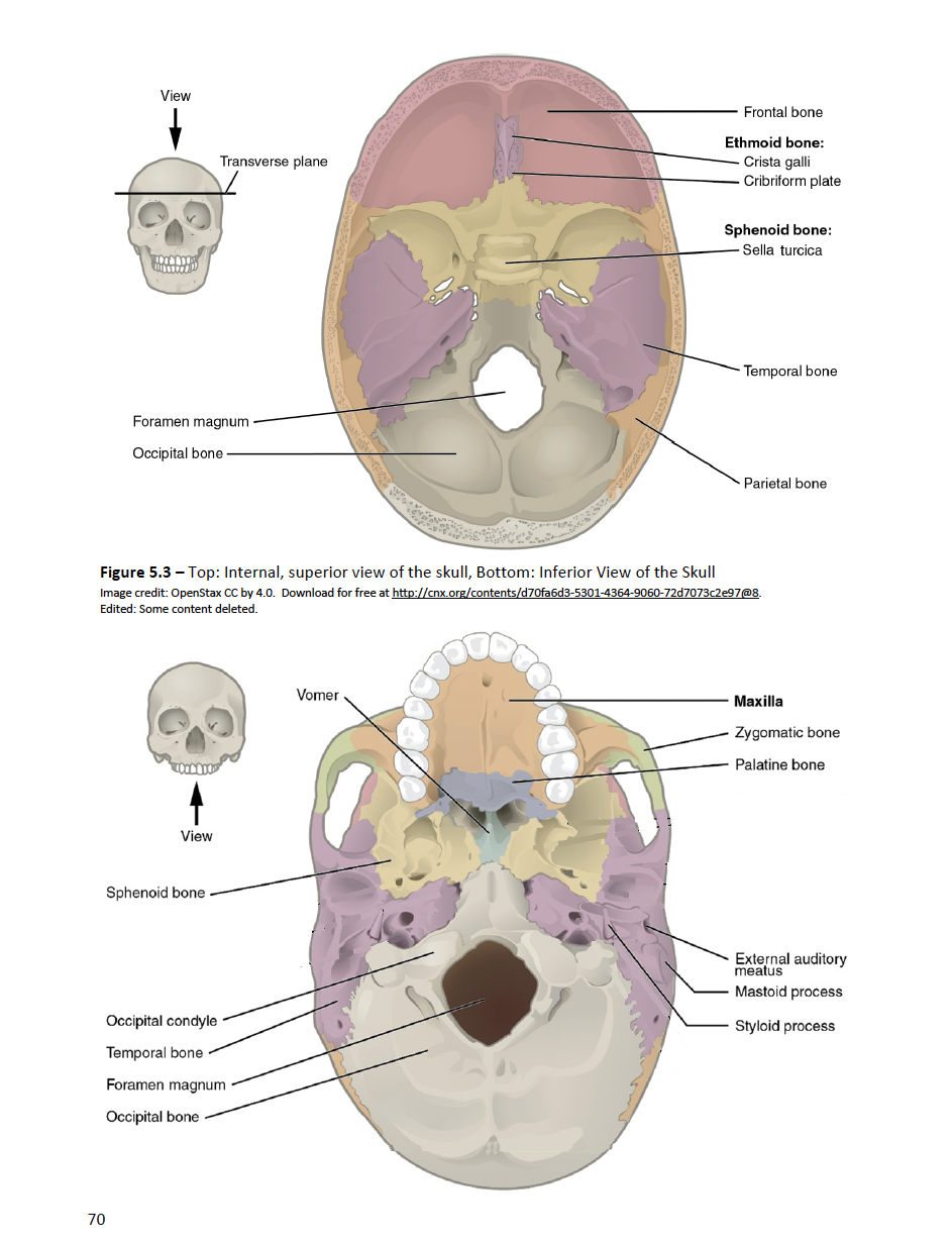
What bones are found on the sphenoid bone?
Sella turcia: a large, saddle-shaped depression inside the cranium. It houses the pituitary gland
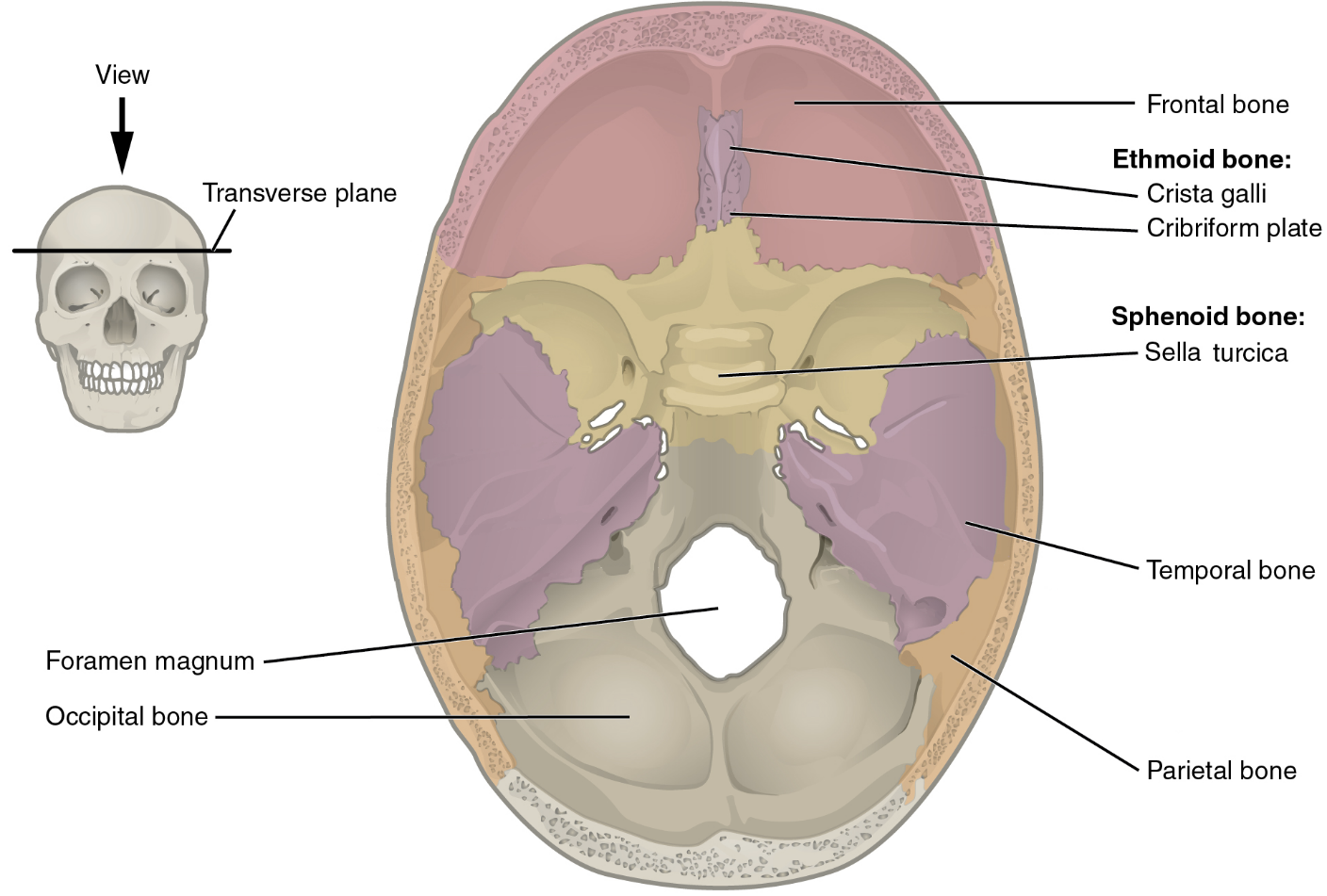
What bones are found in the ethmoid bone?
Crista galli: a raised crest inside the anterior cranium
Cribriform plates: An indented portion of the cranium floor
Cribriform foramina: small holes in the cribriform plate that allows the olfactory (smell) nerves to pass
Perpendicular plate: the anterior, superior part of the nasal septum