Medulla 1/2
1/42
There's no tags or description
Looks like no tags are added yet.
Name | Mastery | Learn | Test | Matching | Spaced | Call with Kai |
|---|
No analytics yet
Send a link to your students to track their progress
43 Terms
Describe the Medulla Oblongata
Structure?
Derived from?
Contains?
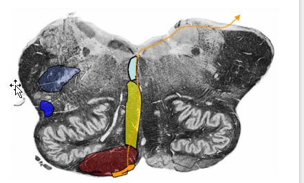
Most caudal brainstem, conical in shape
Derived from the Rhombencephalon
Contains primary nuclei for autonomic control of respiration, heart rate, blood pressure.
What two long tracts decussate in the Medulla Oblongata
two distinct long tract
dorsal column-medial lemniscal system
corticospinal tract
Describe the boundaries of the Medulla Oblongata
Boundaries:
Caudal Ventral Boundary: pyramidal decussation
Rostral Boundary: Obex
No definitive rostral ventral boundary or caudal boundary exist
Closed portion of the medulla containing the central canal.
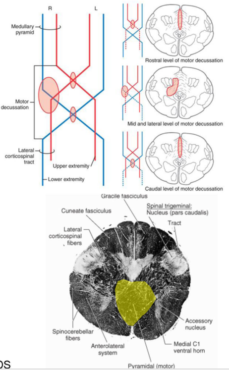
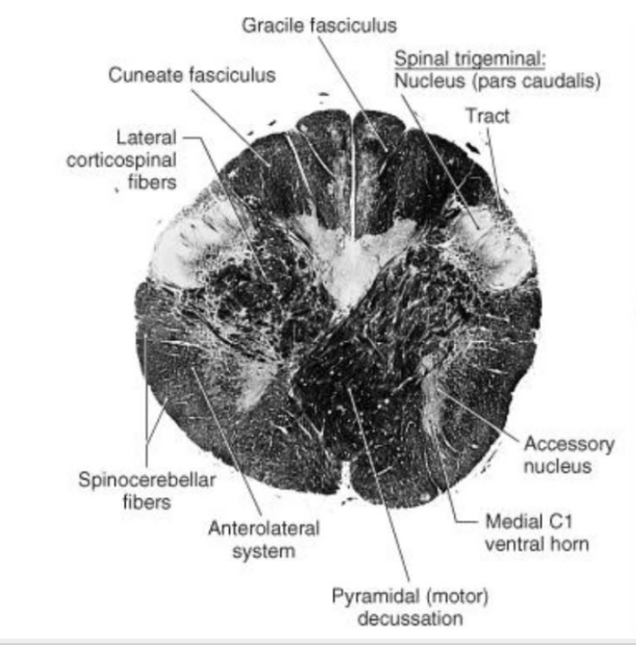
Describe the Pyramidal (Motor) Decussation
Location
Fiber Destiy
Somatotopy of Decussation
Location:
ventral surface of the medulla; disruption of the anterior median fissure
Fibers Destiny Here:
80-90 percent of corticospinal tract fibers decussates (L CST)
8% descend ipsilaterally → anterior funiculus → decussate segmentally (Anterior CST)
2% descend ipsilaterally to contribute to the lateral corticospinal tract (tract of Barnes)
Somatotopy of Decussation:
Upper Extremity fibers: Rostrally
Lower Extremity fibers: Caudally
THUS: iscrete lesions in the pyramidal decussation may produce different and somewhat unusual patterns of weakness.
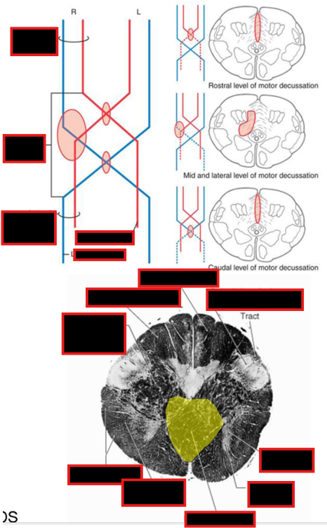
What happens @ the level of Pyramidal Decussation (2) (besides CST decussation)
@ Level Pyramidal Decussation:
Fibers within fasciculus gracilis + fasciculus cuneatus synapse @ Nucleus Gracilis and nucleus cuneatus, respectively.
spinal trigeminal nucleus (pars caudalis) + tract take place of gray matter of dorsal horn and tract of Lissauer, respectively.
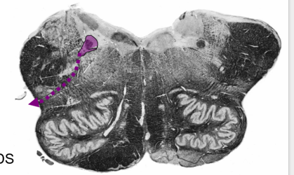
Describe the Spinal Trigeminal Nucleus and Tract
Location
Receives
Pathway of Afferent?
Describe the tuberculum cinereum (or trigeminal tubercle)
Arise from?
Location?
Describe the 3 parts of Spinal Trigeminal Nucleus and Tract
Location
Function
Describe the Somatotopic Organization of the Medulla
Spinal Trigeminal Nucleus and Tract
Location:
within the lateral medulla
Extends caudal pons → upper cervical spinal cord.
Receives:
GSA conveying from Face/External Ear
Pain
Temperature
Crude Touch
Pathway of Trigeminal Afferents:
Enters Brainstem @ level of CNV → Descends in Spinal Trigeminal Tract (located lateral to spinal nucleus) → synapse @ Trigeminal Nucleus along its rostral caudal extent
tuberculum cinereum (or trigeminal tubercle):
Arise from spinal trigeminal nucleus and tract
Located lateral to the cuneate tubercle and tract
3 parts of Spinal Trigeminal Nucleus and Tract
Pars Oralis
Location: rostral pole of hypoglossal N. → caudal end of principal sensory nucleus (Pons))
Function: Tactile input (crude touch)
Pars Interpolaris
Location: level of obex → rostral pole of hypoglossal nucleus)
Function: Dental pain
Pars Caudalis
Location: (C3 → obex)
Function: Pain and temperature
Somatotopic Organization:
V1 synapses ventrally
V2 and V2 synapses progressively more dorsal.
***NOTE: Sensory fibers from the external ear travel in CN VII, IX, and X to synapse in the dorsal most part of the caudal subnucleus (Pars Caudalis)***
Onion Skin Pattern of Somatotopic Organization
Nociceptive afferents from the circumoral (around mouth) region: rostral part of the pars caudalis
More Posterior/Lateral Parts: progressively more caudal parts of the caudal subnucleus
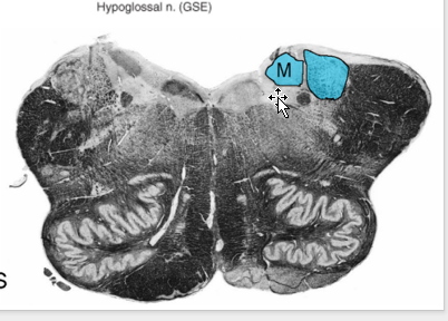
Describe the Accessory Nucleus @ level of Pyramid Dessucation
Location
Route
Innervates
Cortical Input to Neurons
Accessory Nucleus @ level of Pyramid Dessucation:
Location of GSE from Accessory Nucleus:
lateral portion of the ventral horn
SC +medulla junction → C5/6
Route of Rootlets:
exit to form a trunk → ascends through foramen magnum
Innervates: trapezius and SCM.
Cortical Input to Neurons:
To trapezius is crossed
To SCM is ipsilateral
NOTE: not all sources are in agreement
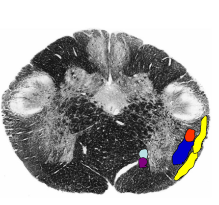
@ level of pyramidal dessucation, What happen to these other tracts:
Medial Long. Fasciculus
Ant./Post. Spinocerebellar Tract
Anterolateral system (spinal lemniscus)
Rubrospinal tract
Other Tracts:
Medial Long. Fasciculus (descending limb - medial vestibulospinal tract) and Tectospinal Tract:
Pushed laterally by the decussating CST fibers.
Ant./Post. Spinocerebellar Tract:
Retain position similar to SC
Anterolateral system (spinal lemniscus)
medial to spinocerebellar tracts;
Retain position like SC
Rubrospinal tract
associated w/ ALS in caudal brainstem.
Retains similar position in the spinal cord.
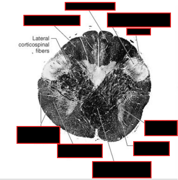
Describe Sensory Decussation of the Dorsal Columns:
Pathway? Locations?
What are the names of the fibers decussating?
Sensory Decussation of Dorsal Column
Dorsal Columns → ascend to caudal medulla → synapse @ gracile/cuneate nucleus
Location: deep to gracile tubercle (clava) and cuneate tubercle
dorsal column nuclei → Decussates @ tegmentum → medial lemniscus → ascend to thalamus (VPL).
Fibers Decussating = internal arcuate fibers
Fibers ascending in medial lemniscus = somatotopically organized
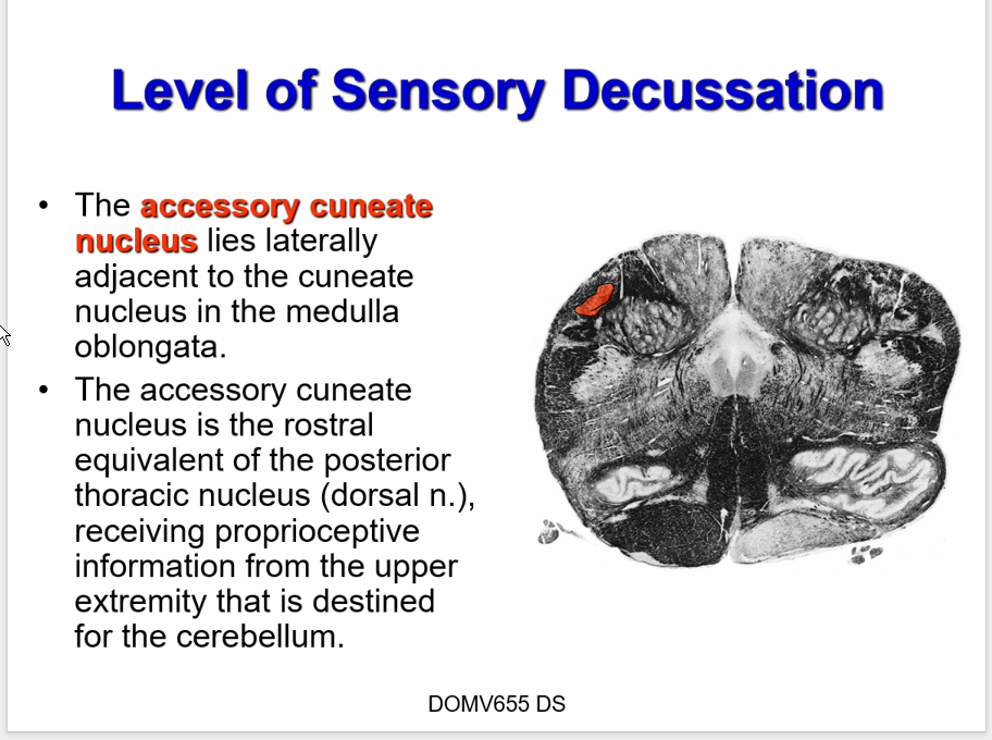
Describe what happens to Accessory Cuneate Nucleus: @ level of Sensory Decussation
Location
Equivalent
Function
Pathway of Fibers
Accessory Cuneate Nucleus:
Location:
Lateral to cuneate nucleus
Equivalent:
rostral equivalent of the posterior thoracic nucleus (AKA, Clarke’s Column) (Dorsal N.)
Function:
proprioceptive information from upper extremity that is destined for the cerebellum
Pathway of Fibers:
Large diameter afferents (proprioceptive + exteroceptive info) → cervical/Upper thoracic (perhaps) → ascend in fasciculus cuneatus → synapse @ Accessory (lateral,external) cuneate nucleus (instead of the posterior thoracic nucleus) → cuneocerebellar tract → cerebellum via inferior cerebellar peduncle (restiform body)
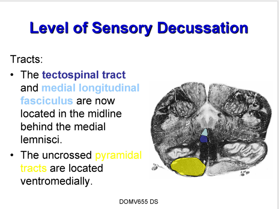
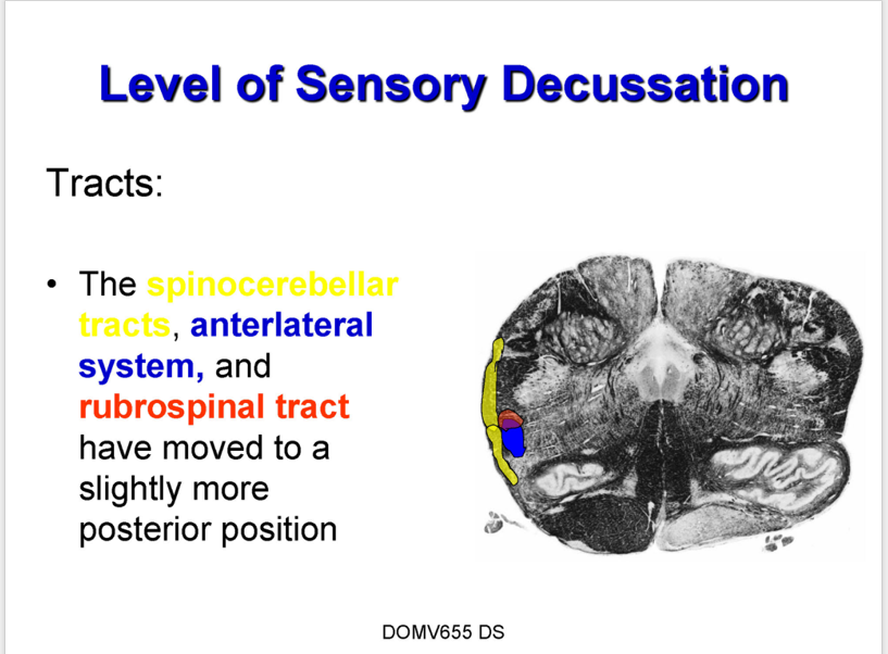
Describe what happens to these tracts @ level of sensory dessucation:
tectospinal tract + medial longitudinal fasciculus
uncrossed pyamidal tracts
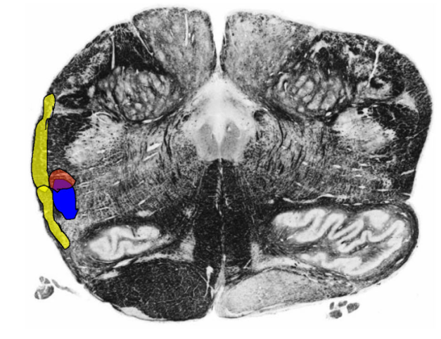
Spinocerebellar Tract, anterlateral system, rubrospinal tract
tectospinal tract + medial longitudinal fasciculus
located in midline behind medial lemniscis
uncrossed pyamidal tracts
located ventromedially.
Spinocerebellar Tract, anterlateral system, rubrospinal tract
moved to a slightly more posterior position
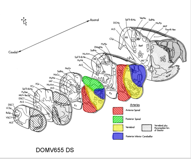
Describe the Blood Supply of the Lower Medulla
anterior spinal A
off Vertebral Artery
Supplies: anteriormedial area of lower medulla
Vertebral Artery
Supplies Lateral Areas
Posterior Spinal Artery:
usually branch of PICA or vertebral Artery
Supplies Dorsal Areas
(ADD PIC LATER)
NOTE: At more Rostral areas, PICA supplies lateral areas
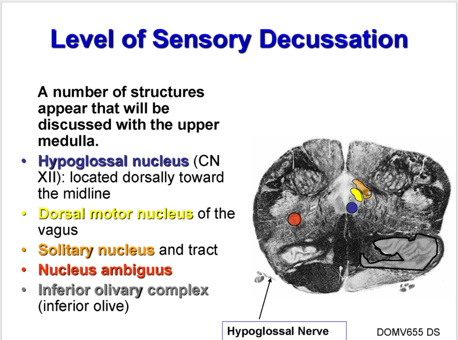
Describe the Upper Medulla:
What is it?
Boundaries?
Describe Development:
Where are Sensory/Motor CN nuclei?
Movement of SVE/GSA column
What forms the inferior Olive?
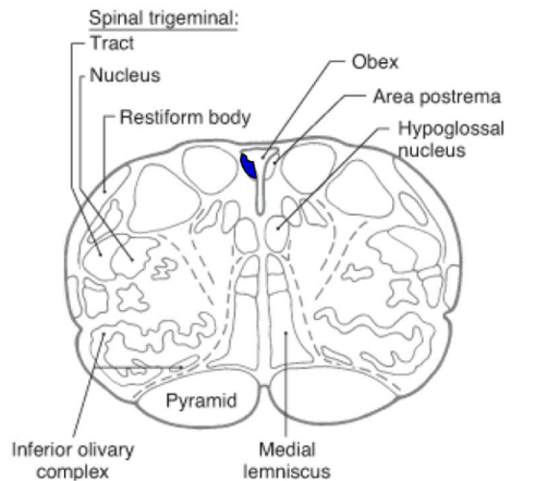
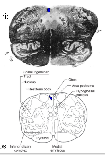
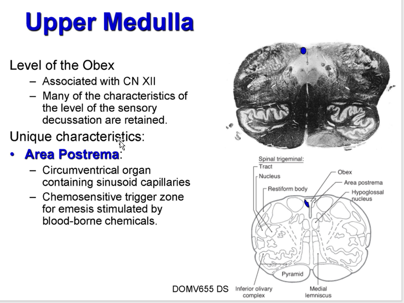
Level of Obex:
associated w/?
Decussation Characteristics?
What is a unique Characteristic @ this level? Function?
What is it?
Open portion of medulla containing caudal half of the fourth ventricle.
Boundaries:
Dorsal Surface:
caudal boundary= obex
rostral boundary is the striae medullares
Ventral Surface:
no definitive caudal boundary
rostral boundary = inferior pontine sulcus
Development:
sensory CN nuclei: dorsolateral position due to dev. of 4th ventricle.
Motor CN nuclei: ventromedially.
SVE/GSA column will migrate anterolaterally.
inferior olive = alar plate derivative.
Level Of Obex:
Associated w/ CN XII
Many of characteristics of level of the sensory decussation are retained.
Unique characteristics: Area Postrema
Circumventrical organ containing sinusoid capillaries
Function:
Chemosensitive trigger zone for emesis stimulated by blood-borne chemicals
Describe what happens @ level of Obex
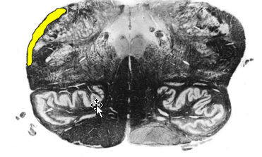
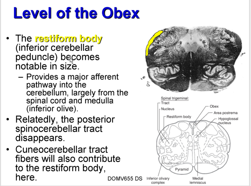
Restiform:
Function?
What dissapears?
What contributes?
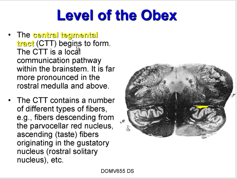
CTT:
Function?
VTT:
Location
Origin
Function?
Restiform Body (inferior cerebellar peduncle)
becomes notable in size.
Function:
Provides major afferent pathway → cerebellum,
largely from SC + medulla (inferior olive).
Relatedly: posterior spinocerebellar tract disappears.
Cuneocerebellar tract fibers will contribute to restiform body
Central Tegmental Tract (CTT)
begins to form
far more pronounced in the rostral medulla and above
Function:
local communication pathway W/in brainstem
contains a # of different types of fibers; EX:
from parvocellar red nucleus,
gustatory nucleus (rostral solitary nucleus),
etc.
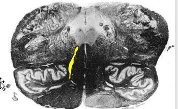
Ventral Trigeminothalamic Tract (VTT):
Begins to form
lateral to the medial lemniscus
Origins:
in the spinal trigeminal nucleus
Function:
carries pain and temperature from the face
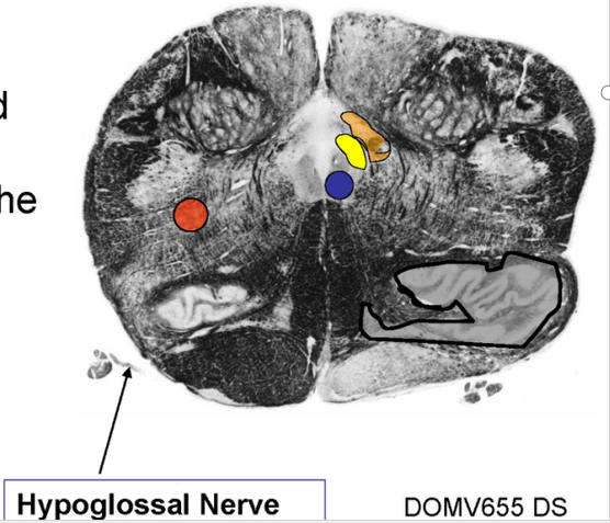
List the Structures/ Pathways retained from Sensorry Decussation?
Structures:
Hypoglossal nucleus and nerve
Nucleus ambiguus
Dorsal motor nucleus of Vagus
Solitary nucleus and tract
Nucleus gracilis and cuneatus
Accessory cuneate nucleus
Inferior olive
Spinal trigeminal nucleus and tract
Pathways:
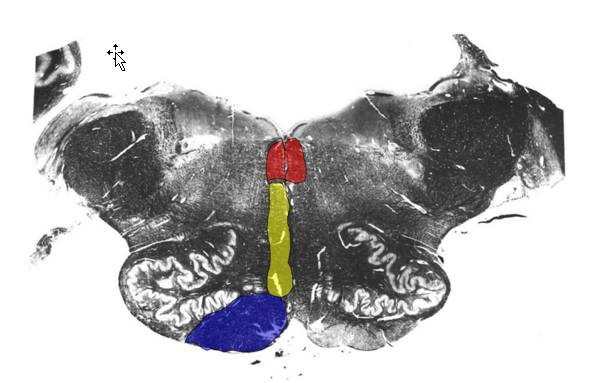
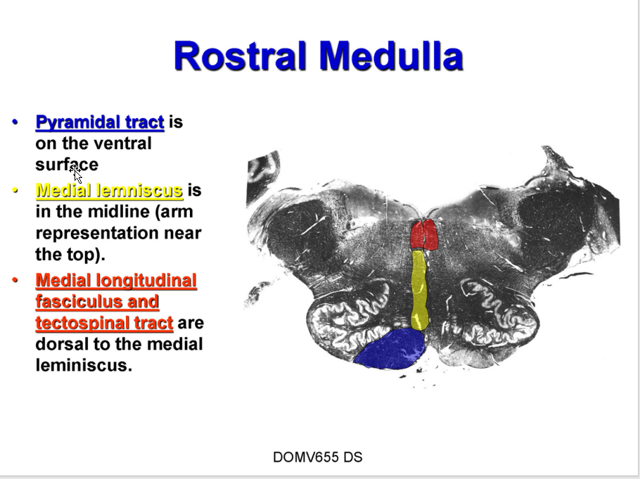
Pyramids
Medial lemniscus
Tectospinal tract
Medial longitudinal fasciculus
Anterior spinocerebellar tract
Anterolateral system
Rubrospinal tract •

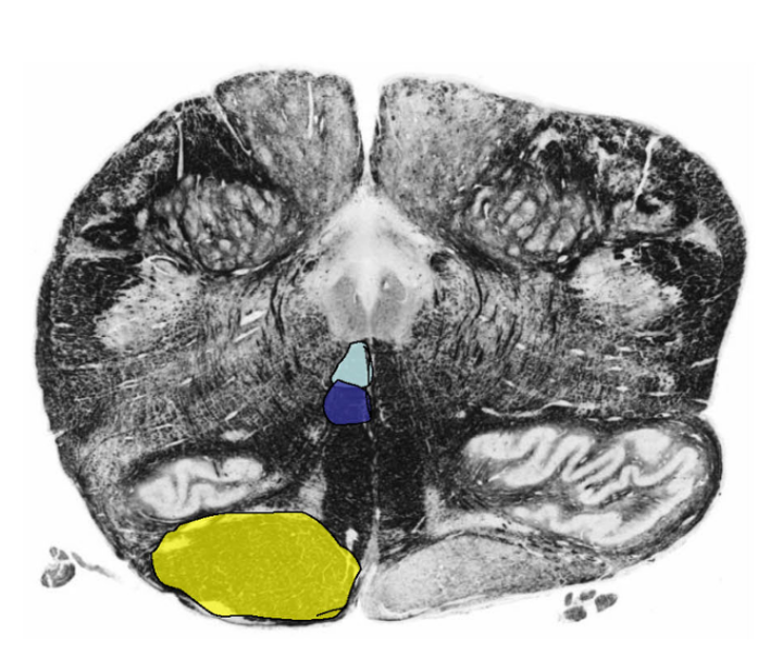
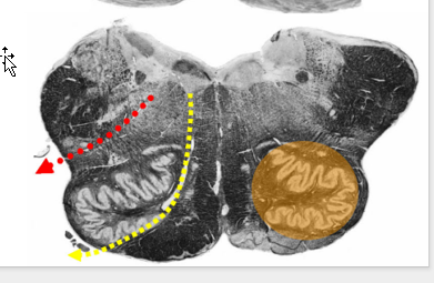
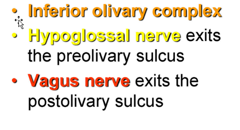
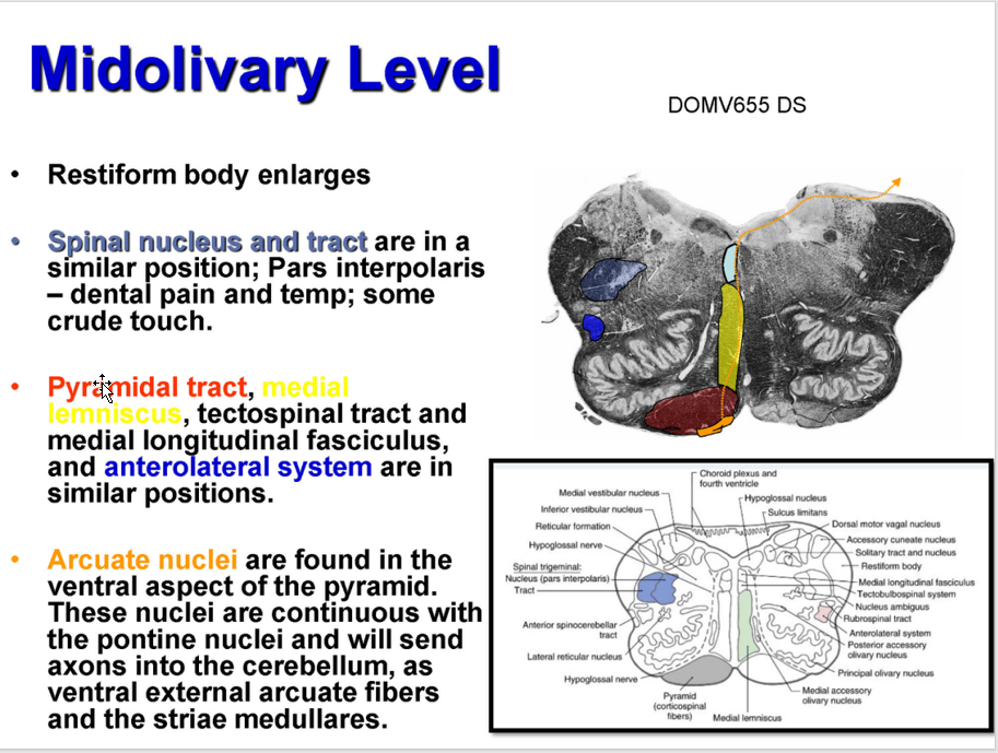
List the three prominent features found at the midolivary level
Prominent Features
Inferior olivary complex
Hypoglossal nerve
exits preolivary sulcus
Vagus nerve
exits postolivary sulcus
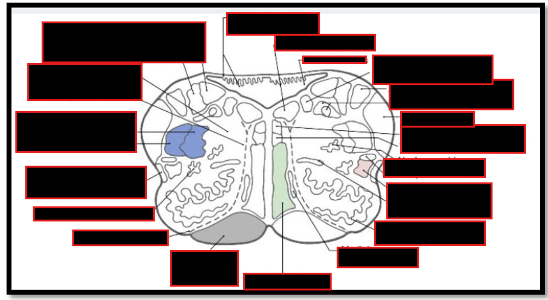
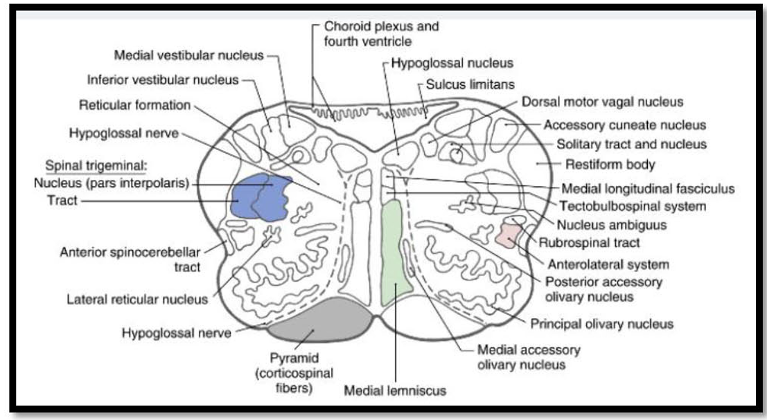
Describe the Structures found @ Midolivary Level
Inferior Olivary Complex
Constituents (2)?
Function?
Afferents?
inferior cerebellar peduncle (AKA?)
Function?
Hypoglossal Nucleus
Location
Function
Axon Route
Cortical Innervation
NA:
Function
Location
Cortical Innervation
DMN:
Location
Function (3)
Axon Route
Solitary nucleus (and tract):
Location
Two zones?
Function
Pathway
Vestibular Nuclei (inferior and medial)
Location
Function (3)
Clinical Importance
Inferior Olivary Complex:
Constituents:
Principle Olivary Nucleus
Function:
Control of planned or skilled voluntary movement
predominantly cortical (and subcortical) afferents
Medial/Dorsal Accessory Olive
Function:
Stereotyped Movement
Receives predominantly spinal afferents
inferior cerebellar peduncle (olivocerebellar tract)
inferior olive → cerebellum (contralaterally)
Reciprocally connected
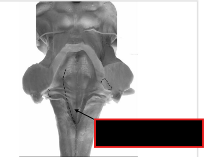
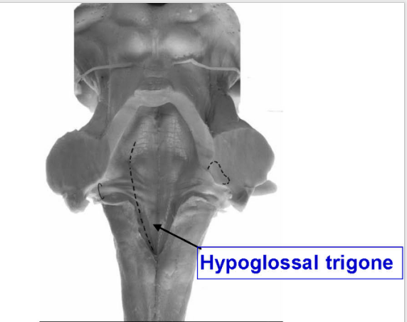
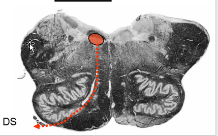
Hypoglossal Nucleus:
Location:
deep to hypoglossal trigone
either side of median sulcus in medullary portion of rhomboid fossa
Function:
(GSE) to most of tongue (except palatoglossus; CN X)
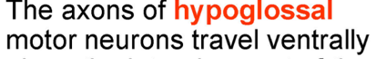
Route of Axons (motor neurons):
travel ventrally lateral to medial lemniscus → brainstem in preolivary sulcus.
Cortical innervation
bilateral w/ contralateral preponderance (more contribution from contra side)
Ipsilateral input allows for functional recovery in the case of a supranuclear lesion.
Nucleus Ambiguus (NA)
Function:
(SVE) to all
(GVE) to cardiac ganglia
Location:
deep w/in medullary reticular formation
btw trigeminal nucleus and inferior olive.
Cortical Innervation:
Bilateral
Dorsal Motor Nucleus of Vagus (DMN)
Location:
deep to vagal trigone
lateral to hypoglossal trigone in the floor of the fourth ventricle.
Function:
GVE component of the vagus nerve
receives input from solitary nucleus → baroreceptor refelx (efferent component)
plays a role in emesis
Route of Axons:
course ventrally and laterally → exit medulla in postolivary sulcus → travel in vagus nerves → synapse on post. gang para. neurons in terminal ganglia
Solitary nucleus (and tract):
Location:
lateral to DMN in the medulla
Two Zones:
rostral (and lateral) gustatory zone:
Function: taste afferents (SVA) from oral cavity and pharynx via CN VII, IX, & X.
Pathway:
gustatory nucleus → Thalamus (VPM) in ipsilateral central tegmental tract → gustatory cortex
caudal cardiorespiratory zone
Function:
receives (GVA) input
CN IX & X: lungs, trachea, larynx, gastrointestinal tract (X)
IX: Carotid Sinus
IX,X: Chemoreceptors
NOTE: Aortic Arch/Bodies also provide input to solitary nucleus via the vagus nerve
baroreceptor reflex (Caudal)
Pathway of these Neurons:
→ DMN, NA, IML of the upper thoracic spinal cord and medullary reticular centers → cardiovascular and respiratory control
Vestibular Nuclei (inferior and medial)
Location:
W/in rostral, dorsolateral medulla
Function:
Receives input from labyrinth of the inner ear
body equilibrium
control of eye movements
Clinical Importance:
Damage = vertigo, nausea, and nystaqmus
Describe what happens to these structures @ midolivary Levels:
Restiform body
Arcuate nuclei
Nucleus gracilis/ cuneatus
Lateral cuneate nucleus
Rubrospinal, reticulospinal and vestibulospinal tracts
anterior spinocerebellar tract
Ventral Trigeminothalamic tract
Similar Position:
Spinal nucleus and tract
Pyramidal tract,
Medial Lemniscus ,
tectospinal tract
medial Ionitudinal fasciculus
anteroateral system are in similar positions.
Midolivary Levels:
Restiform body enlarges
Arcuate nuclei
found in the ventral aspect of the pyramid.
nuclei = continuous w/ pontine nuclei → cerebellum
as ventral external arcuate fibers and the striae medullares.
Nucleus gracilis/ cuneatus
no longer present;
vestibular nuclei adopt their position
Lateral cuneate nucleus
may still be present
Rubrospinal, reticulospinal and vestibulospinal tracts
in lateral position.
anterior spinocerebellar tract
present.
Ventral Trigeminothalamic tract
lateral to medial lemniscus still.
Similar Position:
Spinal nucleus and tract
Pyramidal tract,
Medial Lemniscus ,
tectospinal tract
medial Ionitudinal fasciculus
anteroateral system are in similar positions.


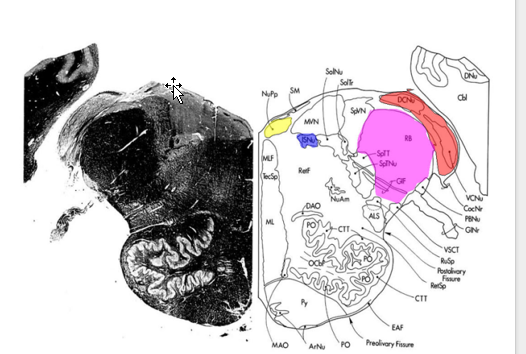
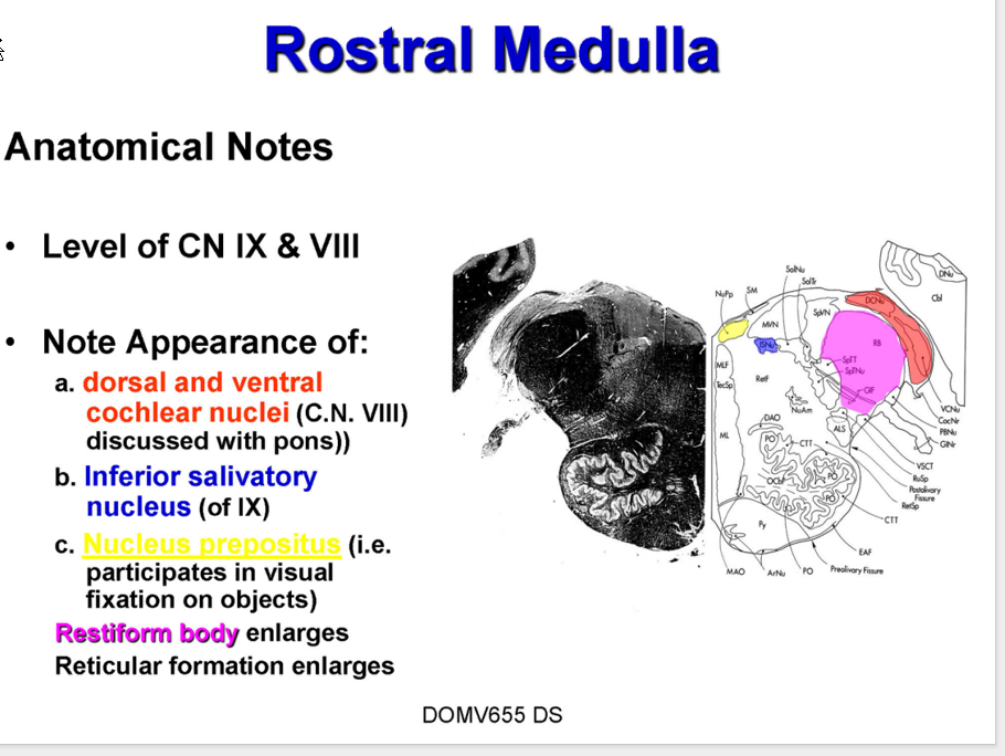
Describe the Rostral Medulla
@ level?
Appearance of?
What enlarges?
Describe Inferior salivatory nucleus (of IX)
Origin of?
Receives influences from
Structures and relationship to restiform body
between restiform body and Inferior olive.
Now Part of Restiform Body:
Reticular Formation Function
@ Level of CN IX & VIII
Appearance
dorsal and ventral cochlear nuclei (C.N. VIII)
Inferior salivatory nucleus (of IX)
Nucleus prepositus:
(i.e. c. participates in visual fixation on objects)
Enlarges:
Restiform body
Reticular formation
Inferior salivatory nucleus (of IX)
Origin of:
Parasymp motor fibers to parotid gland.
Synapse @ Otic
Postgang supply parotid gland
Receives influences from
hypothalamus and olfactory system.
Structures and relationship to restiform body
Structure between restiform body and Inferior olive.
Spinal trigeminal nucleus (and tract),
anterolateral system,
rubrospinal tract
anterior spinocerebellar tract
Now Part of Restiform Body:
Posterior spinocerebellar tract and cuneocerebellar tract
Reticular Formation:
Function:
Wide range including consciousness and sleep.
Describe the Blood Supply of the Medulla:
anterior spinal artery
Medial/Ventral
Vertebral Artery
Intermediate lateral Area
posterior spinal artery
Dorsal (caudal) Region
Posterior inferior cerebellar artery
Dorsolateral