Imaging - Radiologic Evaluation Pelvis and Hip
1/85
There's no tags or description
Looks like no tags are added yet.
Name | Mastery | Learn | Test | Matching | Spaced | Call with Kai |
|---|
No analytics yet
Send a link to your students to track their progress
86 Terms
AP pelvis (pelvis, sacrum, coccyx)
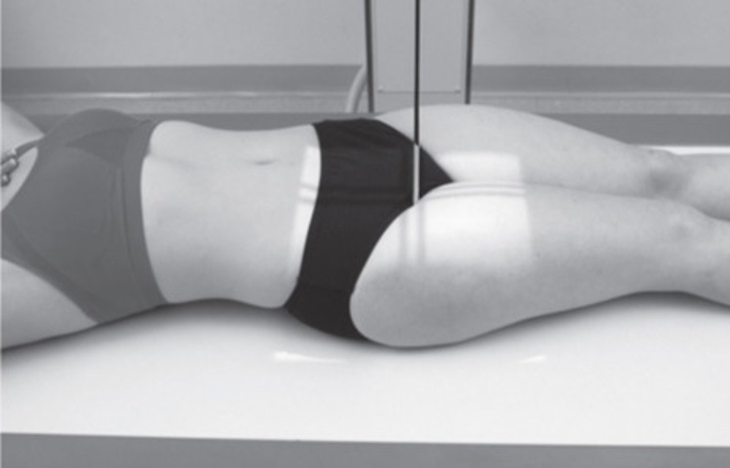
AP hip
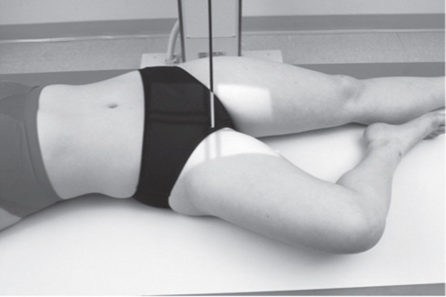
Lateral frog-leg
What are the routine x-ray views of the pelvis and hip?
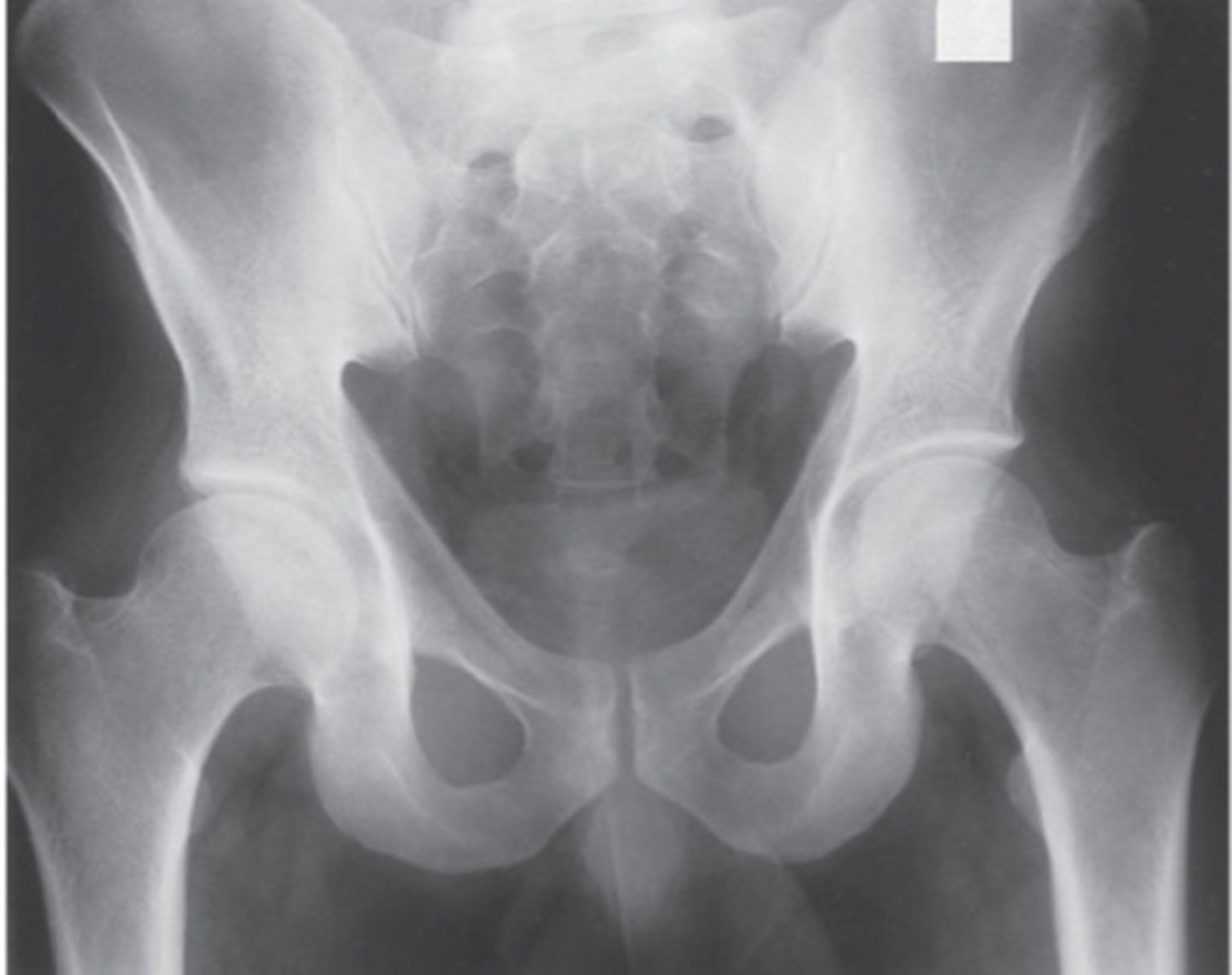
AP pelvis
What view does this show?
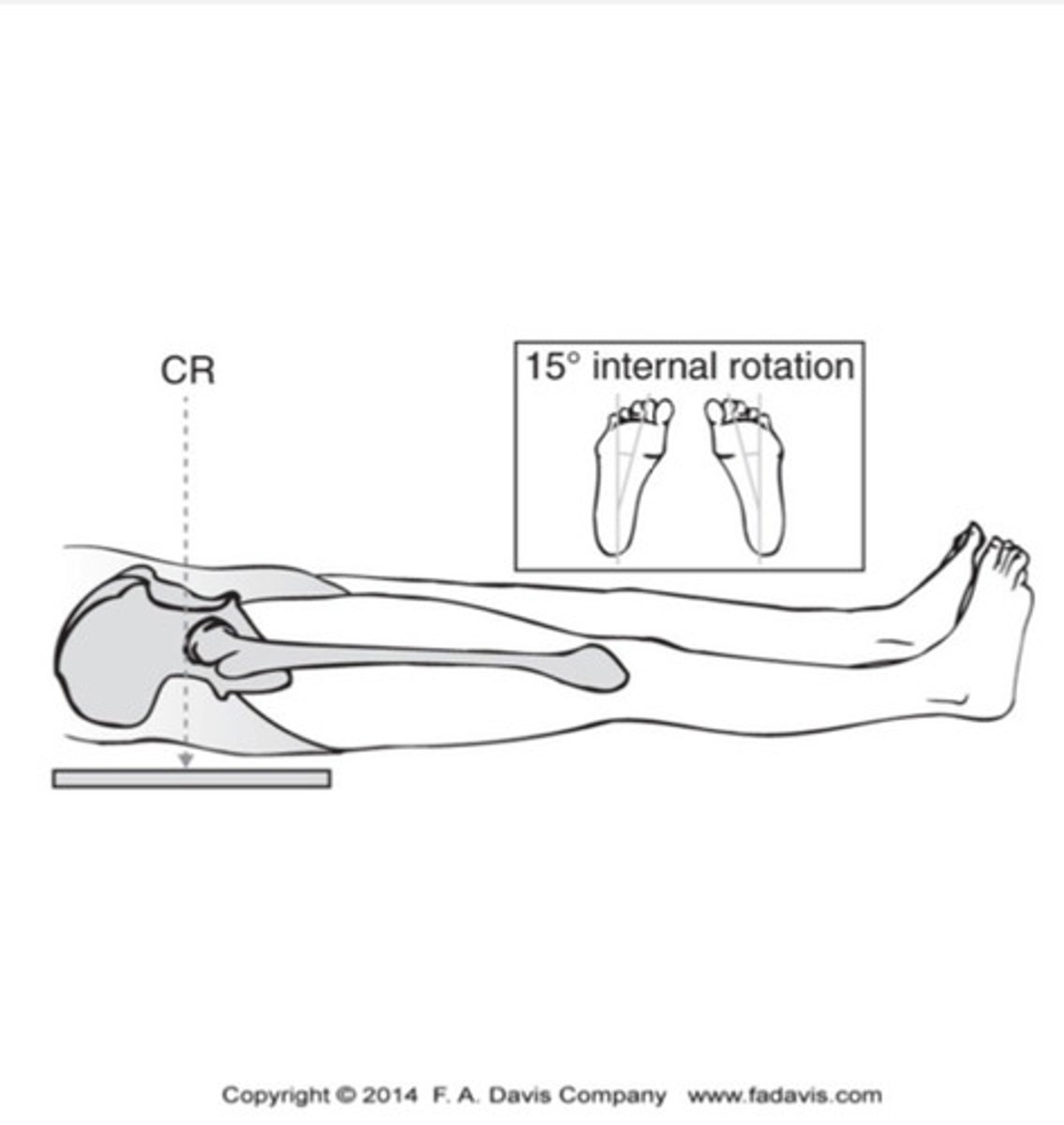
AP pelvis
What view does this show?
AP hip
What view does this show?
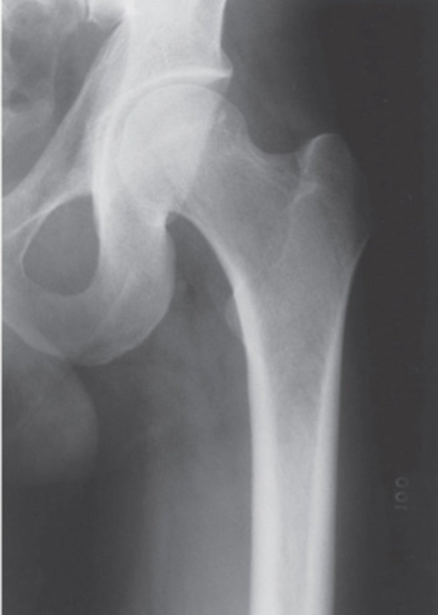
AP hip
What view does this show?
Lateral frog leg
What view does this show?
Lateral frog leg
What view does this show?
Angle of inclination
angle from the head of the femur to the neck of the femur
AP pelvis
How is the angle of inclination best imaged?
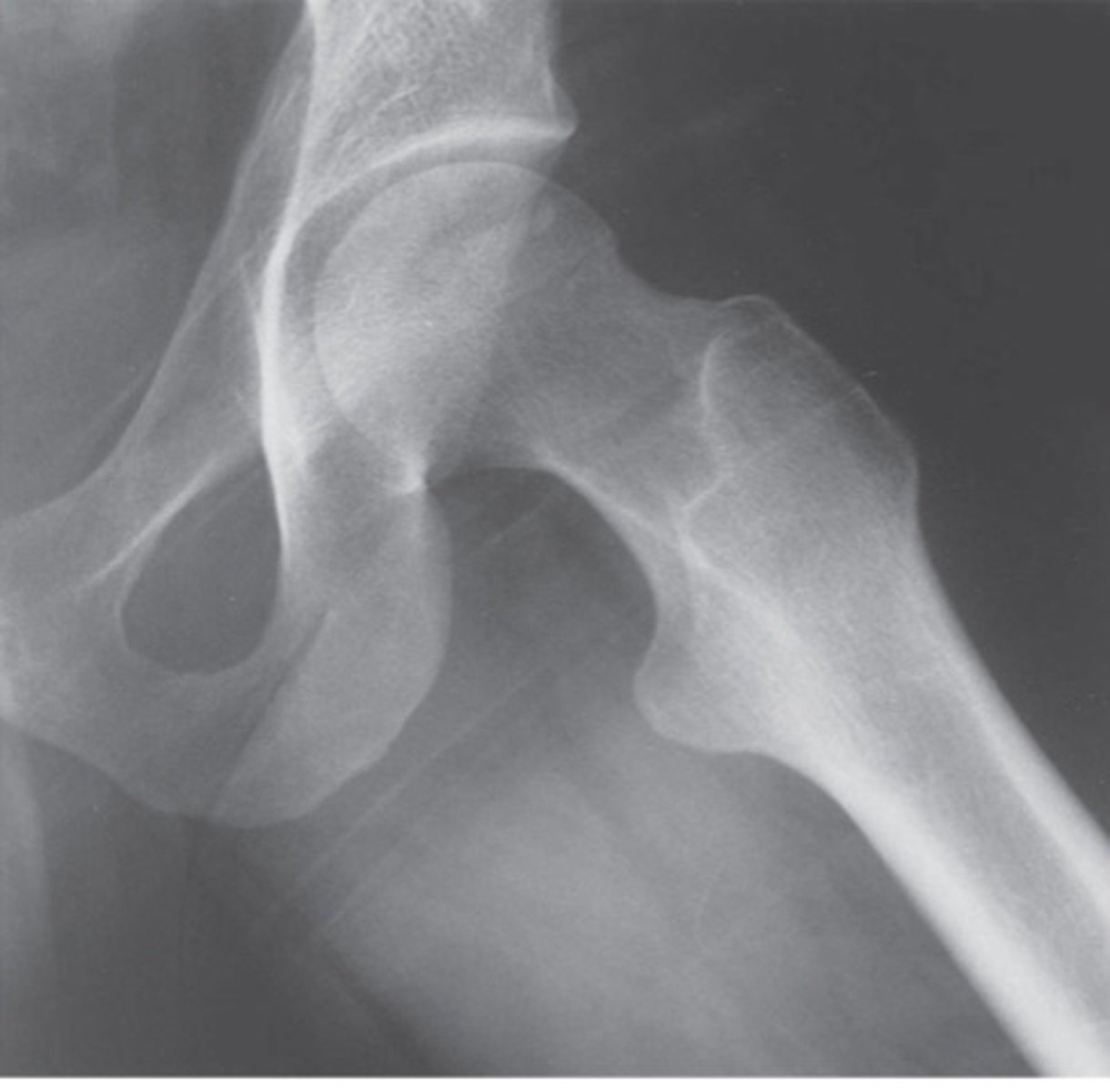
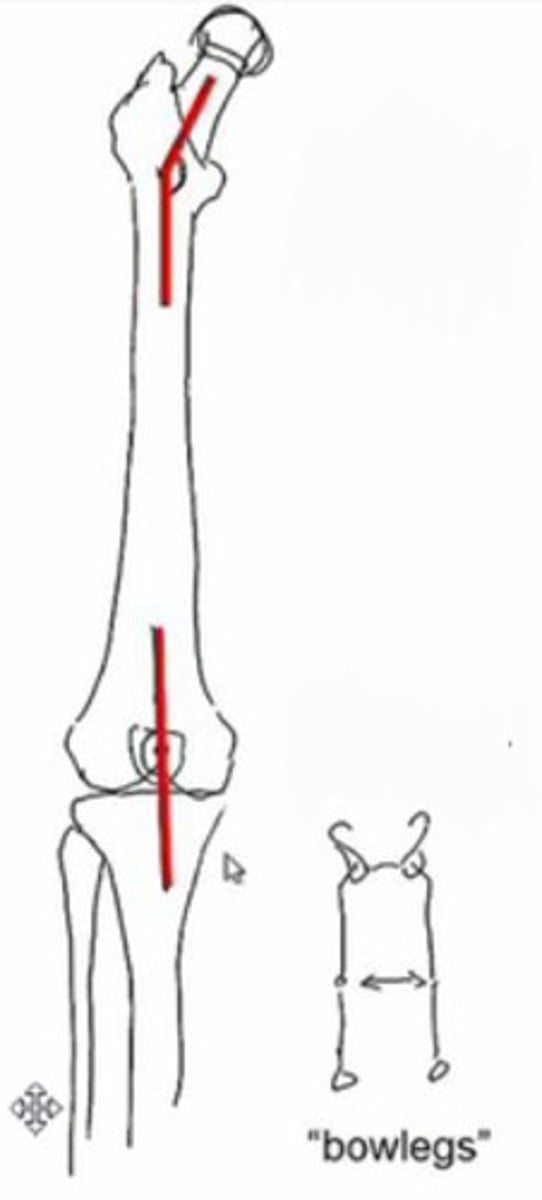
coxa valga and genu varum
What does this image show?
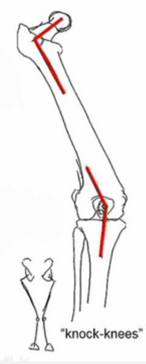
caxa vara and genu valga
What does this image show?
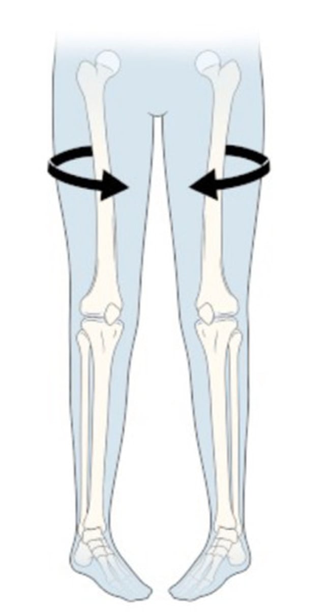
femoral anteversion
What does this image show?
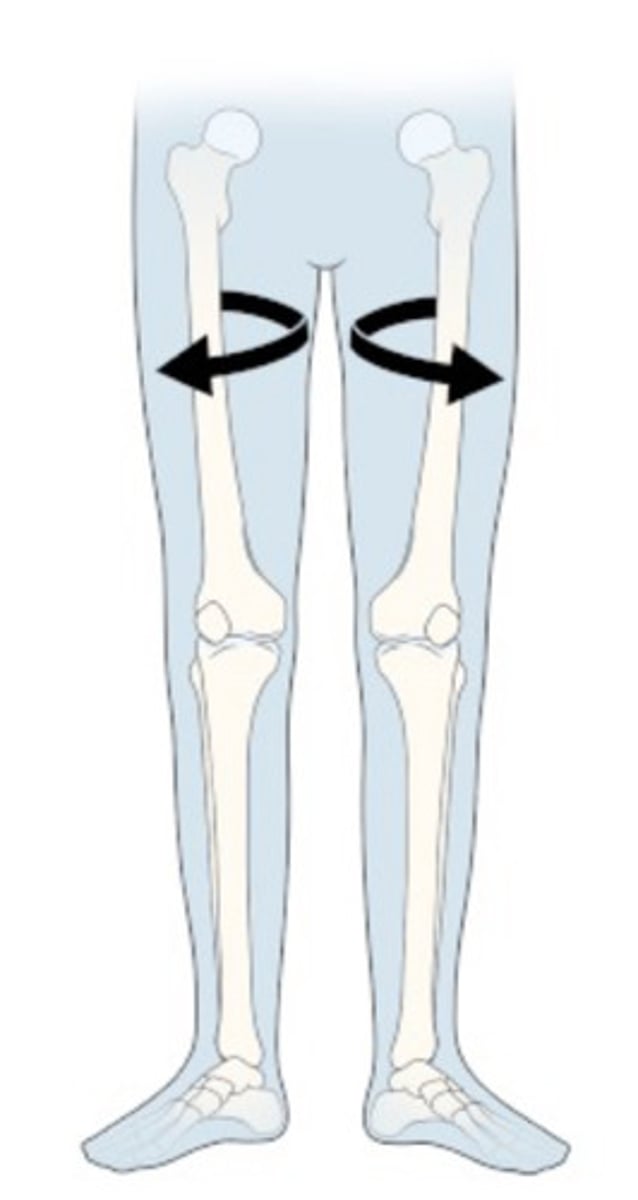
femoral retroversion
What does this image show?
Bilateral sides of pelvis and hips
L5 vertebra
sacrum and coccyx
SI joints
hip joints
Femoral Necks and their angles of inclination
What do you see on a AP pelvis radiograph view?
Proximal femur and acetabulum
Unilateral Femoral Neck
Angle of Inclination femoral neck to shaft
Greater & Lesser Trochanter Intertrochanteric Crest
Cortical outline of femur
What do you see on a AP hip radiograph view?
Lateral frog leg
Femoral head
neck and proximal shaft
Greater & Lesser Trochanters from
medial aspect
lateral frog leg
What radiograph view has the best view of the lesser trochanter?
femoral head in acetabular fossa
medial wall of acetabulum
anterior and posterior rims of acetabulum
Sacrum
Pubic rami
greater and lesser trochanters
What do you see in a CT axial view of the pelvis?
anterical inclination of acetabular cup
acetabular roof
iliopsoas muscle anterior to hip
sacroiliac joints
pubic symphysis
What do you see in a CT saggital view of the pelvis?
bilateral comparison of hip joints
acetabulum
femoral heads, necks, shafts
greater and lesser trochanter
Sacrum, ilia, sacroiliac joints
What do you see in a CT coronal view of the pelvis?
cartilage
Labrum
Presence of FAI
What is an arthrography used to evaluate?
arthrography
contrast material injected into joint space for imaging
Acetabulum
Labrum
Femoral head and neck
Greater and lesser trochanters
Scrum, ilium, sacroiliac joints
pubic symphysis
What do you see in an MRI axial view of the pelvis?
Sphericity of femoral head
Superior aspect of acetabulum and cartilage
What do you see in an MRI sagittal view of the pelvis?
gluteus maximus
hamstrings
What do you see in an posterior MRI axial view of the pelvis?
iliopsoas
sartorius
rectus femoris
vastus medialis
What do you see in an anterior MRI axial view of the pelvis?
Hip joints
Proximal femurs
Sacroiliac joints
ilium
Gluteal muscles
abductors
adductors
What do you see in an MRI coronal view of the pelvis?
avulsions of the ASIS, AIIS, or ischial tuberosity
What does A show?

iliac wing frature
What does B show?
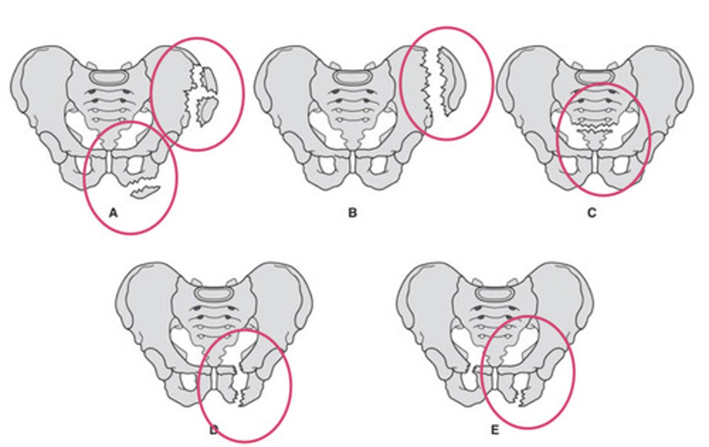
sacral fracture
What does C show?

ipsilateral pubic rami fracture
What does D show?

contralateral pubic rami fracture
What does E show?

sartorius
TFL
Which muscles attach to the ASIS?
Unstable pelvic injury
disruption of 2 or more sites on pelvic ring
internal hemorrhage
What are unstable pelvic injuries associated with?
Vertical shear/Malgaigne fractures
Straddle fractures
Bucket handle fractures
Examples of unstable pelvis injuries
Vertical shear/Malgaigne fractures
lateral fractures of the superior and inferior pubic rami and disruption of the ipsilateral sacroiliac
Straddle fractures
all for ischiopubic rami fractured
Bucket handle fractures
ischial ramus, ipsilateral pubic ramus, and the contralateral sacroiliac joint
SIJ
pubic symphysis
Where do dislocations of the pelvis occur?
Acetabular fracture
from femoral head impacting into acetabulum
CT or advanced imaging
What imaging is used for acetabular fractures?
70% female
40% >85 years old
What is the population that hip fractures occur in?
14%
What percentage of fractures are hip fractures?
Femoral neck
Intertrochanteric
Trochanteric
What are the three types of hip fractures?
intracapsular
What type of hip fractures are femoral neck fractures?
avascular necrosis due to blood supply limitations
What is there a risk of with femoral neck fractures?
AP hip in maximal internal rotation and lateral view (comparison with the uninvolved hip through AP pelvis view)
What imaging is used to diagnose a femoral neck fracture?
ORIF
arthroplasty
How are femoral neck fractures treated?
Trochanteric fracture
isolated fractures of greater and lesser trochanters are typically avulsion fractures caused by forceful muscle contraction
AP and other views
What imaging is used to diagnose a trochanteric fracture?
AP view with LE in ER
What imaging is used to diagnose a lesser trochanteric fracture?
non surgical unless significant displacement
How are trochanteric fractures treated?
gluteus max and min
Triceps coxae
Piriformis
What muscles attach to the greater trochanter?
iliopsoas
What muscles attach to the lesser trochanter?
Subtrochanteric fracture
Proximal femur fracture located from the lesser trochanter to 5cm distal to it that may occur in low energy (elderly) or high energy (young patients) mechanisms
AP and other views
What imaging is used to diagnose a subtrochanteric fracture?
cephalomedullary nail fixation
How are subtrochanteric fractures treated?
joint space narrowing
sclerotic subchondral bone
osteophyte at joint margins
cyst/pseudocyst
How does degenerative joint disease/osteoarthritis appear on imaging?
symmetrical joint space narrowing
loss of bone density in periarticular bone density
articular erosions
synovial cysts
joint effusion
periarticular swelling
How does rheumatoid arthritis appear on imaging?
joint narrowing with sclerosis
osteophyte formation
large subchondral cysts
What are characteristics of OA?
Avascular necrosis of the femoral head
Interruption of blood supply to the femoral head that causes deformities and collapse of the head
intracapsular fracture or displaced fracture interrupt blood supply
What causes Avascular necrosis of the femoral head?
Glucocorticoids
ETOH
post-transplantation
SLE
post-trauma
genetic disorders
What are risk factors of Avascular necrosis of the femoral head?
Legg Calve Perthes
Idiopathic epiphyseal ischemic necrosis at femoral head
hip pain
antalgic gait
What are characteristics of Legg Calve Perthes?
children ages 3-12
What is the population that Legg Calve Perthes occurs in?
standard radiographs
MRI - bone marrow
What is the imaging used to diagnose Legg Calve Perthes disease?
conservative revascularization
core decompression
grafting
THA
What are possible treatments for Legg Calve Perthes disease?
Slipped capital femoral epiphysis (SCFE)
femoral epiphysis slips posteriorly
lateral frog leg to view amount of displacement
What type of imaging is used to diagnose SCFE?
blurring/widening of physis on AP
How is SCFE seen on an image?
surgically
How is SCFE treated?
Femoral acetabular impingement (FAI)
Pathological contact between femoral head-neck junction and acetabular rim during hip movement
CAM
aspherical femoral head - femoral head can not clear the acetabular rim
Pincer
Over-coverage of the femoral head by the acetabulum
Capital
What type of fracture is 1?

Subcapital
What type of fracture is 2?

transcervical
What type of fracture is 3?

Intertrochanteric
What type of fracture is 4?

Subtrochanteric
What type of fracture is 5?
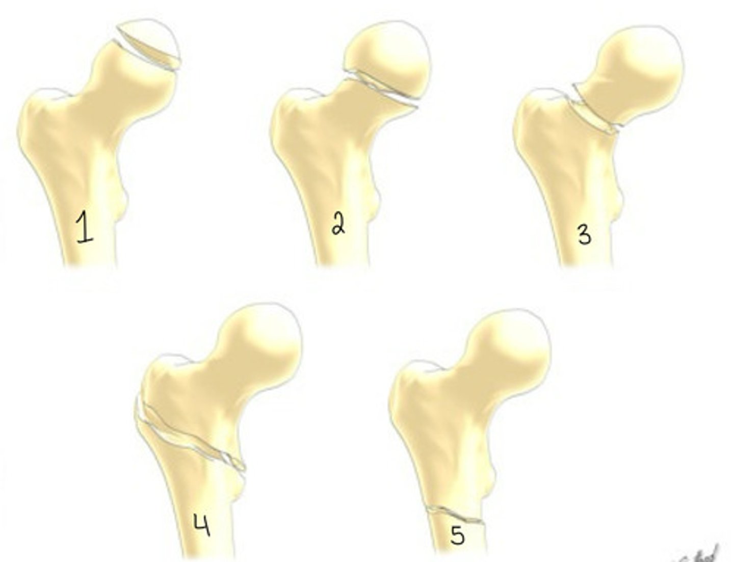
intracapsular
What type of fracture is a capital fracture?
intracapsular
What type of fracture is a subcapital fracture?
intracapsular
What type of fracture is a transcervical fracture?
extracapsular
What type of fracture is a intertrochanteric fracture?
extracapsular
What type of fracture is a subtrochanteric fracture?