L16- Stem cells and cellular plasticity in cancer

0.0(0)
Studied by 0 people
call kaiCall Kai
learnLearn
examPractice Test
spaced repetitionSpaced Repetition
heart puzzleMatch
flashcardsFlashcards
GameKnowt Play
Card Sorting

1/50

encourage image

There's no tags or description

Looks like no tags are added yet.

Last updated 3:30 PM on 5/18/26
Name
Mastery
Learn
Test
Matching
Spaced
Call with Kai

No analytics yet

Send a link to your students to track their progress

51 Terms

1
New cards

what is a stem cell

An undifferentiated cell which is capable of indefinite self-renewal and has the capacity to give rise to other cell types via differentiation.

2
New cards

how are stem cells produced

Cell division produces two daughter cells:

- One maintains stem properties (self- renewal)

- One differentiates to give rise to committed progenitors

<p>Cell division produces two daughter cells:</p><p>- One maintains stem properties (self- renewal)</p><p>- One differentiates to give rise to committed progenitors</p>
3
New cards

what are the types of stem cell

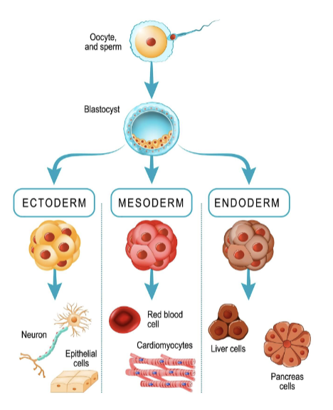
  1. Totipotent (e.g. zygotes)- Differentiate into any cell type

  2. Pluripotent (e.g. embryonic stem cells)- Differentiate into cells from any of the three germ layers

  3. Multipotent/Oligopotent (e.g. tissue- specific stem cells)- Differentiate into a limited number of cell types

  4. Unipotent- Differentiate into a single cell type

4
New cards

what are the 3 germ layers

  1. ectoderm

  2. mesoderm

  3. endoderm

<ol><li><p>ectoderm</p></li><li><p>mesoderm</p></li><li><p>endoderm</p></li></ol><p></p>
5
New cards

what is the history of stem cell research

• 1968 first bone marrow transplant to treat severe combined immunodeficiency

• 1978 Stem cells were discovered in human cord blood

• 1981 First stem cell line developed from mice

• 1995 First embryonic stem cell line derived from a primate

• 1996 Mammal cloned from a hybrid stem cell (nucleus from a mammary gland cell injected into an empty egg cell)

• 1994 First cancer stem cell identified in human leukaemia

6
New cards

How were cancer stem cells first identified in acute myeloid leukaemia (AML) and what are their key features?

  • First identified in 1994 (John Dick’s team) in AML

  • Only a small subset of cells could repopulate leukaemia in immunocompromised mice

  • These cells have a stem-like phenotype (CD34⁺ CD38⁻)

  • Resemble normal haematopoietic stem cells (HSCs)

  • Suggests leukaemia may originate from or mimic normal stem cell populations

7
New cards

wha are the 2 models of carcinogenesis

  1. classical stochastic model

  2. cancer stem cell (CSC) model

8
New cards

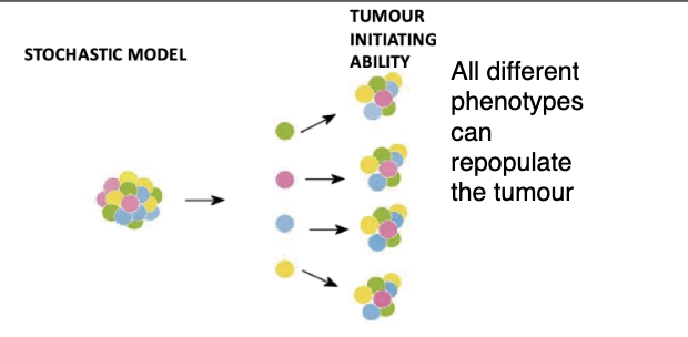
what is the classical stochastic model of carcinogenesis

  • Cancer can arise in any cell and can be propagated by any cell in the population (or most cells at least)

  • Cancer develops through a process of continual mutation and clonal selection of the fittest clones (Darwinian selection).

  • Allows it to populate and continue tumour growth  

<ul><li><p>Cancer can arise in any cell and can be propagated by any cell in the population (or most cells at least)</p></li><li><p>Cancer develops through a process of continual mutation and clonal selection of the fittest clones (Darwinian selection).</p></li><li><p><span style="background-color: inherit; line-height: 22px; color: windowtext;">Allows it to populate and continue tumour growth&nbsp;</span><span style="line-height: 22px; color: windowtext;">&nbsp;</span></p></li></ul><p></p>
9
New cards

what is the cancer stem cell (CSC) model of carcinogenesis, what does ot say about incidence

  • Cancer arises in a cell that has or acquires stem cell properties of self-renewal

  • Tumours display some level of hierarchical organisation with the CSC at the apex of that hierarchy.

  • Incidence of cancer is directly proportional to the number of stem cell divisions in that tissue (Vogelstein model).

<ul><li><p>Cancer arises in a cell that has or acquires stem cell properties of self-renewal</p></li><li><p>Tumours display some level of hierarchical organisation with the CSC at the apex of that hierarchy.</p></li><li><p>Incidence of cancer is directly proportional to the number of stem cell divisions in that tissue (Vogelstein model).</p></li></ul><p></p>
10
New cards

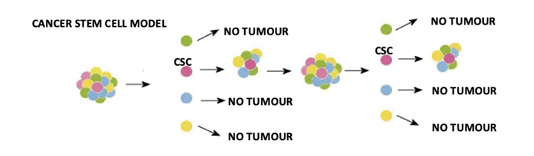
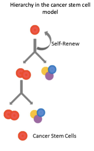
what is the hierarchical structure of the cancer stem cell model

  • Cancer stem cells (CSCs) sit at the top and can self-renew

  • Only CSCs can repopulate and sustain tumour growth

  • Other tumour cells are more differentiated and have limited proliferative capacity

  • Evidence requires serial transplantation showing tumour regrowth from CSCs

non malignant tissues also have a hierarchical structure

<ul><li><p><strong>Cancer stem cells (CSCs)</strong> sit at the top and can <strong>self-renew</strong></p></li><li><p>Only CSCs can <strong>repopulate and sustain tumour growth</strong></p></li><li><p>Other tumour cells are more differentiated and have <strong>limited proliferative capacity</strong></p></li><li><p>Evidence requires <strong>serial transplantation showing tumour regrowth from CSCs</strong></p></li></ul><p>non malignant tissues also have a hierarchical structure </p><p></p>
11
New cards

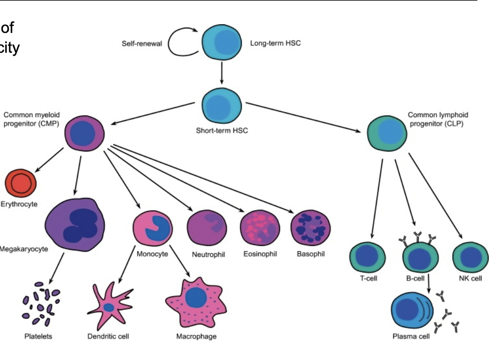
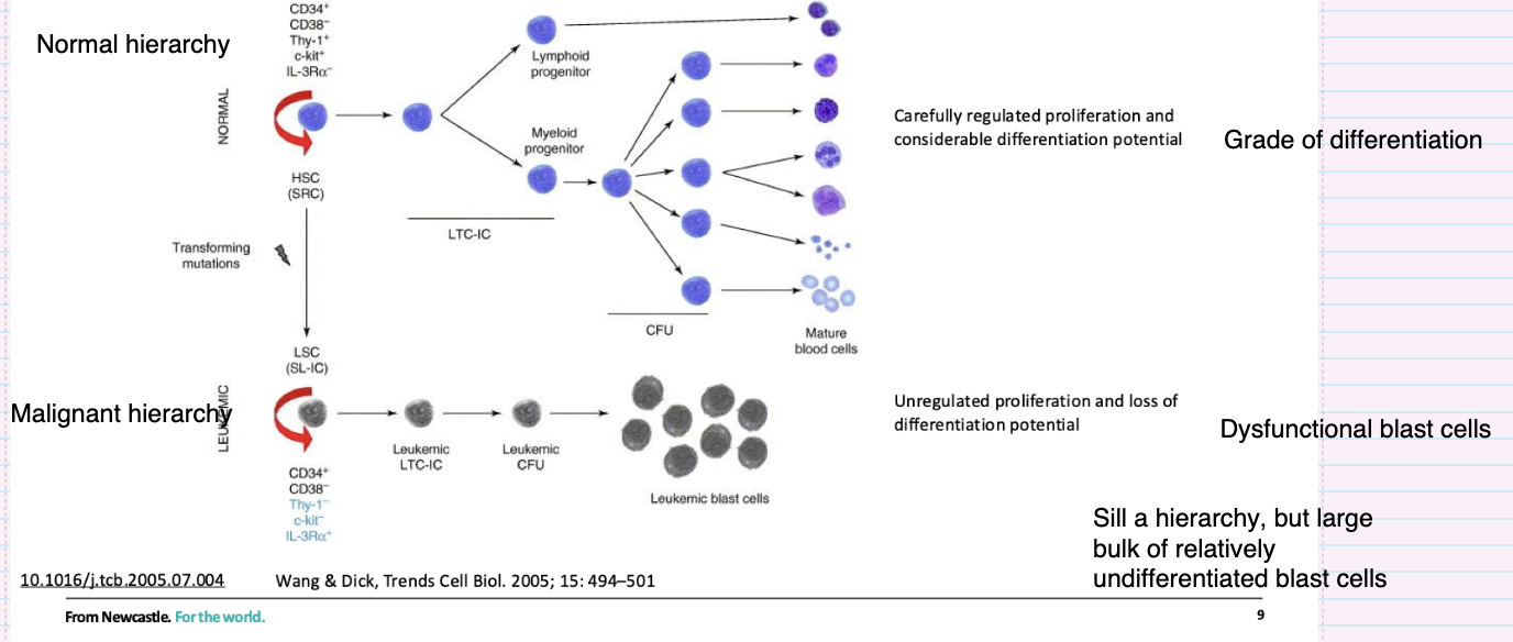
What characterises the normal haematopoietic stem cell hierarchy

  • HSCs at the top with self-renewal ability

  • Give rise to lymphoid and myeloid progenitors

  • Cells undergo controlled proliferation and differentiation

  • Leads to fully differentiated mature blood cells

  • Clear hierarchy based on increasing differentiation

<ul><li><p><strong>HSCs at the top</strong> with self-renewal ability</p></li><li><p>Give rise to <strong>lymphoid and myeloid progenitors</strong></p></li><li><p>Cells undergo <strong>controlled proliferation and differentiation</strong></p></li><li><p>Leads to <strong>fully differentiated mature blood cells</strong></p></li><li><p>Clear <strong>hierarchy based on increasing differentiation</strong></p></li></ul><p></p>
12
New cards

How does the leukemic stem cell hierarchy differ from the normal hierarchy?

  • Originates from transforming mutations in stem/progenitor cells

  • Leukaemic stem cells (LSCs) retain self-renewal

  • Produces a hierarchy but with impaired differentiation

  • Results in accumulation of immature blast cells

  • Characterised by uncontrolled proliferation and dysfunctional cells

<ul><li><p>Originates from <strong>transforming mutations in stem/progenitor cells</strong></p></li><li><p><strong>Leukaemic stem cells (LSCs)</strong> retain self-renewal</p></li><li><p>Produces a hierarchy but with <strong>impaired differentiation</strong></p></li><li><p>Results in <strong>accumulation of immature blast cells</strong></p></li><li><p>Characterised by <strong>uncontrolled proliferation and dysfunctional cells</strong></p></li></ul><p></p>
13
New cards

How have cancer stem cells been identified in solid tumours?

  • Identified by specific surface markers (phenotype) (e.g. CD44⁺, CD133⁺)

  • Defined by ability to initiate tumours in recipient mice (functional test)

  • Demonstrated in prostate and colon cancers

  1. prostate- CD44+, Ɑ2β1high, CD133+

  2. colon- CD133+

  • Supports idea that only a subset of tumour cells drive tumour growth

14
New cards

give examples of different cancers and their marker types
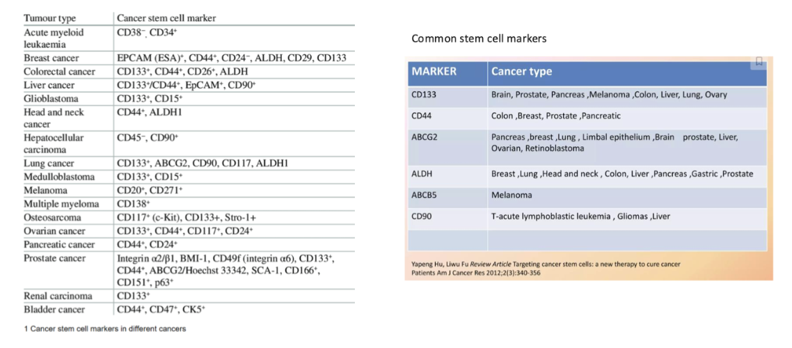
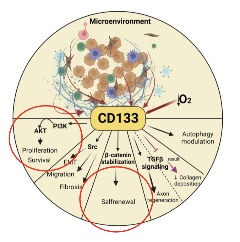
knowt flashcard image
15
New cards

what is CD133

aka Prominin-1 is a transmembrane glycoprotein.

  • Precise function/activity is unknown

  • “Regulates” numerous pathways associated with stem cell characteristics

<p>aka <strong><em>Prominin-1</em></strong> is a transmembrane glycoprotein.</p><ul><li><p>Precise function/activity is unknown</p></li><li><p>“Regulates” numerous pathways associated with stem cell characteristics</p></li></ul><p></p>
16
New cards

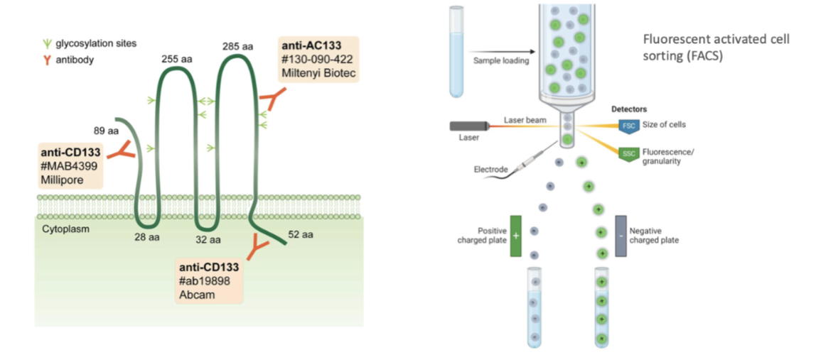
how can canser stem cells be isolated

Use markers to isolate stem cells  

  • Commercial organisations released Abs that bind to CD133, bind to cells and isolaye the cells 

  • Eg through fluorescence activated cell sorting dependent on if they're positive for CD-133 or not  

<p><span style="background-color: inherit; line-height: 22px; color: windowtext;">Use markers to isolate stem cells&nbsp;</span><span style="line-height: 22px; color: windowtext;">&nbsp;</span></p><ul><li><p class="Paragraph SCXO124213417 BCX0" style="text-align: left;"><span style="background-color: inherit; line-height: 22px; color: windowtext;">Commercial organisations released Abs that bind to CD133, bind to cells and isolaye the cells</span><span style="line-height: 22px; color: windowtext;">&nbsp;</span></p></li><li><p class="Paragraph SCXO124213417 BCX0" style="text-align: left;"><span style="background-color: inherit; line-height: 22px; color: windowtext;">Eg through fluorescence activated cell sorting dependent on if they're positive for CD-133 or not&nbsp;</span><span style="line-height: 22px; color: windowtext;">&nbsp;</span></p></li></ul><p></p>
17
New cards

describe the functional identification of cancer stem cells via colony formation in vitro

Colony formation in vitro

• Ability to form colonies when cultured from single cells (property shared by both normal and cancer stem cells)

• Serial passage of colonies and retention of colony formation ability

  • key is if they can form colonies in vitro- serially

<p>Colony formation in vitro</p><p>• Ability to form colonies when cultured from single cells (property shared by both normal and cancer stem cells)</p><p>• Serial passage of colonies and retention of colony formation ability</p><ul><li><p>key is if they can form colonies in vitro- <strong><em>serially</em></strong></p></li></ul><p></p>
18
New cards

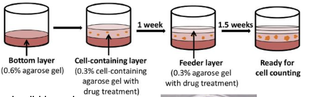
what are the limitations of the functional identification of cancer stem cells (colony formation in vitro)

  • clonogenicity influenced by the tumour micro-environment

  • Expensive cytokines or feeder layers are often required

19
New cards

describe the functional identification of cancer stem cells via serial transplantation in vivo

Another major mechanism to test for cancer stem cells  

  • forms cancer in transplanted animal

  • serial transfer of cancer into subsequent animals

  • Transplant into mice 

  • Leukaemia- normally In bone marrow in femur, see whether cells can form leukaemia in that setting 

  • Called an orthotopic model  

<p><span style="background-color: inherit; line-height: 22px; color: windowtext;">Another major mechanism to test for cancer stem cells&nbsp;</span><span style="line-height: 22px; color: windowtext;">&nbsp;</span></p><ul><li><p>forms cancer in transplanted animal</p></li><li><p>serial transfer of cancer into subsequent animals</p></li></ul><ul><li><p class="Paragraph SCXO121546760 BCX0" style="text-align: left;"><span style="background-color: inherit; line-height: 22px; color: windowtext;">Transplant into mice</span><span style="line-height: 22px; color: windowtext;">&nbsp;</span></p></li><li><p class="Paragraph SCXO121546760 BCX0" style="text-align: left;"><span style="background-color: inherit; line-height: 22px; color: windowtext;">Leukaemia- normally In bone marrow in femur, see whether cells can form leukaemia in that setting</span><span style="line-height: 22px; color: windowtext;">&nbsp;</span></p></li><li><p class="Paragraph SCXO121546760 BCX0" style="text-align: left;"><span style="background-color: inherit; line-height: 22px; color: windowtext;">Called an orthotopic model&nbsp;</span><span style="line-height: 22px; color: windowtext;">&nbsp;</span></p></li></ul><p class="Paragraph SCXO121546760 BCX0" style="text-align: left;"></p>
20
New cards

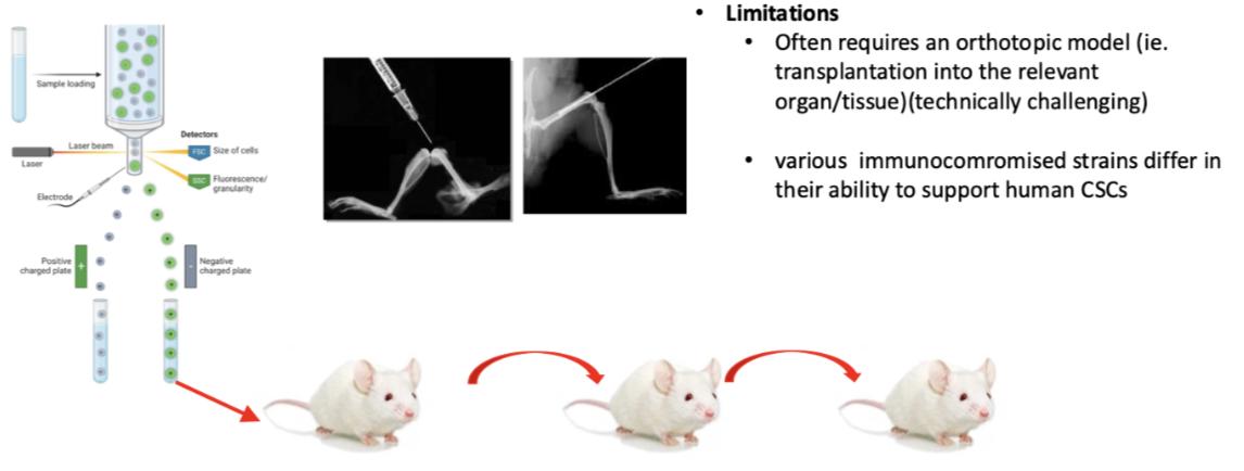
what are the limitations of the functional identification of cancer stem cells ( serial transplantation in vitro)


Hard to set up systems  

  • Easy to get to femur but hard in breast to inject into mammary fat pad and prostate as very small  

  • Often requires an orthotopic model (ie. transplantation into the relevant organ/tissue)(technically challenging)

  • Also need to be in immune compromised mice so foreign human cells aren't recognised- different immunocompromised strains of animals differ in ability to support human CSCs so results don’t necessarily mean there is specific cancer types in sample  

21
New cards

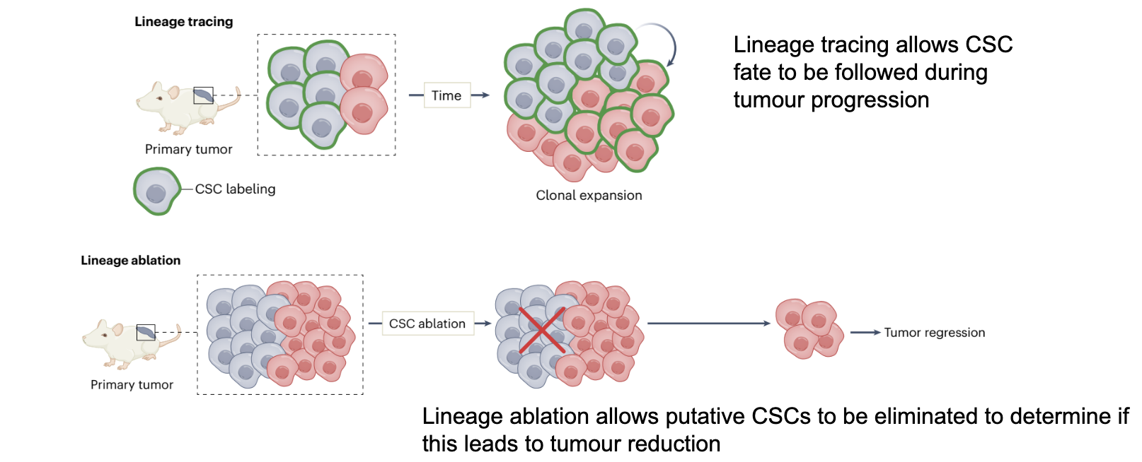
what is Lineage tracing and lineage ablation

Stem cells can also be functionally identified by lineage tracing/ablation experiments

  1. Lineage tracing allows CSC fate to be followed during tumour progression

  2. Lineage ablation allows putative CSCs to be eliminated to determine if this leads to tumour reduction

<p>Stem cells can also be functionally identified by lineage tracing/ablation experiments</p><ol><li><p>Lineage tracing allows CSC fate to be followed during tumour progression</p></li><li><p>Lineage ablation allows putative CSCs to be eliminated to determine if this leads to tumour reduction</p></li></ol><p></p>
22
New cards

What happens when cancer stem cells (CSCs) are specifically targeted in therapy?

  • CSC-targeted therapy removes tumour-initiating cells

  • Tumour cannot sustain itself

  • Leads to gradual tumour degeneration

  • Prevents long-term regrowth

<ul><li><p><strong>CSC-targeted therapy removes tumour-initiating cells</strong></p></li><li><p>Tumour <strong>cannot sustain itself</strong></p></li><li><p>Leads to <strong>gradual tumour degeneration</strong></p></li><li><p>Prevents long-term regrowth</p></li></ul><p></p>
23
New cards

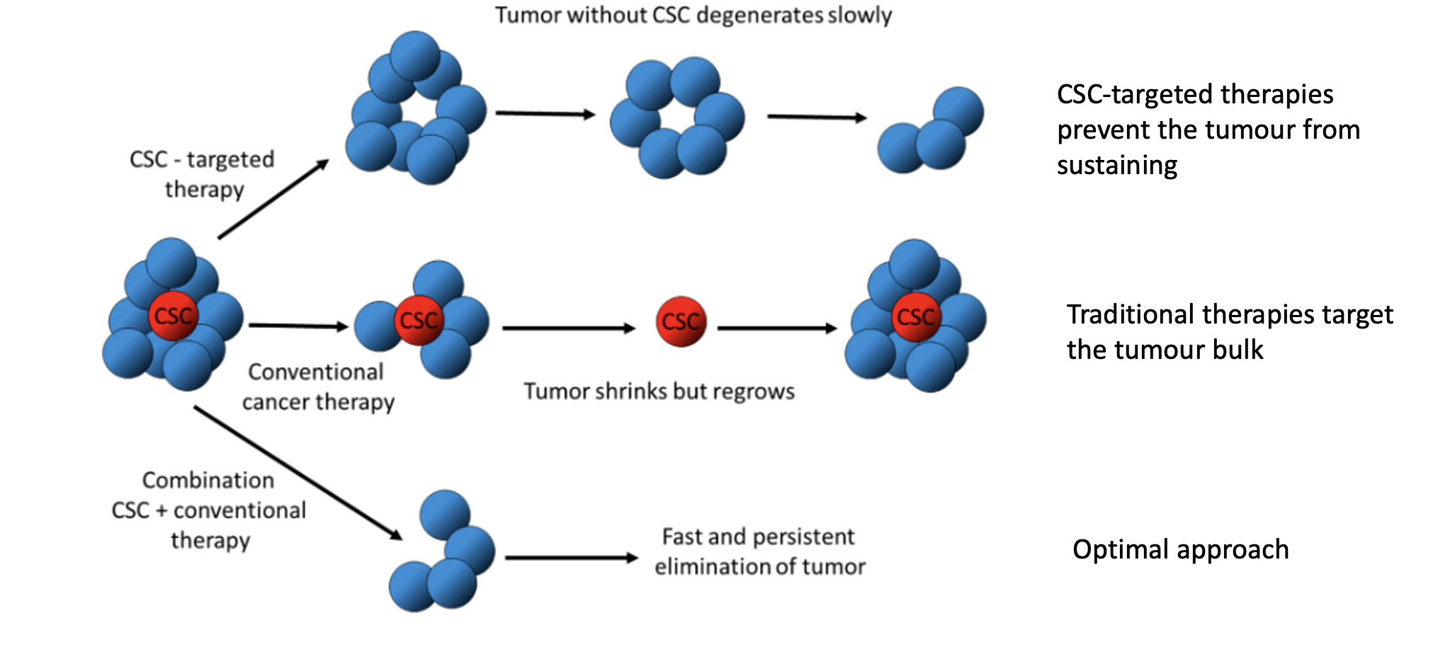
Why can conventional cancer therapies lead to tumour relapse?

  • Target mainly the bulk tumour cells, not CSCs

  • CSCs survive treatment

  • Tumour may initially shrink but regrows from CSCs

  • Explains relapse and treatment resistance

24
New cards

What is the optimal therapeutic strategy for treating cancers with CSCs?

  • Combine CSC-targeted therapy with conventional therapy

  • Eliminates both tumour bulk and stem cells

  • Leads to more complete and sustained tumour eradication

  • Results in faster and more durable response

25
New cards

what is another possible function of CSC markers

they are also attractive therapeutic targets

eg CD44 and CD133

26
New cards

how can CD44 be used as a therapeutic targets

  • CD44 is a major surface hyaluronic (HA) receptor

  • Both HA and CD44 are involved in chemotherapeutic resistance

  • CD44 can interact with P-gp (/P-glycoprotein, a major drug efflux pump) to promote cell migration and invasion of tumor cells

27
New cards

how can CD133 be used as a therapeutic targets

  • CD133, a member of the prominin family, consists of five transmembrane single-chain glycoproteins

  • CD133 can play a dominant role in drug resistance

  • CD133 can enhance the activity of histone deacetylase 6 (HDAC6) via the Wnt/β-catenin signaling pathway

28
New cards

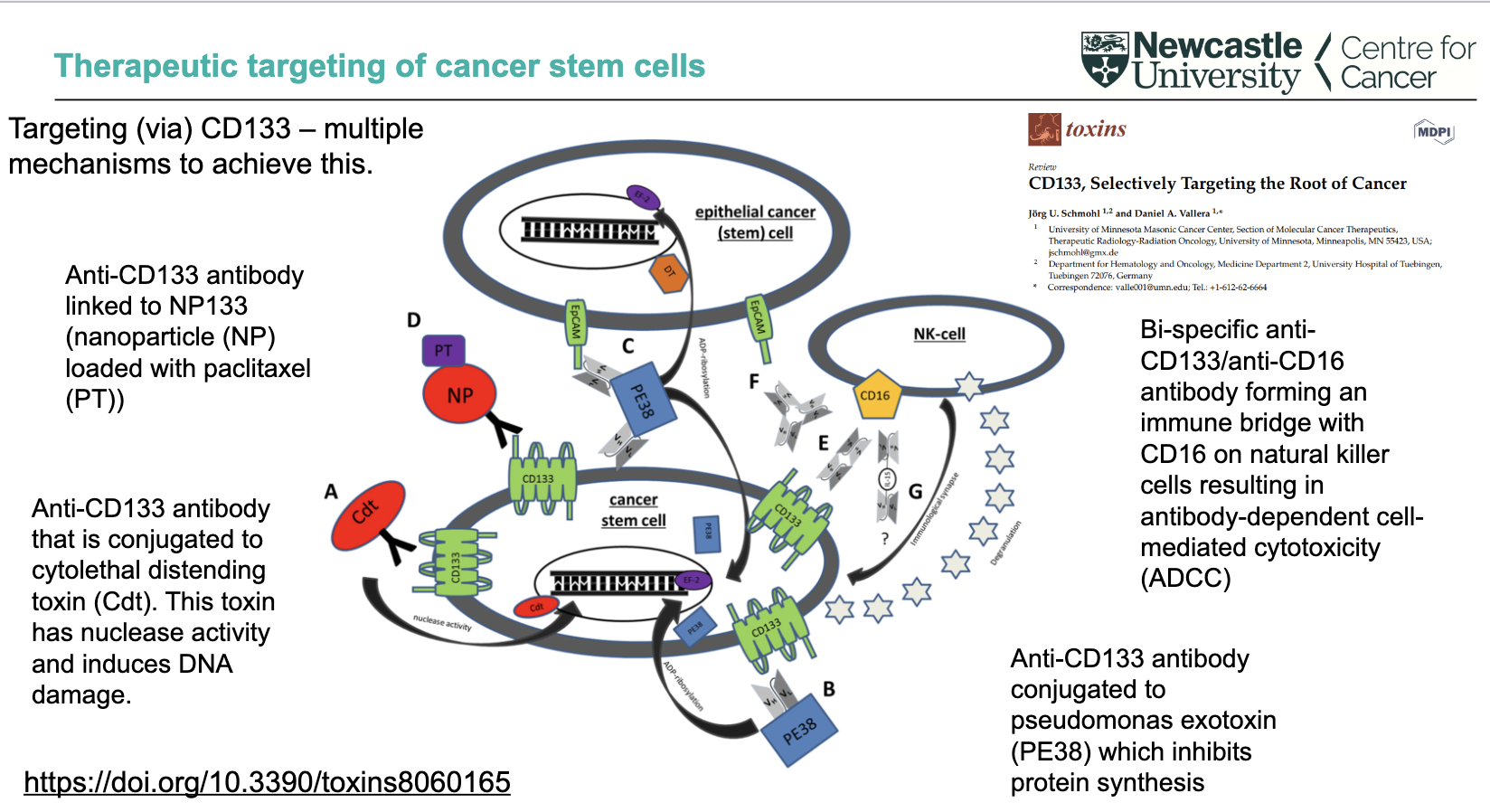
What are direct targeting strategies using CD133 in cancer stem cells?

  • Anti-CD133 antibody linked to nanoparticles (NP) loaded with paclitaxel (PT)

  • Anti-CD133 antibody conjugated to cytolethal distending toxin (Cdt) → nuclease activity → DNA damage

  • Anti-CD133 antibody conjugated to Pseudomonas exotoxin (PE38)inhibits protein synthesis

  • These approaches deliver cytotoxic agents directly to CD133⁺ cancer stem cells

<ul><li><p><strong>Anti-CD133 antibody linked to nanoparticles (NP)</strong> loaded with <strong>paclitaxel (PT)</strong></p></li><li><p><strong>Anti-CD133 antibody conjugated to cytolethal distending toxin (Cdt)</strong> → nuclease activity → <strong>DNA damage</strong></p></li><li><p><strong>Anti-CD133 antibody conjugated to Pseudomonas exotoxin (PE38)</strong> → <strong>inhibits protein synthesis</strong></p></li><li><p>These approaches deliver <strong>cytotoxic agents directly to CD133⁺ cancer stem cells</strong></p></li></ul><p></p>
29
New cards

How can CD133 be targeted via immune-mediated and multi-mechanism approaches?

  • Bi-specific anti-CD133/anti-CD16 antibody

    • Links tumour cells to NK cells (via CD16)

    • Triggers antibody-dependent cell-mediated cytotoxicity (ADCC)

  • CD133 targeting can involve multiple mechanisms simultaneously

  • Overall aim: selectively target and eliminate cancer stem cells (“root of cancer”)

<ul><li><p><strong>Bi-specific anti-CD133/anti-CD16 antibody</strong></p><ul><li><p>Links tumour cells to <strong>NK cells (via CD16)</strong></p></li><li><p>Triggers <strong>antibody-dependent cell-mediated cytotoxicity (ADCC)</strong></p></li></ul></li><li><p>CD133 targeting can involve <strong>multiple mechanisms simultaneously</strong></p></li><li><p>Overall aim: <strong>selectively target and eliminate cancer stem cells (“root of cancer”)</strong></p></li></ul><p></p>
30
New cards

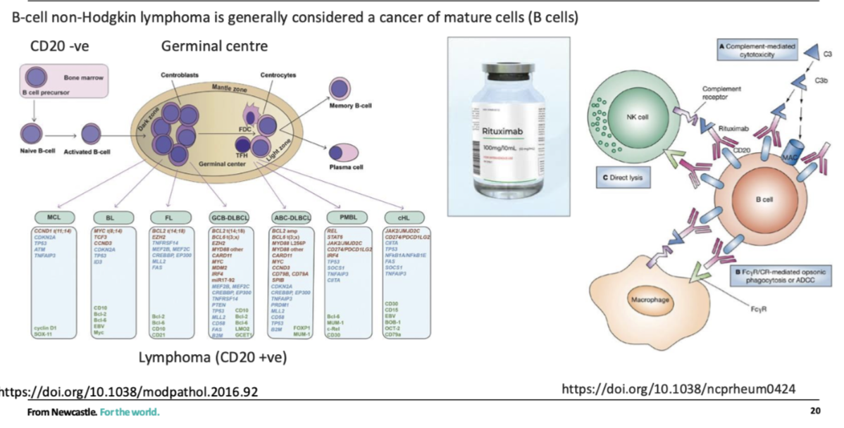
how can normal stem cells be protected during cncer treatment

Protect normal stem cells  

  • Eg need HSCs to repopulate normal haematopoiesis in bone marrow  


Lymphomas- positive for CD20, HSCs In bone marrow are CD20-ve, so can target CD20 +ve  

  • Makes the B cells more visible to NK cells  

<p><span style="background-color: inherit; line-height: 22px; color: windowtext;">Protect normal stem cells </span><span style="line-height: 22px; color: windowtext;">&nbsp;</span></p><ul><li><p class="Paragraph SCXO103470534 BCX0" style="text-align: left;"><span style="background-color: inherit; line-height: 22px; color: windowtext;">Eg need HSCs to repopulate normal haematopoiesis in bone marrow </span><span style="line-height: 22px; color: windowtext;">&nbsp;</span></p></li></ul><p class="Paragraph SCXO103470534 BCX0" style="text-align: left;"><span style="line-height: 22px; color: windowtext;">&nbsp;</span></p><p class="Paragraph SCXO103470534 BCX0" style="text-align: left;"><span style="background-color: inherit; line-height: 22px; color: windowtext;">Lymphomas- positive for CD20, HSCs In bone marrow are CD20-ve, so can target CD20 +ve </span><span style="line-height: 22px; color: windowtext;">&nbsp;</span></p><ul><li><p class="Paragraph SCXO103470534 BCX0" style="text-align: left;"><span style="background-color: inherit; line-height: 22px; color: windowtext;">Makes the B cells more visible to NK cells </span><span style="line-height: 22px; color: windowtext;">&nbsp;</span></p></li></ul><p></p>
31
New cards

what is cancer cell plasticity

the ability of cancer cells to change their phenotype to affect their identity and fate

Often (but not always) this is achieved via non-mutational mechanisms (epigenetic, transcriptomic, proteomic changes)

32
New cards

what can cancer cell plasticity do

• drive transformation to a cancer phenotype

• confer resistance to treatments

• be exploited as a therapeutic option

33
New cards

What is epithelial-to-mesenchymal transition (EMT) and where is it seen?

  • EMT = cells switch from epithelial to mesenchymal phenotype (with reverse process = MET)

  • Occurs naturally during embryogenesis

  • Demonstrates that cell phenotype switching is a normal biological process

  • Important concept for understanding cancer cell plasticity

34
New cards

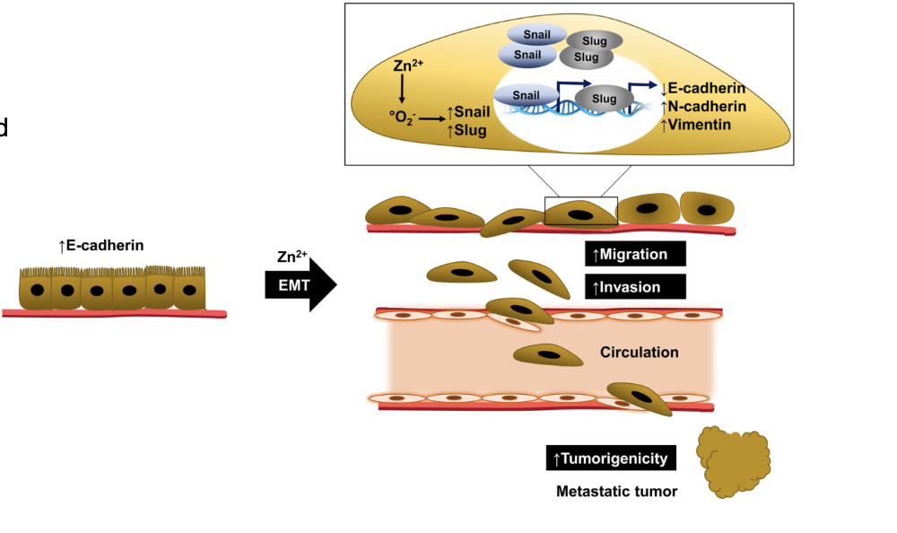
what is EMT (epithelial-mesenchymal transition)

a process whereby polarized epithelial cells (which normally interact with basement membrane) assume a mesenchymal cell phenotype, thereby gaining migratory and invasive properties.

  • And becomes unpolarised, allowing it to migrate  

35
New cards

what does EMT state correlate with

in carcinomas, which arise from epithelial cells, the EMT state correlated with tumour stage, disease aggression and patient outcome.

  • High grade disease – poorly differentiated

  • Low grade disease – well differentiated

36
New cards

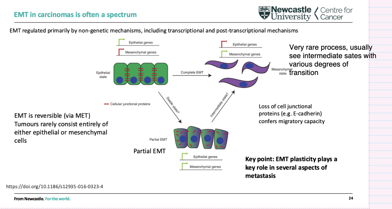
What is EMT regulated by

EMT (& MET) regulated primarily by non- genetic mechanisms, including transcriptional and post-transcriptional mechanisms

<p>EMT (&amp; MET) regulated primarily by non- genetic mechanisms, including transcriptional and post-transcriptional mechanisms</p><p></p>
37
New cards

How can environmental exposures drive cancer through cell plasticity?

  • Cell plasticity = ability of cells to change phenotype

  • Often triggered by environmental exposures (e.g. inflammation, infection, smoking)

  • Involves processes like exposure-driven EMT

  • Leads to metaplasia/transdifferentiation

  • Can initiate pathways toward cancer development

38
New cards

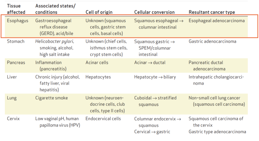
What are examples of cell plasticity leading to cancer in different tissues?

  • Oesophagus (GERD/acid reflux): squamous → columnar → oesophageal adenocarcinoma

  • Stomach (H. pylori, smoking, alcohol, salt): gastric → intestinal → gastric adenocarcinoma

  • Pancreas (inflammation): acinar → ductal → pancreatic cancer

  • Liver (chronic injury): hepatocyte → biliary → cholangiocarcinoma

  • Lung (smoking): cuboidal → squamous → squamous cell carcinoma

  • Cervix (HPV): columnar squamous → cervical cancer

<ul><li><p><strong>Oesophagus (GERD/acid reflux):</strong> squamous → columnar → oesophageal adenocarcinoma</p></li><li><p><strong>Stomach (H. pylori, smoking, alcohol, salt):</strong> gastric → intestinal → gastric adenocarcinoma</p></li><li><p><strong>Pancreas (inflammation):</strong> acinar → ductal → pancreatic cancer</p></li><li><p><strong>Liver (chronic injury):</strong> hepatocyte → biliary → cholangiocarcinoma</p></li><li><p><strong>Lung (smoking):</strong> cuboidal → squamous → squamous cell carcinoma</p></li><li><p><strong>Cervix (HPV):</strong> columnar <span data-name="left_right_arrow" data-type="emoji">↔</span> squamous → cervical cancer</p></li></ul><p></p>
39
New cards

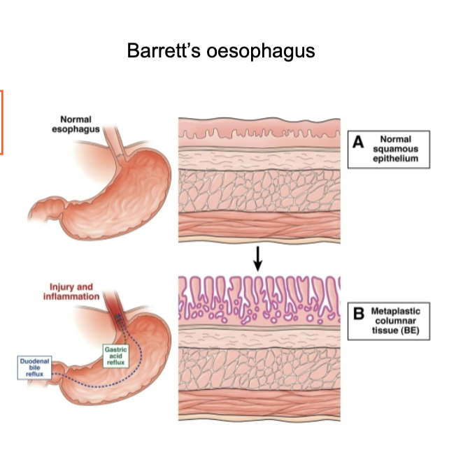
What is Barrett’s oesophagus and why is it clinically important?

  • Caused by chronic acid reflux (GERD)injury and inflammation

  • Normal squamous epithelium transforms to columnar epithelium (metaplasia)

  • Represents exposure-driven EMT / phenotype switching

  • Acts as a precursor to oesophageal adenocarcinoma

  • Key feature: transition from squamous → columnar epithelium → important for diagnosis and risk monitoring

<ul><li><p>Caused by <strong>chronic acid reflux (GERD)</strong> → <strong>injury and inflammation</strong></p></li><li><p>Normal <strong>squamous epithelium transforms to columnar epithelium</strong> (metaplasia)</p></li><li><p>Represents <strong>exposure-driven EMT / phenotype switching</strong></p></li><li><p>Acts as a <strong>precursor to oesophageal adenocarcinoma</strong></p></li><li><p>Key feature: <strong>transition from squamous → columnar epithelium</strong> → important for diagnosis and risk monitoring</p></li></ul><p></p>
40
New cards

What are the key features of EMT in carcinomas?

  • EMT exists as a spectrum, with many intermediate (partial EMT) states

  • Rarely complete—tumours usually contain a mix of epithelial and mesenchymal features

  • Reversible process (via MET)

  • Driven mainly by non-genetic mechanisms (transcriptional/post-transcriptional)

  • Loss of cell junction proteins (e.g. E-cadherin) → increased migration

  • EMT plasticity is important for metastasis

<ul><li><p>EMT exists as a <strong>spectrum</strong>, with many <strong>intermediate (partial EMT) states</strong></p></li><li><p><strong>Rarely complete</strong>—tumours usually contain a mix of epithelial and mesenchymal features</p></li><li><p><strong>Reversible process</strong> (via MET)</p></li><li><p>Driven mainly by <strong>non-genetic mechanisms</strong> (transcriptional/post-transcriptional)</p></li><li><p><strong>Loss of cell junction proteins (e.g. E-cadherin)</strong> → increased migration</p></li><li><p>EMT plasticity is <strong>important for metastasis</strong></p></li></ul><p></p>
41
New cards

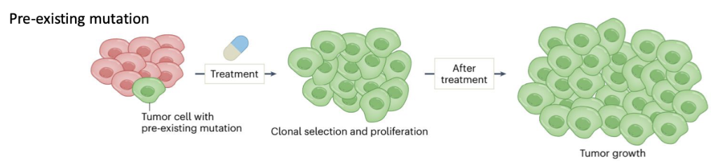
How do pre-existing mutations cause cancer therapy resistance?

  • Some tumour cells already have a pre-existing resistance mutation before treatment

  • Treatment kills sensitive cells but selects resistant clones

  • These cells undergo clonal selection and proliferation → tumour regrowth

  • Key idea: mutation is already present prior to therapy

<ul><li><p>Some tumour cells already have a <strong>pre-existing resistance mutation</strong> before treatment</p></li><li><p>Treatment kills sensitive cells but <strong>selects resistant clones</strong></p></li><li><p>These cells undergo <strong>clonal selection and proliferation</strong> → tumour regrowth</p></li><li><p>Key idea: mutation is <strong>already present prior to therapy</strong></p></li></ul><p></p>
42
New cards

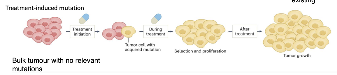
How do treatment-induced mutations lead to cancer therapy resistance?

  • Tumour initially has no relevant resistance mutations

  • Exposure to treatment (agents) induces mutations in one or more cells

  • Mutations occur in key genes → resistant phenotype

  • These cells are then selected and proliferate during treatment

  • Leads to tumour regrowth after therapy

<ul><li><p>Tumour initially has <strong>no relevant resistance mutations</strong></p></li><li><p><strong>Exposure to treatment (agents)</strong> induces mutations in one or more cells</p></li><li><p>Mutations occur in <strong>key genes → resistant phenotype</strong></p></li><li><p>These cells are then <strong>selected and proliferate during treatment</strong></p></li><li><p>Leads to <strong>tumour regrowth after therapy</strong></p></li></ul><p></p>
43
New cards

How does cellular plasticity contribute to therapy resistance (non-genetic)?

  • Resistance often associated with a mesenchymal state

  • Driven by non-genetic mechanisms (transcriptional changes)

  • Multiple resistance mechanisms (e.g. ↓ drug influx transporters)

  • EMT plasticity linked to immunotherapy resistance

    • Resistance to dendritic and cytotoxic T-cell killing

    • Creation of an immunosuppressive microenvironment

<ul><li><p>Resistance often associated with a <strong>mesenchymal state</strong></p></li><li><p>Driven by <strong>non-genetic mechanisms</strong> (transcriptional changes)</p></li><li><p>Multiple resistance mechanisms (e.g. <strong>↓ drug influx transporters</strong>)</p></li><li><p>EMT plasticity linked to <strong>immunotherapy resistance</strong></p><ul><li><p>Resistance to <strong>dendritic and cytotoxic T-cell killing</strong></p></li><li><p>Creation of an <strong>immunosuppressive microenvironment</strong></p></li></ul></li></ul><p></p>
44
New cards

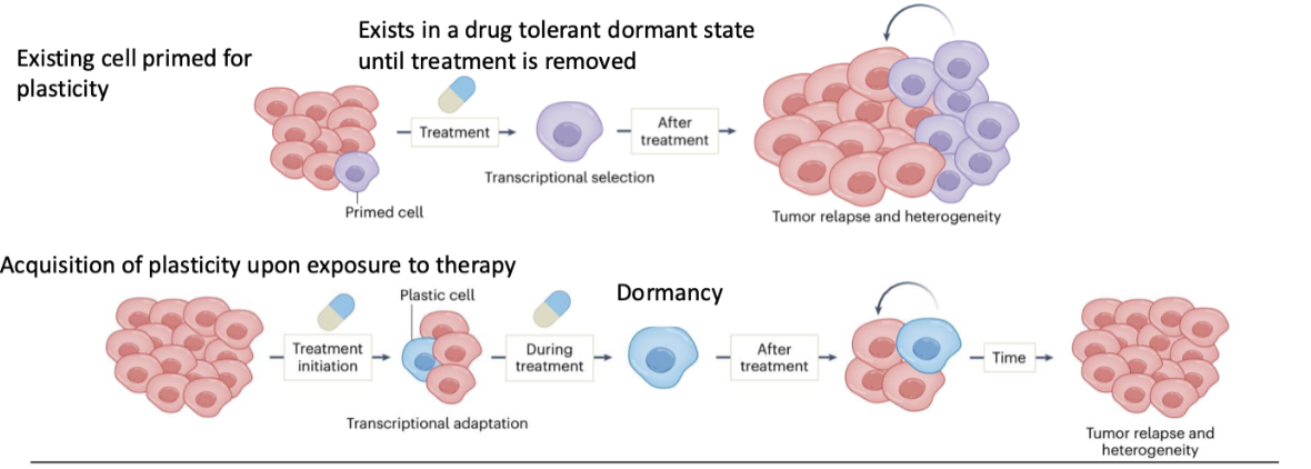
How can pre-existing plasticity lead to therapy resistance?

  • Some cells are already primed for plasticity

  • Under treatment → transcriptional selection

  • Cells enter a drug-tolerant dormant state

  • Survive until treatment stops

  • Then re-expand → tumour relapse and heterogeneity

<ul><li><p>Some cells are <strong>already primed for plasticity</strong></p></li><li><p>Under treatment → <strong>transcriptional selection</strong></p></li><li><p>Cells enter a <strong>drug-tolerant dormant state</strong></p></li><li><p>Survive until treatment stops</p></li><li><p>Then <strong>re-expand → tumour relapse and heterogeneity</strong></p></li></ul><p></p>
45
New cards

How can plasticity be acquired during therapy and lead to relapse?

  • Cells adapt during treatment (transcriptional adaptation)

  • Gain plastic phenotype in response to therapy

  • Enter dormancy during treatment

  • After treatment → cells reactivate and proliferate

  • Leads to tumour relapse and increased heterogeneity

<ul><li><p>Cells <strong>adapt during treatment</strong> (transcriptional adaptation)</p></li><li><p>Gain <strong>plastic phenotype</strong> in response to therapy</p></li><li><p>Enter <strong>dormancy</strong> during treatment</p></li><li><p>After treatment → cells <strong>reactivate and proliferate</strong></p></li><li><p>Leads to <strong>tumour relapse and increased heterogeneity</strong></p></li></ul><p></p>
46
New cards

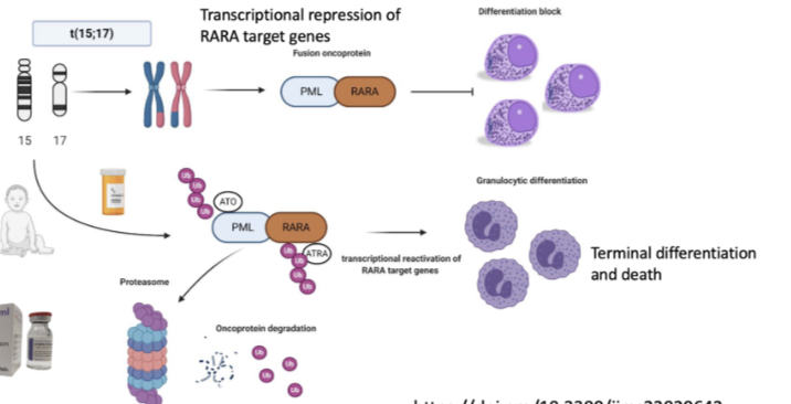
How does acute promyelocytic leukaemia (APL) arise and why is it dangerous?

  • Caused by t(15;17) translocation → fusion of PML + RARA genes

  • Forms PML-RARA fusion oncoprotein

  • Leads to transcriptional repression of RARA target genes

  • Causes a block in differentiation (promyelocytes cannot mature)

  • Results in accumulation of immature cells

  • Associated with extremely high risk of fatal bleeding → requires immediate treatment

<ul><li><p>Caused by <strong>t(15;17) translocation</strong> → fusion of <strong>PML + RARA genes</strong></p></li><li><p>Forms <strong>PML-RARA fusion oncoprotein</strong></p></li><li><p>Leads to <strong>transcriptional repression of RARA target genes</strong></p></li><li><p>Causes a <strong>block in differentiation</strong> (promyelocytes cannot mature)</p></li><li><p>Results in accumulation of immature cells</p></li><li><p>Associated with <strong>extremely high risk of fatal bleeding → requires immediate treatment</strong></p></li></ul><p></p>
47
New cards

How does APL demonstrate transcriptional reprogramming in cancer?

  • Fusion event alters transcriptional control (not just DNA mutation effect)

  • Target protein is transcriptionally changed → reprogrammed state

  • Causes differentiation block in myeloid cells

  • Shows cancer can be driven by changes in cell state (plasticity), not just mutations

<ul><li><p>Fusion event <strong>alters transcriptional control</strong> (not just DNA mutation effect)</p></li><li><p><strong>Target protein is transcriptionally changed → reprogrammed state</strong></p></li><li><p>Causes <strong>differentiation block</strong> in myeloid cells</p></li><li><p>Shows cancer can be driven by <strong>changes in cell state (plasticity), not just mutations</strong></p></li></ul><p></p>
48
New cards

How is APL treated and what does this show about targeting cellular plasticity?

  • Treated with all-trans retinoic acid (ATRA) and arsenic trioxide (ATO)

  • These therapies degrade the PML-RARA fusion oncoprotein (via proteasome)

  • Leads to reactivation of RARA target genes

  • Cells undergo granulocytic differentiation → terminal differentiation and death

  • Cell returns to a more normal transcriptional state

  • Key point: therapy does not reverse the genetic mutation, but changes the transcriptional state to treat cancer

<ul><li><p>Treated with <strong>all-trans retinoic acid (ATRA)</strong> and <strong>arsenic trioxide (ATO)</strong></p></li><li><p>These therapies <strong>degrade the PML-RARA fusion oncoprotein</strong> (via proteasome)</p></li><li><p>Leads to <strong>reactivation of RARA target genes</strong></p></li><li><p>Cells undergo <strong>granulocytic differentiation → terminal differentiation and death</strong></p></li><li><p>Cell returns to a more <strong>normal transcriptional state</strong></p></li><li><p>Key point: therapy <strong>does not reverse the genetic mutation</strong>, but <strong>changes the transcriptional state to treat cancer</strong></p></li></ul><p></p>
49
New cards

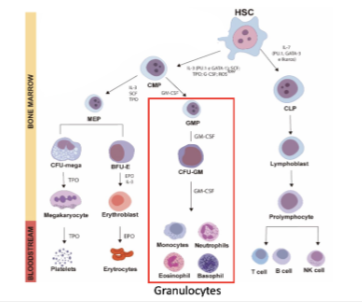
How does acute promyelocytic leukaemia (APL) relate to normal haematopoietic differentiation?

  • Occurs within the myeloid lineage (granulocyte pathway)

  • Normal pathway:

    • HSC → CMP → GMP → CFU-GM → granulocytes (neutrophils, eosinophils, basophils)

  • APL causes a block at the promyelocyte stage (failure of differentiation)

  • Demonstrates that cancer can arise from disruption of normal differentiation hierarchy

  • Key concept: cellular plasticity + differentiation block → disease

<ul><li><p>Occurs within the <strong>myeloid lineage (granulocyte pathway)</strong></p></li><li><p>Normal pathway:</p><ul><li><p><strong>HSC → CMP → GMP → CFU-GM → granulocytes (neutrophils, eosinophils, basophils)</strong></p></li></ul></li><li><p>APL causes a <strong>block at the promyelocyte stage</strong> (failure of differentiation)</p></li><li><p>Demonstrates that cancer can arise from <strong>disruption of normal differentiation hierarchy</strong></p></li><li><p>Key concept: <strong>cellular plasticity + differentiation block → disease</strong></p></li></ul><p></p>
50
New cards

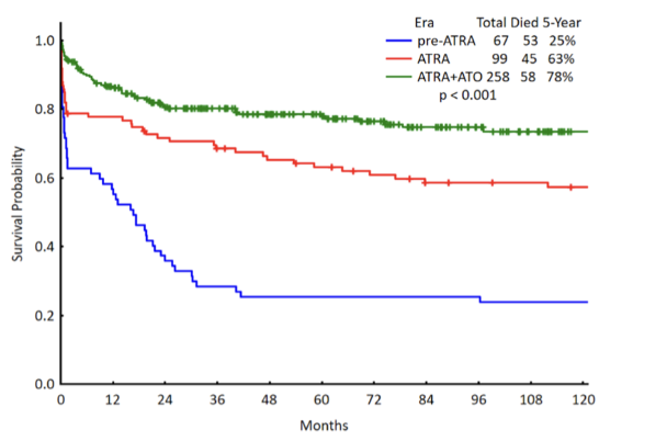
How has treatment of APL improved survival outcomes?

  • Historical (pre-ATRA): very poor survival

  • ATRA alone improves outcomes

  • ATRA + arsenic trioxide (ATO) gives best outcomes

  • Dramatic increase in long-term survival probability

  • Current complete cure rate ≈ 94%

  • Demonstrates success of targeting cellular plasticity (differentiation therapy)

<ul><li><p>Historical (pre-ATRA): <strong>very poor survival</strong></p></li><li><p><strong>ATRA alone</strong> improves outcomes</p></li><li><p><strong>ATRA + arsenic trioxide (ATO)</strong> gives best outcomes</p></li><li><p>Dramatic increase in <strong>long-term survival probability</strong></p></li><li><p><strong>Current complete cure rate ≈ 94%</strong></p></li><li><p>Demonstrates success of <strong>targeting cellular plasticity (differentiation therapy)</strong></p></li></ul><p></p>
51
New cards

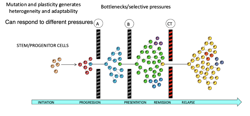
How does tumour heterogeneity contribute to cancer adaptability and progression?

  • Mutation + plasticity → tumour heterogeneity

  • Tumours contain diverse subclones that can respond to different selective pressures

  • Disease progression involves bottlenecks/selective pressures (A, B, CT)

  • Only certain clones survive and expand through each stage

  • Leads to stages: initiation → progression → presentation → remission → relapse

  • Key idea:

    • Heterogeneity allows cells to adapt, survive, and repopulate tumours

    • Enables cells to pass through bottlenecks and drive aggressive cancer phenotypes

<ul><li><p><strong>Mutation + plasticity → tumour heterogeneity</strong></p></li><li><p>Tumours contain diverse subclones that can <strong>respond to different selective pressures</strong></p></li><li><p>Disease progression involves <strong>bottlenecks/selective pressures</strong> (A, B, CT)</p></li><li><p>Only certain clones <strong>survive and expand</strong> through each stage</p></li><li><p>Leads to stages: <strong>initiation → progression → presentation → remission → relapse</strong></p></li><li><p>Key idea:</p><ul><li><p>Heterogeneity allows cells to <strong>adapt, survive, and repopulate tumours</strong></p></li><li><p>Enables cells to <strong>pass through bottlenecks and drive aggressive cancer phenotypes</strong></p></li></ul></li></ul><p></p>