Brain + Spinal Cord
1/56
There's no tags or description
Looks like no tags are added yet.
Name | Mastery | Learn | Test | Matching | Spaced | Call with Kai |
|---|
No analytics yet
Send a link to your students to track their progress
57 Terms
Top to bottom
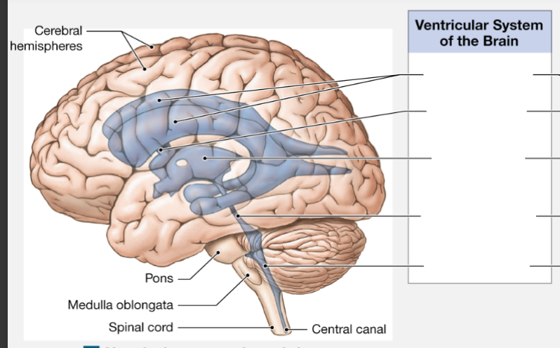
lateral ventricles
interventricular foramen
third ventricle
cerebral aqueduct
fourth ventricle
Cervical Vertebrae
Nerves
7 bones
C1 - C8
Thoracic Vertebrae
Nerves
12 bones
T1-T12
Lumbar Vertebrae
Nerves
5 bones
L1 - L5
Sacrum
Nerves
Just one bone
S1 - S5
Epineurium
Covers the peripheral nerve
superficial
Perineurium
Around one fascicle of the peripheral nerve
between the epineurium and the endoneurium
Endoneurium
inside the fascicle, covering the myelinated axon of the peripheral nerve
deep
Spinal Nerves
Mixed nerves. Contain both afferent (sensory) and efferent (motor) fibers
Anterior (ventral) Root
Posterior (dorsal) Root
Spinal (dorsal root) ganglion
Anterior (ventral) Root
axons of motor neurons
Posterior (dorsal) Root
Axons of sensory neurons
Spinal (dorsal) ganglion
cell bodies of sensory neurons just outside of the spinal cord
T or F: the spinal cord is not as long as vertebral column
True
Dermatome
the region of the skin innervated by nerves that originate from a single spinal nerve pair.
Meninges (Superficial to deep) in the spinal cord
Dura mater: thick and adheres to the vertebrae
Arachnoid mater: web-like, delicate layer
Pia mater: adheres directly spinal cord
Reflex arc
The wiring of a single reflex that includes 5 steps
arrival of stimulus and activation of receptor
activation of a sensory neuron
information processing in the CNS
activation of a motor neuron
Response by a peripheral effector
Reflex Organization (4 categories)
Development: innate or required
Response: somatic or visceral (autonomic)
Complexity: monosynaptic or polysynaptic
Processing site: cranial reflex or spinal reflex
Stretch Reflex
Monosynaptic reflex that control the most rapid motor responses of the nervous system. Provides automatic regulation of skeletal muscle length using muscle spindles
Muscle Spindles
inner part made of intrafusal fibers associated with special sensory neurons
outer part made of extrafusal fibers
Withdrawal Reflex
Makes you move away from pain.
Reciprocal Inhibition: flexors stimulated; extensor inhibited or vice versa
Crossed Extensor Reflex
If you were to step on something painful. Makes both side of the body work
Ipsilateral: the side receiving the painful stimulus. Pulls away
Contralateral: the other side of the body used for stabling
Primary Brain Vesicles
Prosencephalon: forebrain
Mesencephalon: midbrain
Rhombencephalon: hindbrain
At 3 weeks
Secondary Brain Vesicles
Prosencephalon splits into telencephalon (cerebrum) and the diencephalon
Mesencephalon (midbrain)
Rhombencephalon splits into metencephalon (cerebellum and pons) and myelencephalon (medulla oblongata)
Meninges in the brain (superficial to deep)
Periosteal cranial dura
Dural sinus
Meningeal cranial dura
arachnoid mater
pia mater
1 and 3 make up the dura mater
Dural Folds
Falx cerebri: dura mater growing down separating the left and right hemispheres
Falx cerebelli: growing between the right and left hemispheres of cerebellum
Tentorium cerebelli: growing between top and bottom of brain separating cerebellum from cerebrum
Superior sagittal sinus
wraps around the midline perimeter of brain
Functions of CSF
supports the brain
cushions the brain and spinal cord against physical trauma
transports nutrients, chemical messages, and wastes
Choroid Plexus
Makes the CSF inside the 3rd and 4th ventricles. Made up of ependymal cells anchores by tight junctions.
Arachnoid granulations
where arachnoid mater punctures through the dura mater creating a valve where CSF fluid can drain into the superior sagittal sinus
Blood supply to brain
internal carotid and vertebral arteries. Brain is protected by blood brain barrier made by astrocytes.
Reflex centers of the Medulla Oblongata
Reticular formation: Regulate autonomic functions like
breathing, blood pressure, thermoregulation. Regulates
some endocrine functions. Body posture, somatic reflexes,
alertness, sleep.
Cardiovascular centers: Regulates heart rate, force of
cardiac contractions, and peripheral blood flow.
Respiratory rhythmicity centers: regulate breathing rate
Decussation of pyramids
Somatic sensory information leaving the medulla oblongata to go to the thalamus, cross over to the opposite sides of the brain
Medulla oblongata cranial nerves
8-12
CN. VIII
vestibulocochlear nerve
Sensory: vestibular (balance) and cochlear (hearing) systems of the ear
CN IX
glossopharyngeal
sensory: taste from posterior 1/3 of tongue
motor: some salivary glands and one muscle of the throat
CN X
Vagus nerve
Sensory and motor: most muscles of the throat for swallowing and speaking, smooth muscle in most visceral organs
CN XI
Accessory nerve
Some overlap with CN X.
Motor: controls of some muscles in the neck (trapezius)
CN XII
Hypoglossal nerve
Motor: control of tongue
Pons
Relays sensory info to the thalamus and cerebellum
White matter: relays info to other brain regions
Gray matter: functions overlap with other brain regions
Apneustic and pneumotaxic centers: adjust activities of the respiratory rhythmicity in the medulla oblongata
Pons cranial nerves
5-8
CN V
Trigeminal nerve
Sensory: face
Motor: muscles of jaw for chewing
CN VI
Abducens nerve
Motor: muscle that abducts the eyes
CN VII
Facial nerve
Sensory: taste from the anterior 2/3 of tongue
Motor: facial expressions and some salivary glands
Midbrain
Corpora quadrigemina
Superior colliculi: control reflex movement of eyes, head, neck in response to visual stimuli
Inferior Colliculi: control reflex movement of head, neck, and trunk in response to auditory stimuli
Substantia nigra: Appears black from presence of melanin. Secretes dopamine. Inhibits activity of basal nuclei in cerebrum, which control subconscious muscle tone and learned movements.
Midbrain cranial nerves
3 and 4
CN III
Oculomotor nerve
motor: muscles that move the eye and constrict the pupil
CN IV
Trochlear nerve
Motor: muscle that moves the eye
Functions of the cerebellum
1. Adjusting the postural muscles of the body
Alters muscle tone to maintain balance
Modifies activity of motor centers in brain stem
2. Fine-tuning movement controlled at conscious and subconscious level
Refines learned movement patters
Compares motor commands with proprioceptive information and makes
adjustments so movements are smooth
Works indirectly by regulating activity of motor pathways in other brain regions
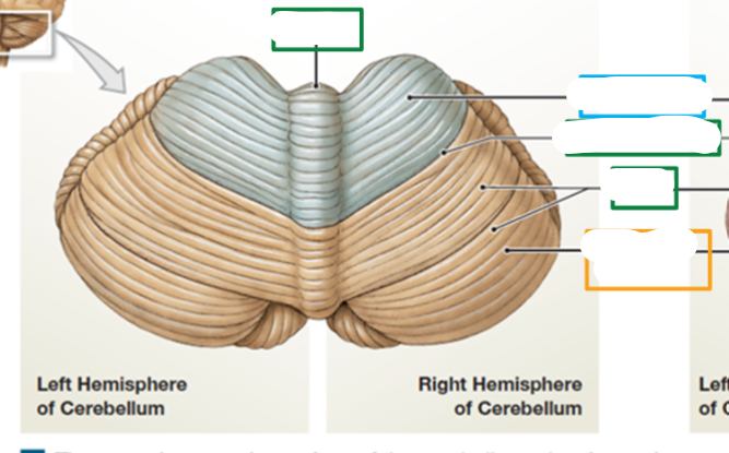
top to bottom
vermis
anterior lobe
primary fissures
folia
posterior lobe