OB 2 Wk 9 Quiz
 Call Kai
 Learn
Learn Practice Test
 Spaced Repetition
 Match
Match Flashcards
 Knowt Play
1/123

There's no tags or description
Looks like no tags are added yet.
Name | Mastery | Learn | Test | Matching | Spaced | Call with Kai |
|---|
No analytics yet
Send a link to your students to track their progress
124 Terms
acronym to help with sonographic detection of heart disease
CHRISTMAS
most heart diseases are commonly associated with trisomy
21
- ASD
- tetralogy of fallot
- double outlet RV
- coarctation of AO
- hypoplastic left heart
cardiac anomalies associated with chromosomal defects
the most common severe congenital abnormality
congenital heart disease
normal heart positioning
levocardia
heart is in right chest, with the apex pointing to the right of thorax
dextrocardia
heart is in right side of chest with cardiac apex pointing medially or to the left
dextroposition
dextroposition is normally found with ________ factors
intrinsic
heart is displaced further towards the left chest
levoposition
cardiac apex is pointing towards the midline of the chest
mesocardia
disease of myocardial tissue in the heart, due to exposure to virus or bacteria, errors of metabolism, and endocardial fibroelastosis
cardiomyopathy
characterized by necrosis and destruction of myocardial cells and inflammatory infiltrate
myocarditis
with viral myocarditis, all 4 chambers will be
dilated with thin walls
abnormal collection of fluid surrounding the epicardial layer of the heart
pericardial effusion
normal hypoechoic area in the peripheral part of the epicardial/pericardial interface should be
2mm or less
pericardial effusion is separation greater than
2mm
defect that creates a communication between the right and left atrium
atrial septal defect
most common atrial septal defect, but most difficult to see
osteum secundum
atrial septal defect associated with trisomy 21
osteum primum
least common atrial septal defect, located near entrance of SVC into RA
sinus venosus
failure of the ________ to close may cause an atrial septal defect
foramen ovale
most common congenital lesion of the heart
ventricular septal defect
ventricular defects smaller than ______ are not detected by US
2mm
VSD has a ______ prognosis
good
AKA endocardial cushion defect or AV malformation
atrioventricular septal defect
defect in the atrial and ventricular septum --resulting in a defect in then crux of the heart
AVSD
caused by failure of the common AV orifice to separate into the mitral and tricuspid valves
AVSD
there is an increased incidence of
- down syndrome
- asplenia
- polysplenia
AVSD
interruption of the growth of tricuspid leaflets causing the valve to be hypoplastic or atresic
tricuspid atresia
in the 4 chamber view, there will be:
- large, dilated LV
- small, underdeveloped RV
- mitral valve is dominant
- echogenic tricuspid annulus
tricuspid atresia
abnormal displacement of the septal leaflet of the tricuspid valve, towards the apex of the RV
Ebsteins anomaly
with ebstein's anomaly, the ________ will be massively dilated
right atrium
- malformed tricuspid valve
- atrialized portion of RV
- reduced capacity of pumping portion of RV
3 factors causing abnormal function of right side of heart
defect where right heart is underdeveloped, due to obstruction of RVOT secondary to pulmonary stenosis
hypoplastic right heart
on US:
small RV and RA
small tricuspid valve
hypoplastic right heart
defect where the left heart is underdeveloped
hypoplastic left heart
there will be a hypertrophied left ventricle with a hypoechoic, enlarged appearance
hypoplastic left heart
a mother who had a child with a left heart abnormality has a significantly ______ risk of delivering another child with same abnormality
higher
the risk of left heart abnormalities ________ significantly with each pregnancy
increases
most common form of cyanotic heart disease in infants and children
tetralogy of fallot
tetralogy of fallot consists of _____ abnormalities
4
- VSD
- over-riding AO
- pulmonary stenosis
- RV hypertrophy
4 abnormalities associated with tetralogy of fallot
large septal defect with mild to moderate pulmonary stenosis
acyanotic disease
large septal defect with severe pulmonary stenosis, will have "blue baby" at birth
cyanotic disease
if the aorta overrides over 50%, it is termed
double-outlet right ventricle
this means that both the great vessels arise from the right side of the heart
double-outlet right ventricle
most common form of RVOT obstruction
pulmonary stenosis
abnormal pulmonic cusps become thickened and the main pulmonary artery may become hypoplastic
pulmonary stenosis
when the AO is abnormally connected to the right ventricle and the MPA is abnormally connected to the left ventricle
transposition of the great arteries
this view is key in imaging the great arteries and their relationship
RVOT
in normal RVOT, the pulmonary artery is seen _______ to AO
anterior
in transposition, the relationship between AO and MPA is
not present
in LVOT, you should see a normal criss crossing pattern--with transposition this is not possible and the great arteries are seen ________
parallel
a heart lesion in which only 1 great artery arises from the base of the heart
truncus arteriosus
on US:
an abnormal, large, single vessel arising from the ventricle
truncus arteriosus
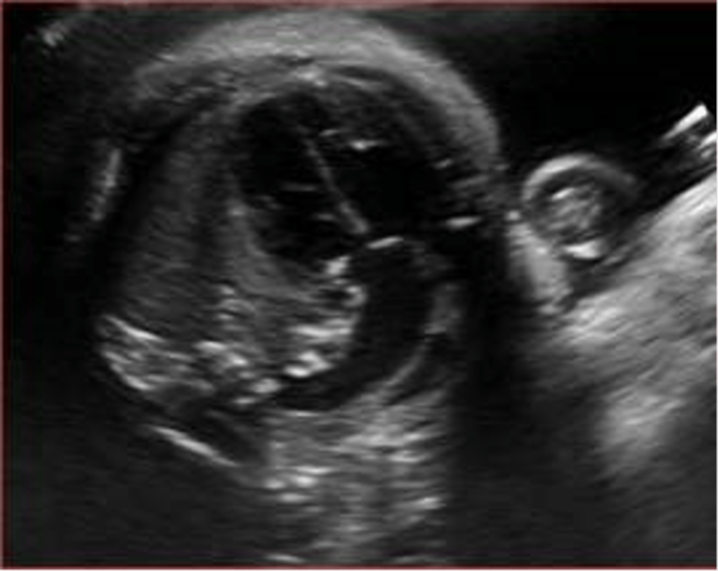
shelf like lesion in the isthmus of the aortic arch
coarctation of the AO
narrowing or kink in the AO
coarctation of the AO
- aortic stenosis
- aortic insufficiency
- septal defects
- transposition
- truncus arteriosus
- double outlet RV
associated intracardiac malformations seen with AO coarctation
most cardiac tumors are
benign and isolated
less than 10% of cardiac tumors are
malignant
most common heart lesion
rhabdomyoma
second most common heart lesion
teratoma
rhabdomyoma is associated with
Tuberous sclerosis
rhabdomyoma is best seen in _________ view
4 chamber
consists of 2 atria and 1 ventricle
single ventricle defect
1 ventricle receives both the mitral and tricuspid valves
single ventricle defect
abnormal development of the heart outside of the chest
ectopia cordis
seen as an echogenic structure within the heart as bright as bone
echogenic intracardiac focus
echogenic intracardiac focus is seen in __________ of heart
left ventricle
echogenic intracardiac focus can be normal or associated with
trisomy 21
develops from caudal region of the primitive heart
sinus venosus
receives all blood returning to the heart from the common cardinal veins, vitelline veins, and umbilical veins
sinus venosus
develops into right and left atria
primitive atrium
develops into left ventricle
primitive ventricle
develops into right ventricle
bulbus cordis
dilates to form aortic sac from which the aortic arches arise
truncus arteriosus
devision of 4 chambers occurs during the ________ weeks if fetal development
4th, 5th
develop in the atrioventricular region of heart and fuse to divide into the atrioventricular canal
endocardial cushions
allows communication from the right atrium to the left atrium
foramen ovale
foramen oval opens towards the
L atrium
allows fetal circulation to bypass the lungs
ductus arteriosus
allows for communication from the pulmonary artery to the aortic arch
ductus arteriosus
allows fetal circulation to bypass the liver
ductus venosus
blood passes from the umbilical vein, through the ductus venosus, into the
IVC
at birth, placental blood flow is interrupted causing oxygen levels to ________ and carbon dioxide levels to _______
decrease , increase
this causes the newborn to take the first breath and begin neonatal lung function
decrease in oxygen and increase in carbon dioxide
after first breath, the lungs fill with air vs fluid and allows for a _________ in vascular resistance and an _________ in pulmonary blood flow
decrease , increase
pressure in the left atrium becomes _______ than in the right atrium and causes the foamed ovale to close
higher
if the foramen ovale does not close at birth, it is termed
patent foramen ovale
with a patent foramen ovale, flow is often _______ or can become ________
reversed
mixed
the ductus arteriosus becomes the _________ in the neonate
ligamentum arteriosum
normal fetal HR
120-160
bradycardia is heart rate below
100
tachycardia is heart rate above
200
- IUGR
- cardiac arrhythmias
- abnormal amniocentesis
- abnormal AF collections
- abnormal HR
- thickened NT
- hydrops
fetal indicators for an echo
- previous child with heart disease
- parent with heart disease
- diabetes
- lupus
- drug/alcohol abuse
maternal indicators for an echo
apex of heart should be at a ______ angle to midline
45
- 4 chamber heart
- RVOT and LVOT
- aortic arch
- ductal arch
normal cardiac study views
most commonly acquired view of the fetal heart
4 chamber
moderator band is seen in
right ventricle