proteins,enzymes,nucleic acids,DNA exam qs
 Call Kai
 Learn
Learn Practice Test
 Spaced Repetition
 Match
Match Flashcards
 Knowt Play
1/98

There's no tags or description
Looks like no tags are added yet.
Name | Mastery | Learn | Test | Matching | Spaced | Call with Kai |
|---|
No analytics yet
Send a link to your students to track their progress
99 Terms
describe the induced fit model of enzyme action and how enzymes act as a catalyst
Substrate binds to active site/enzyme, enzyme-substrate complexes are formed, active site changes shape to be complementary to the substrate
suggest and explain a procedure scientists can use to stop an enzyme reaction
boil/add strong acid/alkali, enzyme is denatured
what is a reason an enzyme reaction graph may plateau?
all active sites are occupied
a competitive inhibitor decreases the rate of an enzyme-controlled reaction. explain how
inhibitor similar shape to substrate, binds to active site, prevents enzyme-substrate complexes forming
Describe how the structure of a protein depends on the amino acids it contains.
structure determined by position of amino acids/R group
primary structure is sequence of amino acids
secondary structure is formed by hydrogen bonding between amino acids.
tertiary structure is formed by interactions between R groups, creates an active site in enzymes
Explain how the induced fit in an active site of an enzyme causes a high rate of reaction.
lowers activation energy- induced fit causes active site of enzyme to change shape, so formation of an enzyme-substrate complex causes these bonds to break
describe a biochemical test to confirm the presence of proteins
add biurets solution, positive result is purple/lilac
A dipeptide consists of two amino acids joined by a peptide bond. Dipeptides may differ in the type of amino acids they contain. Describe two other ways in which all dipeptides are similar and one way in which they might differ.
Similar: amine/NH2 group at end, carboxyl/COOH group at end but variable groups are different
How many R groups do all dipeptides have?
2
Describe how a peptide bond is formed between two amino acids to form a dipeptide.
condensation reaction between amine and carboxyl group
The secondary structure of a polypeptide is produced by bonds between amino acids. Describe how.
Hydrogen bonds between NH and C=O
Two proteins have the same number and type of amino acids but different tertiary structures. Explain why.
different sequence of amino acids forms hydrogen, ionic and disulphide bonds in different places
Formation of an enzyme-substrate complex increases the rate of reaction. Explain how.
reduces activation energy due to bending bonds
what is an R group?
variable side chain-determines what amino acid it is
how many amino acids in human body?
20
what 3 things can the R group be
hydrogen atom, carbon chain, more complex stucture
what 4 elements are amino acids generally made up of?
carbon hydrogen oxygen nitrogen
what element do only some amino acids contain?
sulphur
basic formula of amine group
nh2
formula of carboxyl group
COOH
what reaction causes formation of dipeptides?
condensation reaction
how many h20s does one formation of a dipeptide make?
1
what is formed from a dipeptide bond?
OH and H forms water, C and N bond
are hydrophilic molecules charged or uncharged?
uncharged
are hydrophobic molecules charged or uncharged?
charged
primary structure of a protein?
The sequence of amino acids in the polypeptide chain, determined by the DNA base sequence of the gene
secondar structure of a protein?
The amino acid chain spontaneously forms one of two secondary structures- alpha helices and beta pleated sheets
what is a secondary structure protein stabilised by
Hydrogen bonds between C=O and NH sections
A tertiary protein structure is established by 3 types of bond. Describe them and put them in the order of strength
Hydrogen bonds- attractions between slightly positive and slightly negative R-groups.
Ionic bonds between positive and negative R-groups.
Disulphide bridge- covalent bonds between R-groups that contain sulphur.
Bacteria living in hot water springs (thermophiles) have more cysteine + methionine. Why?
They have more disulphide bridge covalent bonds which are very strong, so it’s less likely the proteins will denature
what is a quaternary protein structure?
More than one polypeptide chain held together by hydrogen, ionic and disulphide bonds.
2 examples of quaternary structure proteins
haemoglobin, antibodies
what are the bonds in antibodies?
disulphide bonds
how many heavy chains and how many light chains in antibody molecule
2 heavy chains, 2 light chains
what does antibody molecule look like
Y
what is haemoglobin as a quaternary protein structure?
2 alpha chains and 2 beta chains each containing a haem group to which O2 can bind
what causes molecules to always fold in the same shape in a protein?
protein has a primary structure therefore bonds form in the same position
what are enzymes
globular proteins that act as biological catalysts
why is tertiary structure of an enzyme important
active site must be complementary in shape to the substrate
each enzyme is ….. to one particular substrate or functional group
specific
why is activation energy required
energy needed to start a reaction- some bonds in reactants need to be broken even in an exothermic reaction, to form new bonds in the products
how do enzymes speed up metabolic reactions?
reduce activation energy
lock and key theory
Substrate has a complementary shape and so it fits perfectly into the active site of the enzyme, to form an enzyme-substrate complex.
induced fit theory
Enzyme has to alter its tertiary structure slightly to form an enzyme-substrate complex. This weakens the bond that needs to be broken in the substrate so activation energy is reduced.
explain why bond is weakened in induced fit theory
active site has changed shape to fit the substrate, which puts stress on this bond and weakens it. so, if the reactants collide, there’s a greater chance of a chemical reaction.
What factors affect the rate of an enzyme controlled reaction?
temperature, PH, substrate concentration
how does temperature affect enzyme action
as temp increases, enzyme +substrate molecules = more KE
more collisions occur, more enzyme-substrate complexes are formed, ROR increases.
at even higher temperatures, polypeptide chain vibrates so much that hydrogen/ionic bonds pulled apart, enzyme loses its tertiary structure, denatured
active site changes shape , so no enzyme-substrate complexes can form.
explain how PH affects enzyme action
each enzyme has optimum PH where tertiary structure is stable.
If the PH moves away from optimum, excess H+ or OH- ions can disrupt ionic + hydrogen bonds in the enzyme, loses its tertiary structure.
active site changes shape, no enzyme-substrate complexes can be formed, ROR decreases
how doe substrate concentration affect enzyme action?
as substrate conc increases, ROR increases proportionally as more enzyme-substrate complexes are formed.
at high substrate concentrations, enzymes working at maximum rate, adding more substrate =no effect on ROR, something else has to become the limiting factor.
how does enzyme concentration affect enzyme action
As enzyme conc increases, ROR increases proportionally. Double enzyme conc means double reaction rate because twice the enzyme-substrate complexes are formed.
Why are enzymes never in excess?
They are reusable, therefore present in small numbers
how to perform iodine test?
Put 2cm cubed test solution into test tube, add 2 drops iodine solution, positive result = blue/black
what tests for starch
iodine test
test for sugars
benedicts test
how to test for reducing sugars
2cm cubed test solution into test tube, add equal quantity benedict’s reagent, shake + heat for a few minutes in 95 degree water bath, positive result = green, brown or red
how to test for non reducing sugars
2cm cubed test solution into test tube, add equal quantity of dilute HCL, boil for a few mins to hydrolyse glycosidic bonds, neutralise- add small amounts of solid sodium-hydrogen-carbonate until it stops fizzing, positive result = green, brown or red
what does emulsion test test for
lipids
how to perform emulsion test
Shake some test material with 4cm cubed ethanol, filter liquid into test tube of water, leaving any solids behind, positive result: lipids precipitate in water, cloudy-white emulsion
what does biurets test test for?
proteins
how to perform biurets test
2cm cubed test solution into test tube, add equal volume biuret solution down side of test tube, POSITIVE RESULT = blue ring at surface, disappears upon shaking, solution turns lilac-purple
What are competitive inhibitors and what do they do?
molecule, has shape similar to enzyme’s normal substrate, can fit into the active site and form an enzyme-inhibitor complex
Is the action of a competitive inhibitor reversible?
Yes, the inhibitor can leave the active site
Describe what a competitive inhibitor looks like on a graph when added to a reaction, compared to no inhibitor
Increases at a slower rate, however eventually reaches the same point as no inhibitor
What is a non-competitive inhibitor and what does it do?
binds to the enzyme in a place other than the active site. this changes the tertiary structure of the molecule, also altering the shape of the active site
Is the action of a non-competitive inhibitor reversible?
no- no more enzyme-substrate complexes can be formed because the tertiary structure has been altered
When using a non-competitive inhibitor, why does the reaction plateau at a lower rate?
active sites no longer complementary, so increasing the substrate concentration has no effect as they are still not complementary
Describe how monomers join to form the primary structure of a protein.
Condensation reaction between amino acids form peptide bonds, creating specific sequences of amino acids
nucleic acids
polymers of repeating nucleotide monomers
what do nucleotides consist of?
pentose sugar, phosphate, nitrogenous base
how are nucleotides joined together and what does it make?
phosphodiester bonds formed by a condensation reaction between the -OH on C3 of one nucleotide and an OH- on the phosphate, forming a polynucleotide chain
What does DNA contain?
the pentose deoxyribose and one of four bases: cytosine, guanine, adonine, thymine
in dna, what are 2 polynucleotide chains held together by?
hydrogen bonds between base pairs to form a double helix. these are antiparallel (parallel but run in opposite directions)
what is eukaryotic dna associated with?
histones- proteins which protect the fragile dna and have a role in the control of dna expression
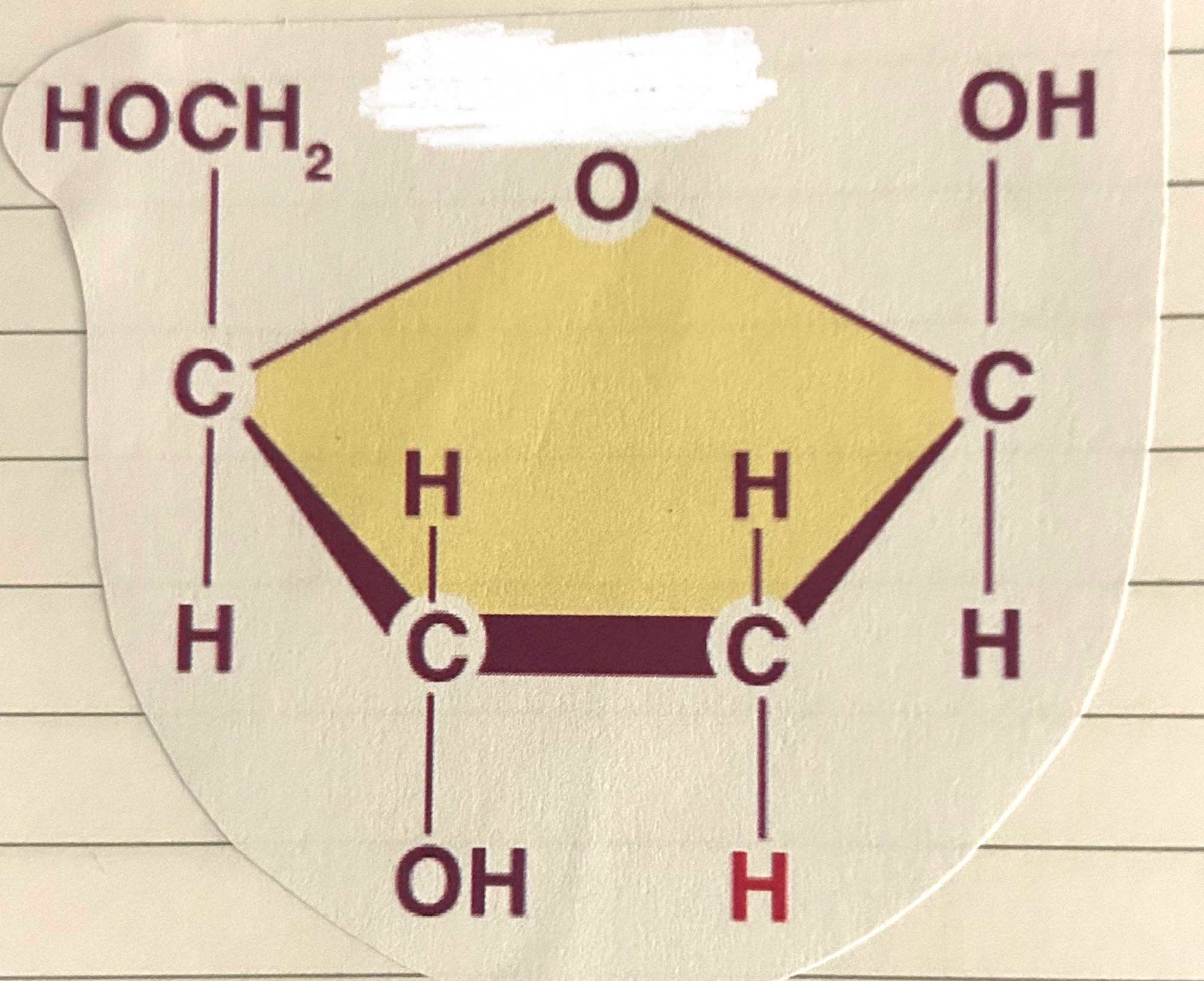
what is this?
deoxyribose
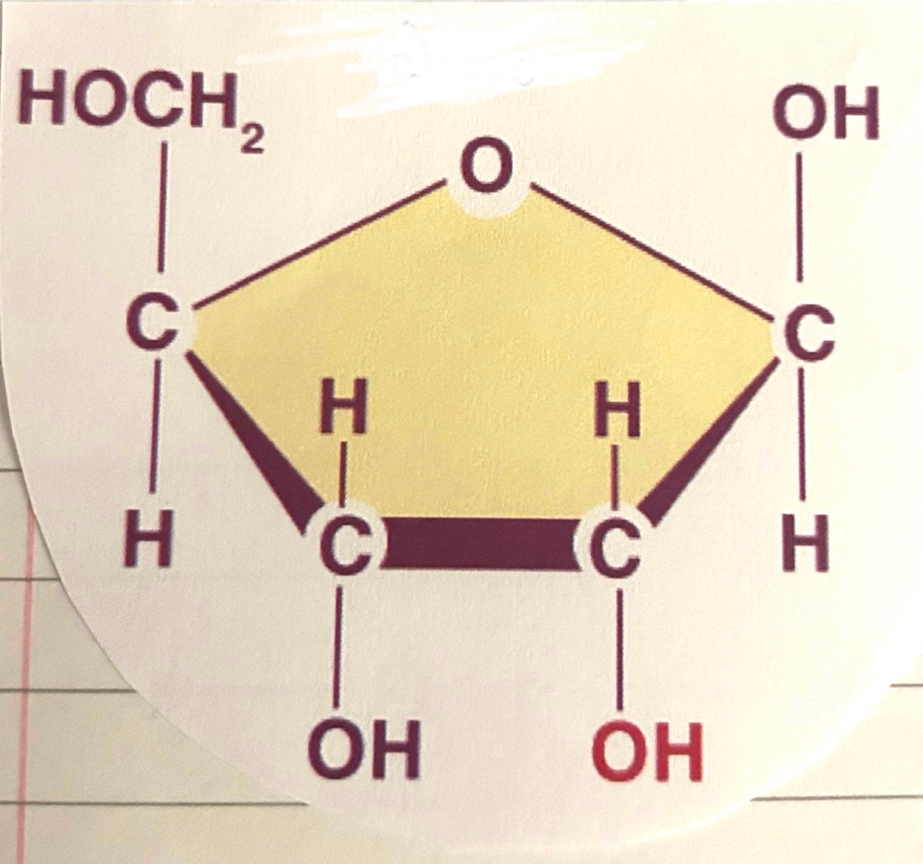
what is this?
ribose
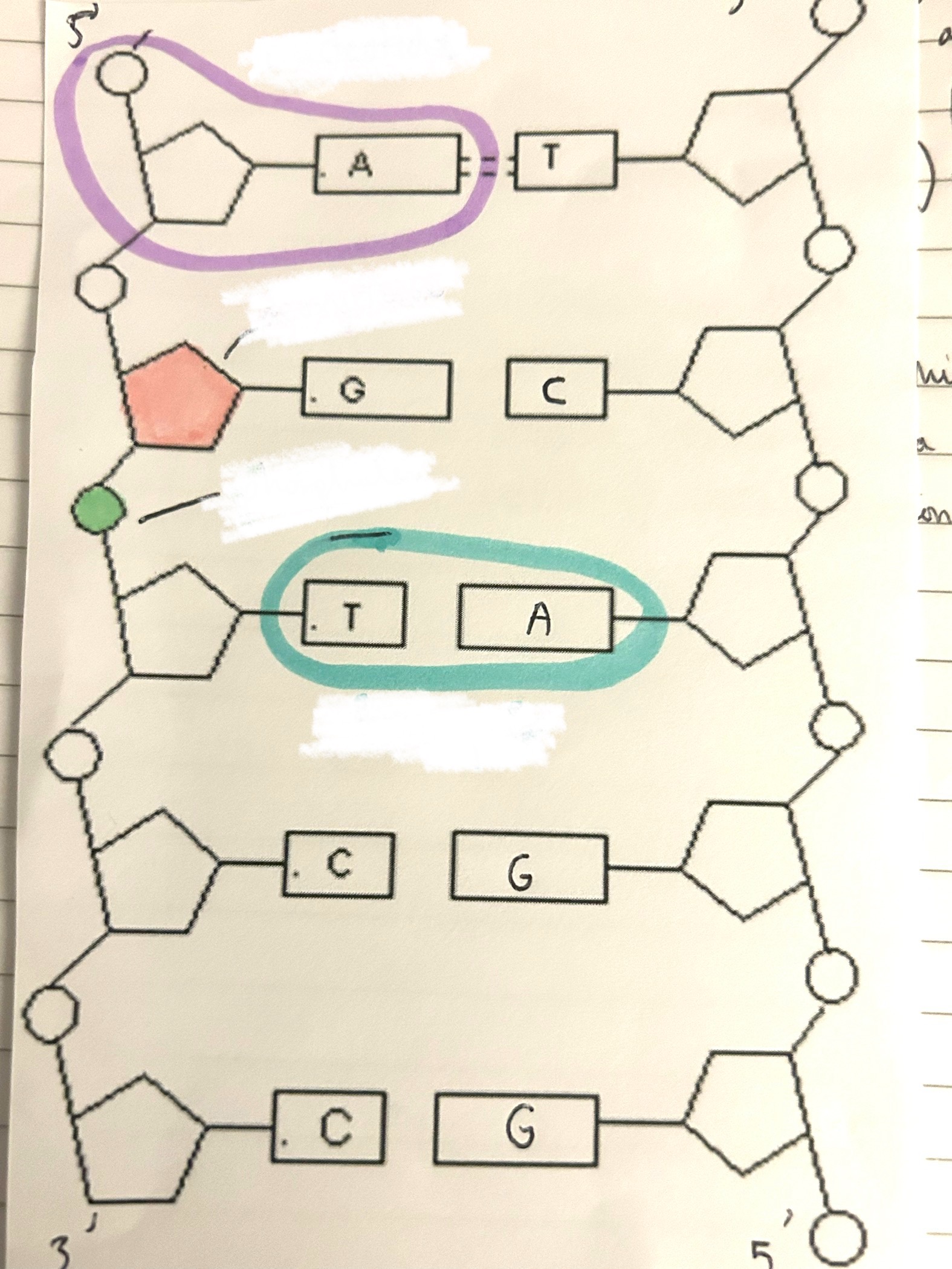
what is the purple part?
nucleotide
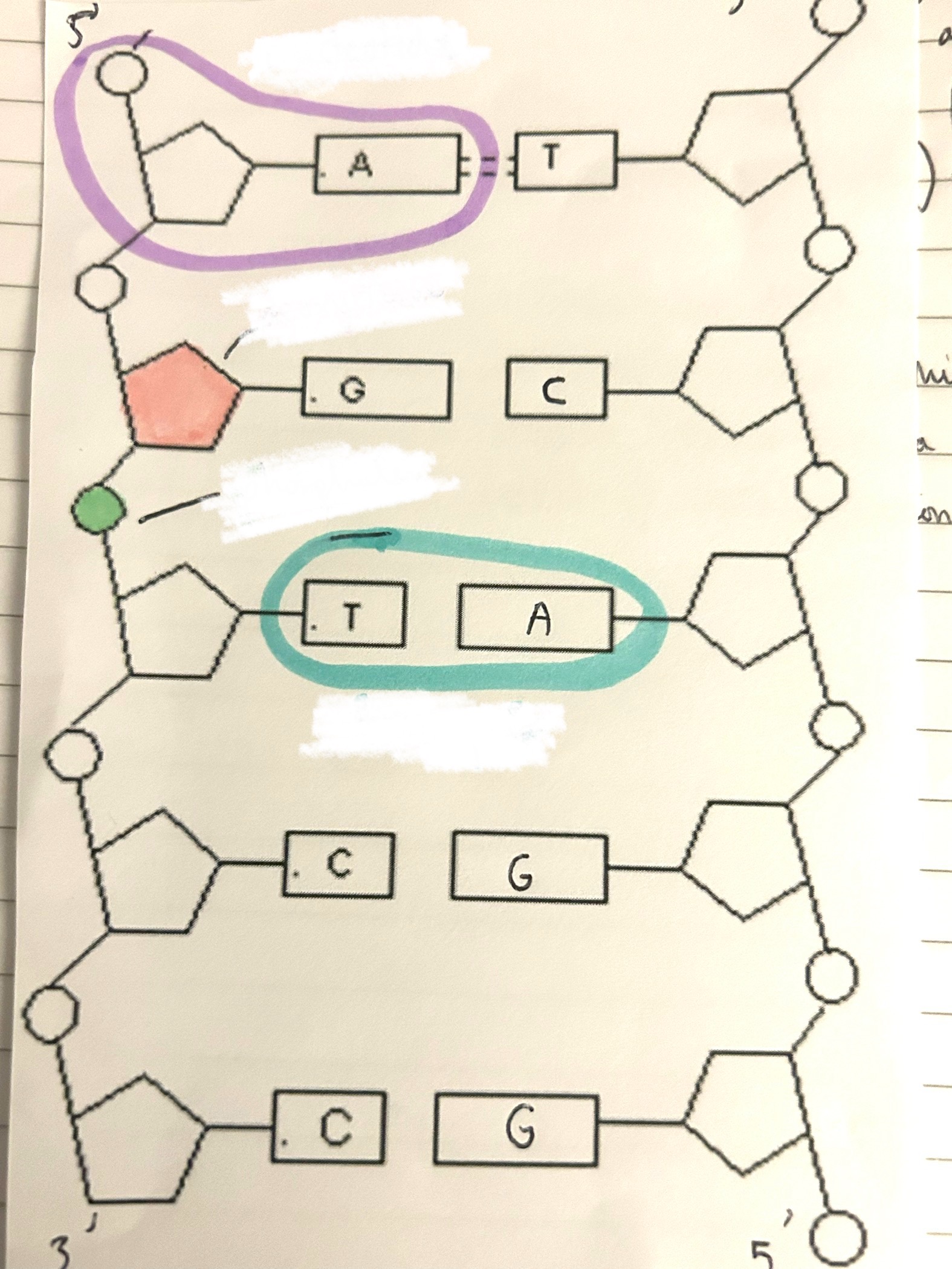
what is the pink part?
deoxyribose
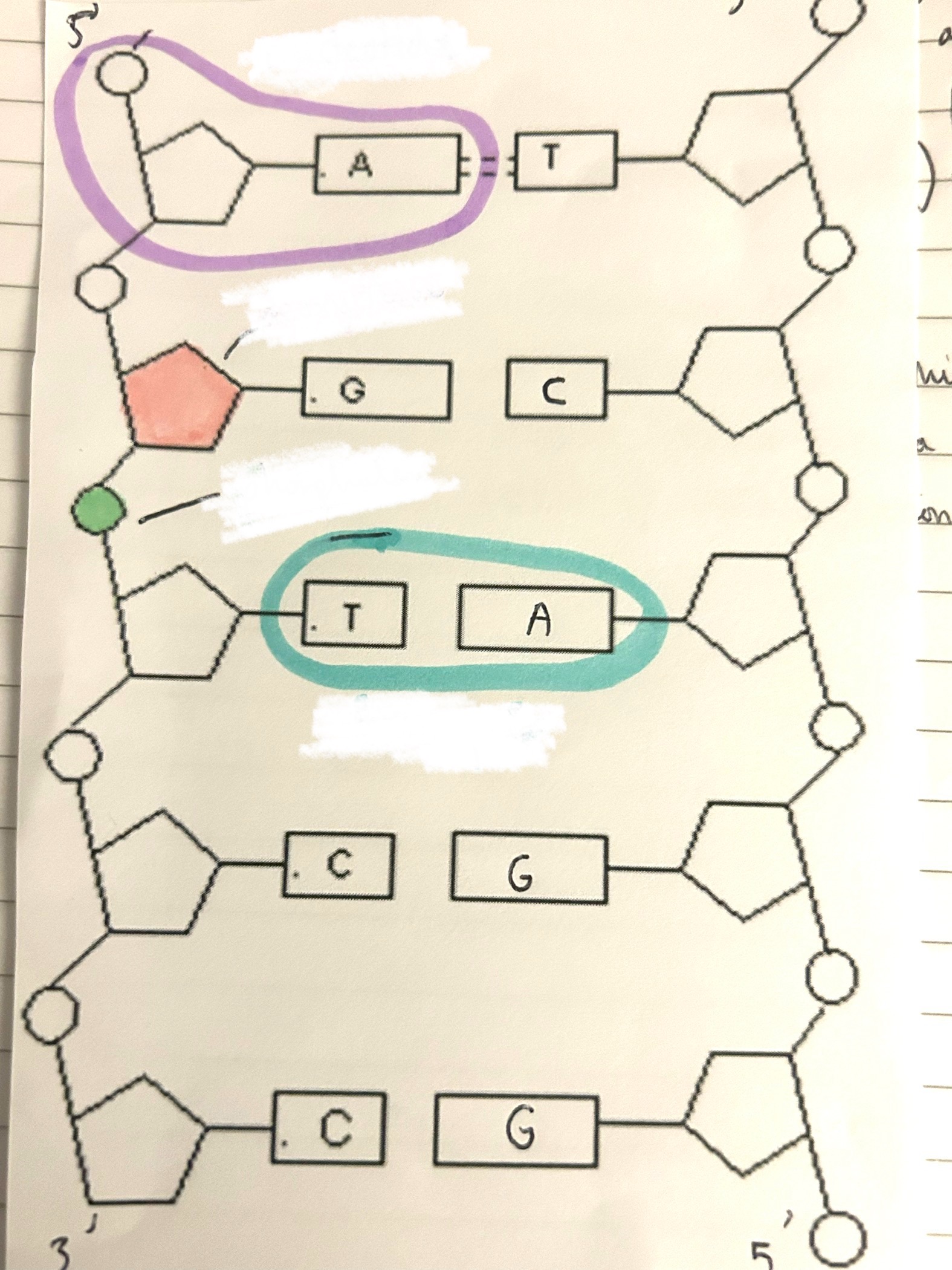
what is the green part?
phosphate
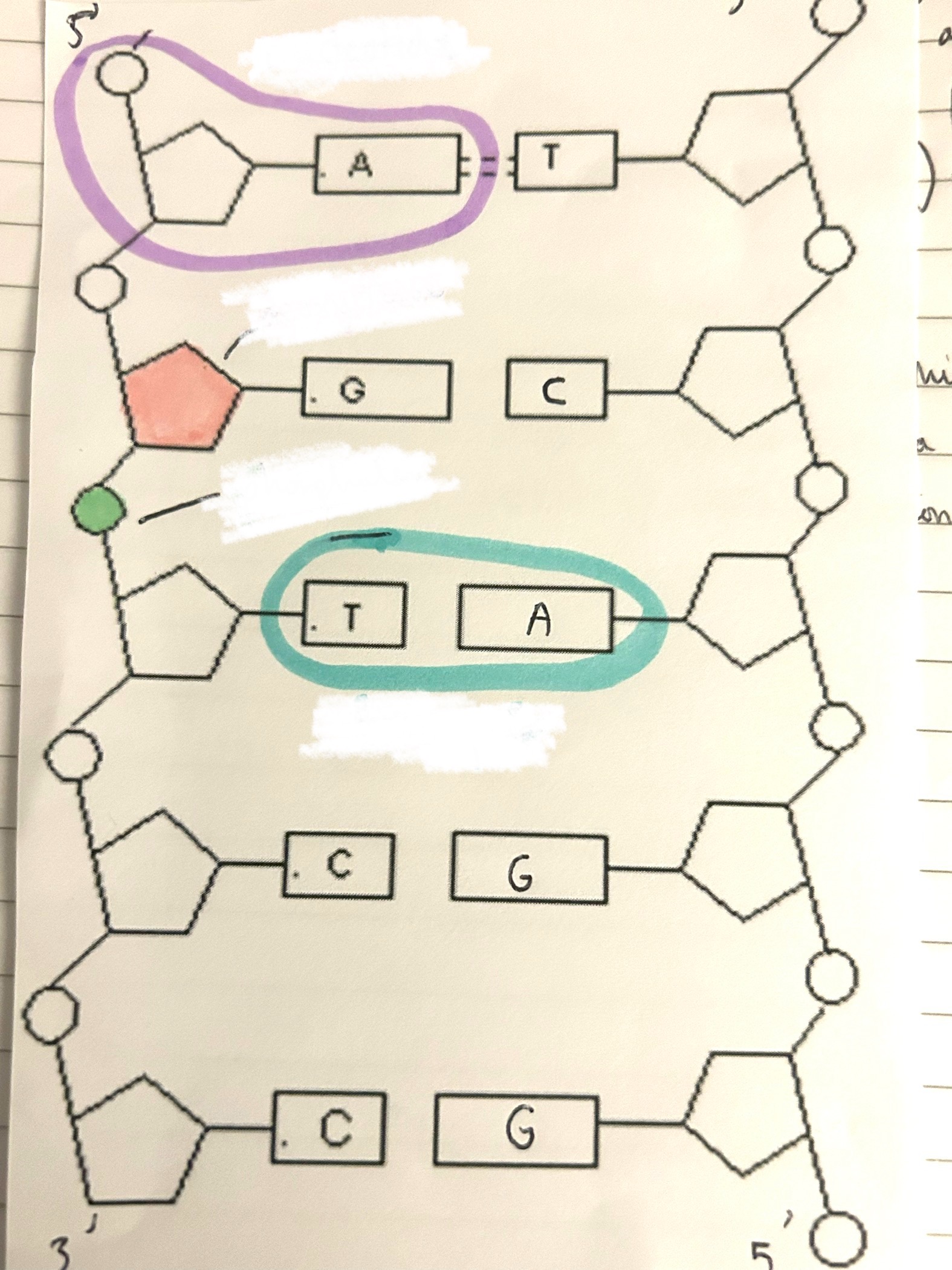
what is the blue part?
base pair
what are the 4 base pairs and which goes with which?
cytosine and guanine, thymine and adenine
what 3 things are nucleotides made up of?
pentose sugar, phosphate group, nitrogenous base (cysotine, thymine, uracil, adenine, guanine
a pentose sugar, phosphate group and an organic base use what reaction to form a mononucleotide?
condensation
how do 2 mononucleotides join together and what bond does it form?
condensation reaction
between deoxyribose sugar of one nucleotide, phosphate group of another
forming a dinucleotide
forms a phosphodiester bond
what does the continued linking of mononucleotides make?
a long chain called a polynucleotide
what is RNA and what is it made up of?
a single, relatively short polynucleotide chain (polymer). made of pentose sugar ribose and organic bases are adenine, guanine, cytocine and uracil
explain how organic bases help to stabilise the structure of dna
hydrogen bonds between base pairs hold polynucleotide chains together to form double helix
held in antiparallel directions
what is the pentose of dna?
deoxyribose
what is the sugar in dna?
deoxyribose
what is the sugar in rna?
ribose
4 differences between DNA and RNA.
in DNA, bases are CGAT whereas in RNA, bases are CGAU
DNA double stranded, RNA is single stranded.
in DNA, deoxyribose is the pentose whereas in RNA, ribose is the pentose.
DNA is long molecule, whereas RNA is shorter.
dna replication (4 stages)
dna helicase unzips dna, exposing free base.
free complementary nucleotides bind to the exposed bases and DNA polymerase makes a complementary polynucleotide chain, adding 1 nucleotide at a time.
other strand uses another dna polymerase because this new dna strand needs to be made in the opposite direction.
eventually, each strand has been copied and 2 new identical strands of DNA are produced
what are the 2 stages of protein synthesis?
transcription, translation
what bonds are the two strands in dna joined by?
hydrogen bonds formed by certain bases
are matching base pairs always in the same quantity?
yes, however the ratio of adenine and thymine to cytosine and guanine varies from species to species
what makes up the structural backbone of a dna molecule?
phosphate and deoxyribose wind around each other alongside the base pairs to form a double helix
why is dna a stable molecule? 3 reasons
phosphodiester backbone protects the chemically reactive organic bases inside the double helix
hydrogen bonds link the organic base pairs forming bridges (rungs) between the phosphodiester uprights
three hydrogen bonds between cytosine + guanine, so higher the proportion of C-G pairings, the more stable the DNA molecule
what is dna
hereditary material responsible for passing genetic information from cell to cell and generation to generation.
5 ways dna is adapted to its function (number them)
stable structure,normally passes from generation to generation without significant change. most mutations are repaired, so persistent mutations are rare
2 separate strands joined by hydrogen bonds so can separate for dna replication/ protein synthesis.
large molecule- carries lots of genetic information.
base pairs in helical cylinder of deoxyribose-phosphodiester backbone, so genetic information protected from outside chemical/physical forces
base pairing leads to dna being able to replicate and transfer information as mRNA