Lab study guide: spinal cord and nerves
1/83
There's no tags or description
Looks like no tags are added yet.
Name | Mastery | Learn | Test | Matching | Spaced | Call with Kai |
|---|
No analytics yet
Send a link to your students to track their progress
84 Terms
Cervical enlargement

A region of the spinal cord that is enlarged to accommodate the nerves supplying the upper limbs
Lumbar enlargement

A region of the spinal cord that is enlarged to accommodate the nerves supplying the lower limbs
Cervical spinal nerves

Nerves that emerge from the cervical region of the spinal cord, specifically C1-C8
Thoracic spinal nerves

Nerves that emerge from the thoracic region of the spinal cord, specifically T1-T12
Lumbar spinal nerves

Nerves that emerge from the lumbar region of the spinal cord, specifically L1-L5
Conus medullaris

The tapered end of the spinal cord, typically located around the L1-L2 vertebrae
Cauda equina

A bundle of spinal nerves and spinal nerve rootlets that arise from the lower part of the spinal cord
Filum terminale

A fibrous extension of the pia mater that anchors the spinal cord to the coccyx
Spinal dura mater

The outermost layer of the meninges surrounding the spinal cord
Arachnoid mater

The middle layer of the meninges, located between the dura mater and pia mater
Pia mater

The innermost layer of the meninges that closely adheres to the surface of the spinal cord
Ventral/ anterior median fissure

A deep groove along the anterior midline of the spinal cord
Dorsal/ posterior median sulcus

A shallow groove along the posterior midline of the spinal cord
Central canal

A small channel in the center of the spinal cord that contains cerebrospinal fluid
Dorsal/ posterior horns

Regions of gray matter in the spinal cord that contain sensory neurons
Ventral/anterior horns

Regions of gray matter in the spinal cord that contain motor neurons
Lateral horns

Regions of gray matter in the spinal cord associated with the autonomic nervous system
Ventral root

The root of a spinal nerve that contains motor fibers
Dorsal root

The root of a spinal nerve that contains sensory fibers
Dorsal root ganglion
A cluster of sensory neuron cell bodies located in the dorsal root of a spinal nerve
Spinal nerve

A mixed nerve that carries both sensory and motor fibers
Dorsal/posterior white columns/funiculi

Regions of white matter in the spinal cord that carry sensory information to the brain
Ventral/anterior white columns/funiculi

Regions of white matter in the spinal cord that carry motor information from the brain
Epineurium

The outermost layer of connective tissue surrounding a nerve
Perineurium
The connective tissue that surrounds a fascicle of nerve fibers
Fascicle (nerve)
A bundle of nerve fibers within a nerve
Endoneurium
The innermost layer of connective tissue surrounding individual nerve fibers
Axon

A long, slender projection of a neuron that conducts electrical impulses away from the neurons cell body
Myelin sheath

A fatty layer that insulates axons and increases the speed of electrical impulses
Cervical plexus

A network of nerves formed by the anterior rami of C1-C5
Brachial plexus

A network of nerves formed by the anterior rami of C5-T1
Lumbar plexus

A network of nerves formed by the anterior rami of L1-L4
Sacral plexus

A network of nerves formed by the anterior rami of L4-S4
Phrenic nerve

innervates the diaphragm
Musculocutaneous nerve

Nerve that innervates the flexor muscles of brachium
Median nerve

Nerve that innervates the flexor muscles of antebrachium
Ulnar nerve

Nerve that innervates flexors of antebrachium and hand
Radial nerve

Nerve that innervates extensor of antebrachium and brachium
Axillary nerve

Nerve that innervates the deltoid and teres minor muscles
Femoral nerve

Innervates quadriceps,sartorius, pectineus, and iliacus
Obturator nerve

Innervate adductor muscles and gracilis
Tibial nerve

Innervates muscles in posterior thigh (except short head of biceps femoris), leg and foot
Common fibular nerve

Innervates short head of biceps femoris, tibialis anterior, fibularis, extensor of toes
Cerebral hemispheres

The right and left halves of the cerebrum
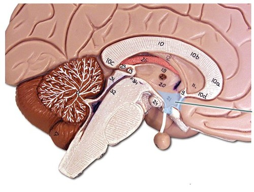
Diencephalon

Thalamus, hypothalamus, epithalamus
Cerebellum

the “little brain” at the rear of the brainstem; functions include processing sensory input and coordinating movement output and balance
Brain stem

midbrain, pons, medulla oblongata
grey matter

neuron cell bodies and short, unmyelinated axons
White matter

Whitish nervous tissue of the CNS consisting of myelinated axons
Spinal cord white matter

Whitish nervous tissue of the CNS consisting of myelinated axons
spinal cord grey matter

neuron cell bodies and short, unmyelinated axons
Lateral ventricles

A set of paired ventricles lying within the cerebral hemispheres
Third ventricle

The midline ventricle that conducts cerebrospinal fluid from the lateral ventricles to the fourth ventricle
Cerebral aqueduct

connects the third and fourth ventricles
Fourth ventricle

Small triangular chamber between the pons and cerebellum
Gyri (gyrus)

Ridges of the brain
Sulci (sulcus)

Shallow grooves in the brains surface
Longitudinal fissure

Indentation that separates the cerebrum into right and left hemispheres
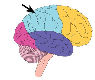
Frontal lobe

The lobe at the front of the brain associated with movement, speech, and impulse behavior
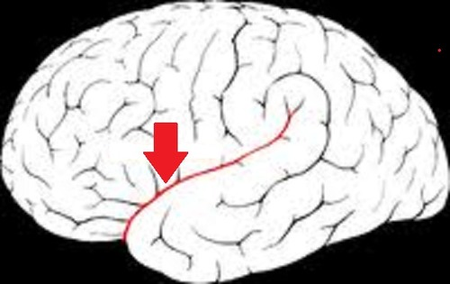
Central sulcus
separates frontal and parietal lobes
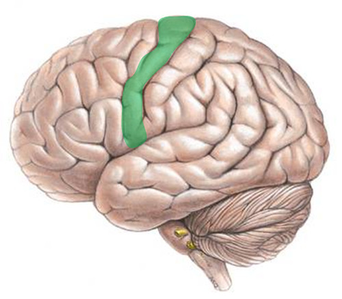
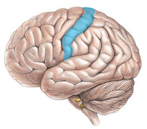
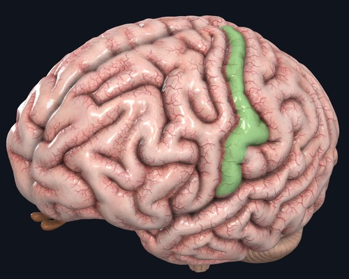
Precentral gyrus
anterior to the central sulcus
Parietal lobes
portion of the cerebral cortex lying at the top of the head and toward the rear
postcentral gyrus
posterior to the central sulcus
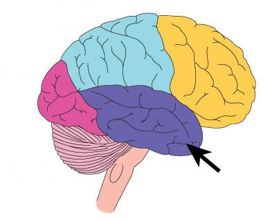
Occipital lobe
A region of the cerebral cortex that processes visual information
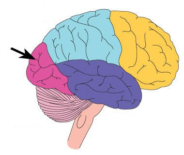
temporal lobes
portion of the cerebral cortex lying roughly above the ears; includes the auditory areas, each receiving information primarily from the opposite ear
Lateral sulcus
separates temporal lobe from parietal and frontal lobes
Primary motor cortex (precentral gyrus)
the section of the frontal lobe responsible for voluntary movement

Primary somatosensory cortex (postcentral gyrus)
Site involved with processing of tactile and proprioceptive information
Visual/vision area (occipital lobe)

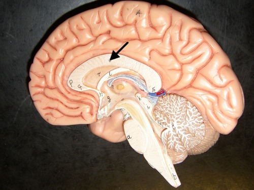
corpus callosum
the large band of neural fibers connecting the two brain hemispheres and carrying messages between them
Thalamus
the brains sensory switchboard, located on top of the brainstem; it directs messages to the sensory receiving areas in teh cortex and transmits replies to the cerebellum and medulla
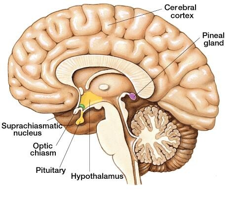
Hypothalamus
A neural structure lying below the thalamus; it directs several maintenance activities (eating, drinking, body temperature), helps govern the endocrine system via the pituitary gland, and is linked to emotion and reward
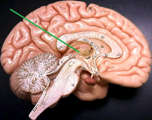
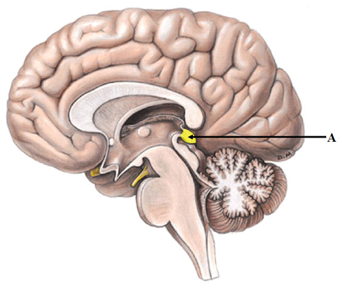
Pituitary gland
The endocrine system’s most influential gland. Under the influence of the hypothalamus, the pituitary regulates growth and controls other endocrine glands
Pituitary gland
The endocrine system’s most influential gland. Under the influence of the hypothalamus, the pituitary regulates growth and controls other endocrine glands

Epithalamus
Forms roof of 3rd ventricle has connections between limbic system and other parts of brain contains the pineal body (which secretes melatonin)
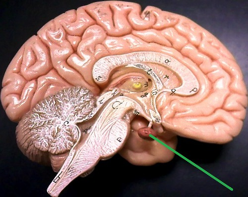
Pineal gland
Secretes melatonin
Midbrain
A small part of the brain above the pons that integrates sensory information and relays it upward
Pons
A brain structure that relays information from the cerebellum to the rest of the brain
Medulla oblongata
Part of the brainstem that controls vital life-sustaining unctions such as heartbeat, breathing, blood pressure, and digestion
Folia
Folds of the cerebellum