L1: Principles of Vertebrate development
1/34
There's no tags or description
Looks like no tags are added yet.
Name | Mastery | Learn | Test | Matching | Spaced | Call with Kai |
|---|
No analytics yet
Send a link to your students to track their progress
35 Terms
Gastrulation
when blastula transforms from ball of cells into multi-layered strucuture
composed of three distinct germ layers
Requires the control of
differentiation
Morphogenesis
Reductionist vs integrative approach to developmental biology
Reductionist
break down into smaller manageable parts
Integrative
info from multiple independent experiments
performed in different experimental systems
Body plan
basic organization and arrangement of an animal’s body
inc:
symmetry, segementation and arragement of organs and tissues
Gastrulation
crucial early stage of embryonic development
one layered blastula (blastocyst in mammals) is reorganized into 2 or 3 layers gastrula
Blastula
hollow sphere of cells formed during early embryonic development in aniamls
Germ layers
fundamental cell layers that form during early embryonic development in most animals
lead to formation of various tissues and organs
In tripoblastic animals:
ectoderm
mesoderm
endoderm
Differentiation
process by which cells become specialised
acquire distinct strucutures and functions
Morphogenesis
biological process by which a cell, tissue or organism develops its shape and structure
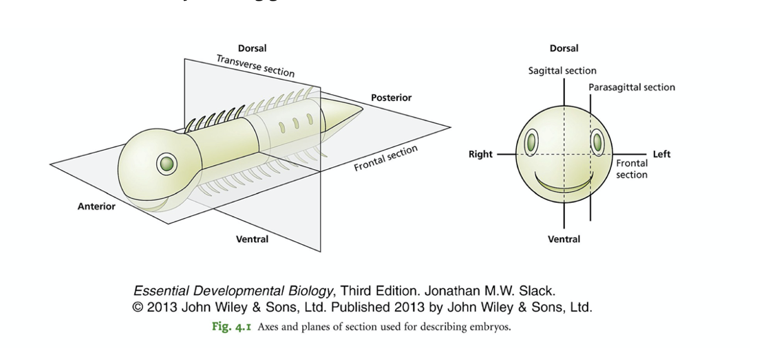
Principle anatomic axes
Dorsal vs ventral
Anterior vs posterior
left vs right
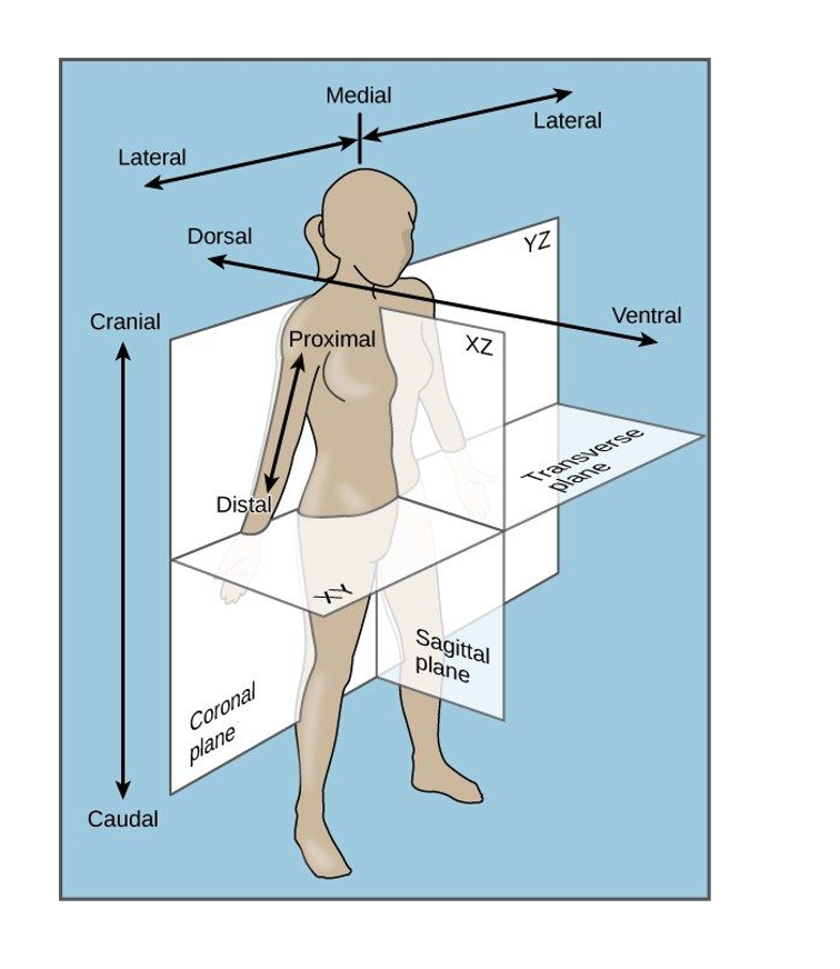
Principal axis in a human (additional axis)
Proximal distal
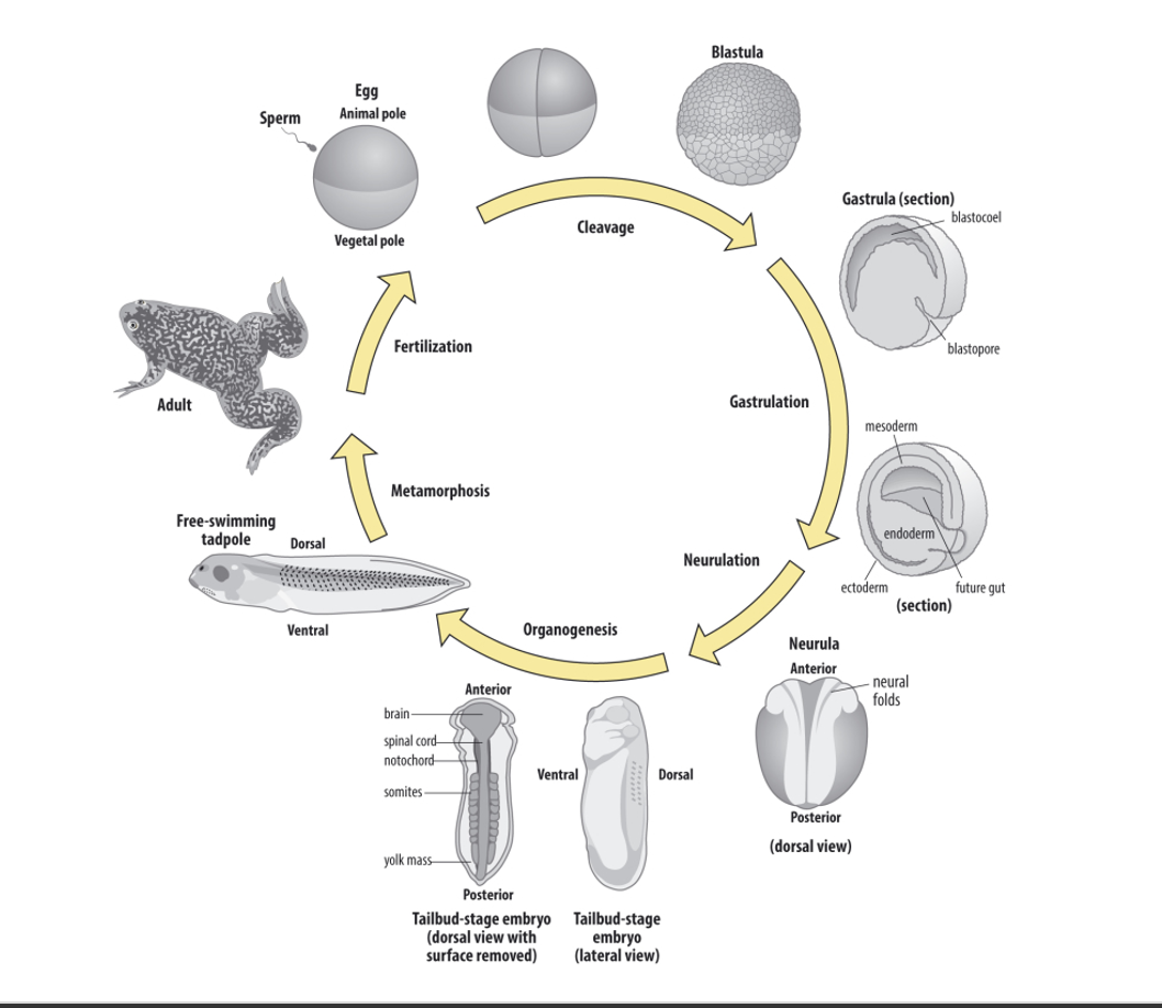
Overview of Xenopus African clawed frog life cycle
Cleavage
really fast so no cell growth
Gastrulation (large scale morphogenesis (tissue shape changes))
Ball/disc—> 3D
Neuralation
Ectoderm → Neural tube
Organogenesis
Metamorphosis (in frogs)
Why a good model + cons (idk)
Ease of experimental embryology
can get lots of proteins from it in biochem analysis
Cons
Polyplody
metamorphosis?
What happens during cell differentiation
cells progressively restricted in array of cell types they can become
(their cell fate)
determined by signalling and other ascpects of the cell’s environment
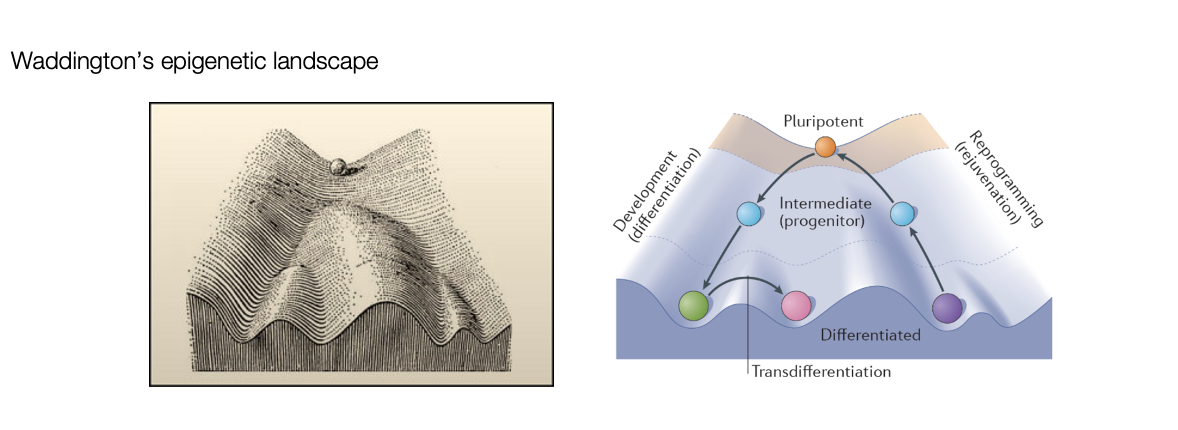
What is this modelled by
Conrad Waddington’s Epigenetic landscape
cells as a ball rolling down a series of valleys
guided to one fat or another

Key terms of potenecy
Totipotent→ generate all embryonic and extraembryonic cells
Pluripotent→ generate all embryonic cells
Multipotenet→ all cells of multiple lineages
Unipotentent→ all cells of a single lineage
Can you go up the landscape
Yes→ transdifferentiation
experimentally
e.g induced pluripotency with Yamanaka factors
back up the landscape
Three types of evidence needed to proove a mechanism in dev biology and how these are exp tested
Is it at right place/time (CORRELATION)
EXP: e.g gene expression: find correlation between gene and space/time (imaging)
Is it necessary for controlling the process? (REQUIREMENT)
EXP: knockout/ loss of function
Is it enough to drive the process? (SUFFICIENT)
EXP: gain of function/ over express the gene
Developmental biology uses many techniques and collaberations
Other sciences
physistics
mathematicians (make models)
computer scientists
increasingthe norm for dev biology
Techniques used by developmental biologists to uncover the mechanisms of development and what questions they try to answer
Experimental embryology→ ‘cut and paste’ (classic technique)
cut and past tissues
Q: what requiremnts of tissue interactions
(+ descriptive of just looking which came before experimental embryology)
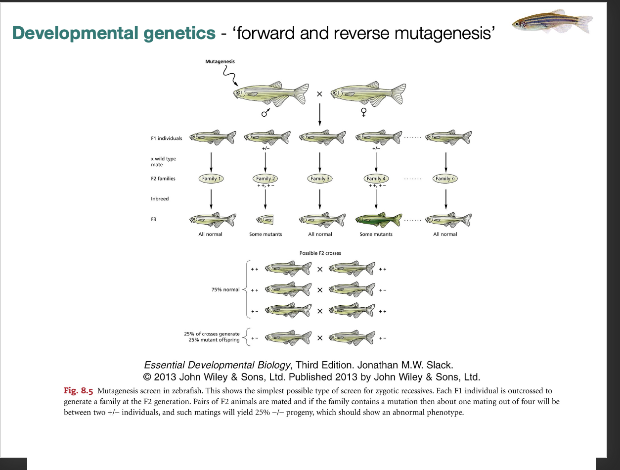
Developmental genetics→ ‘forward and reverse mutagenesis’
Q: what is role of gene
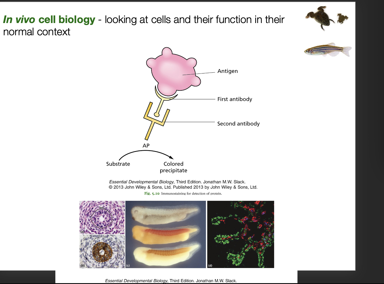
In vivo cell biology→ looking at cells and their function in their normal context
add context to in vitro studies
Q: is the gene made in the right time and place?
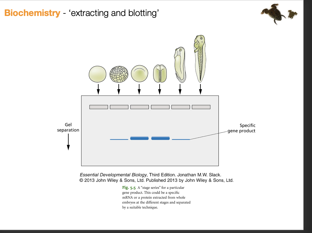
Biochemistry→ ‘extracting and blotting’
Q: what does the protein it codes for actually do
Neuroscience→ how is the nervous systm formed and wired
Q: development of neural connections must explain neurosceince function
neural development underpins the logic of how the brain must work
Stem cell biology→ recapitualting aspects of developent in vitro
What we learn from developmental biology we can apply to stem cell biology
Evolutionary developmental biology→ ‘Evo-Devo’ how so evolutionary changes in gene expression or function generate new phenotypes?
can only understand how evo works if you understand how dev works and how dev can be altered to form organsims that can change in time
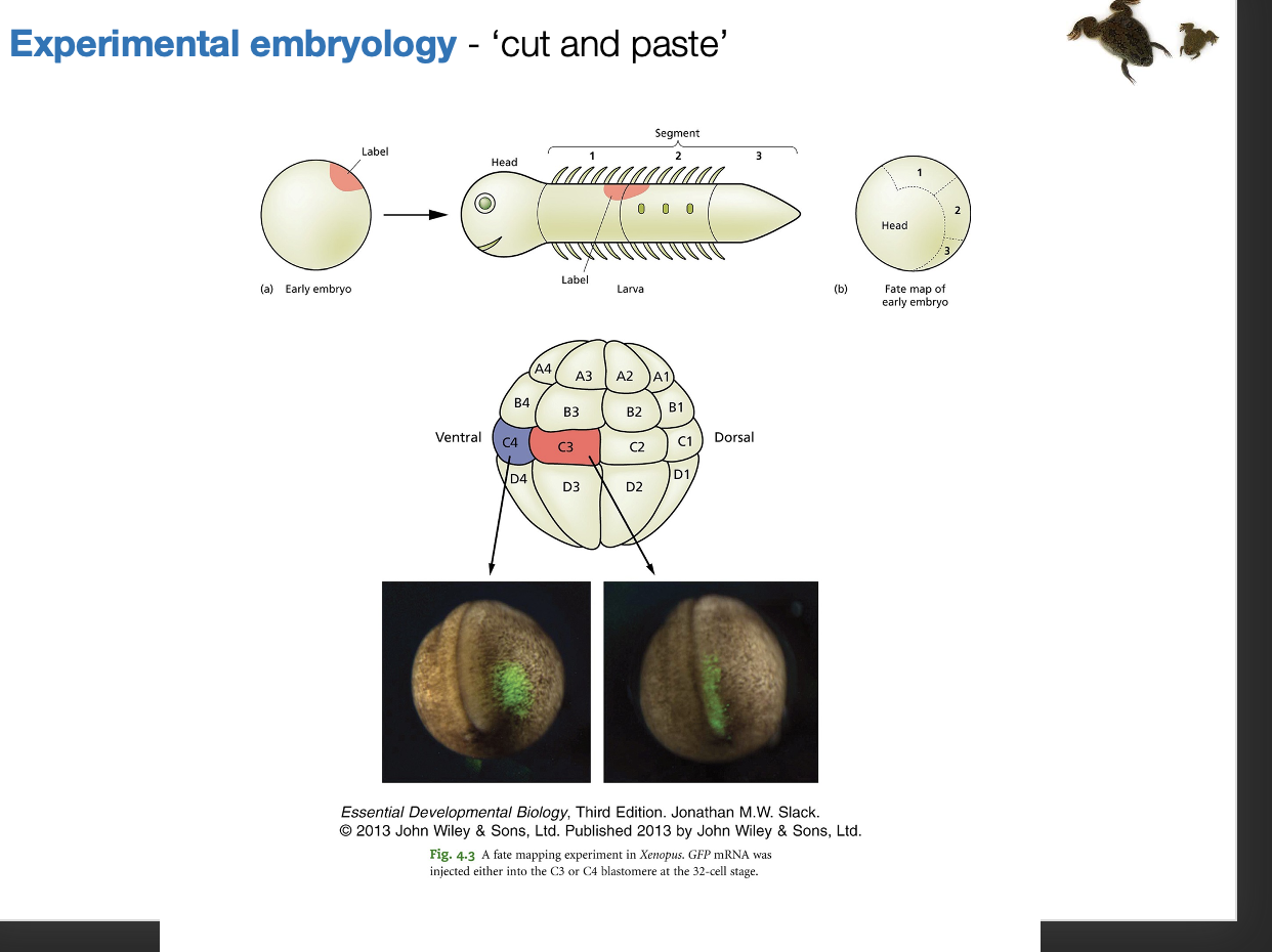
Experimental embryology→ ‘cut and paste’: Key technique 1
Making Fate maps
mark one cell early in development
follow through develop
find ultimate fate
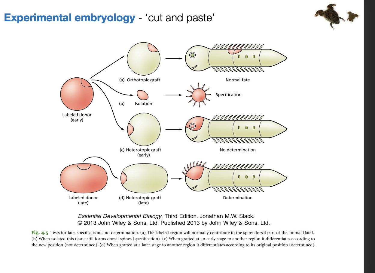
Experimental embryology→ ‘cut and paste’: Key technique 2
Testing Specification vs determination/competence
SPECIFICATION
take cell out
put in neural environment
NOTE: still caveats (pertubation and may still be other signals)
the cell fate observed must be what it is specified to be
THEREFORE: the cell already has received info to make this cell fate before it left
DETERMINATION
Take out cell
put in different environment
Receives these signals
IF IT DOES NOT DIFFERENTIATE to cells expected of that environment→ the cell must be determined already
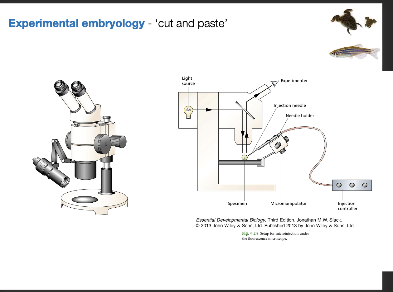
Equipment needed for this manipulations
binocular microscope
microsurgery
microinjector
inject specific mRNA DNA or cells
Common for Xenopus or Zebrafish
Use eyelash on a capillary tube to act as a knife
Developmental genetics→ ‘forward and reverse mutagenesis’ TECHNIQUES
Forward
find a phenotype
find the gene responsible
Reverse
Known gene
alter with CRISPR (knockout)
find what phenotype it is responsible for
TECHNIQUE: ZEBRAFISH have much mutant data
many mutagenesis screens
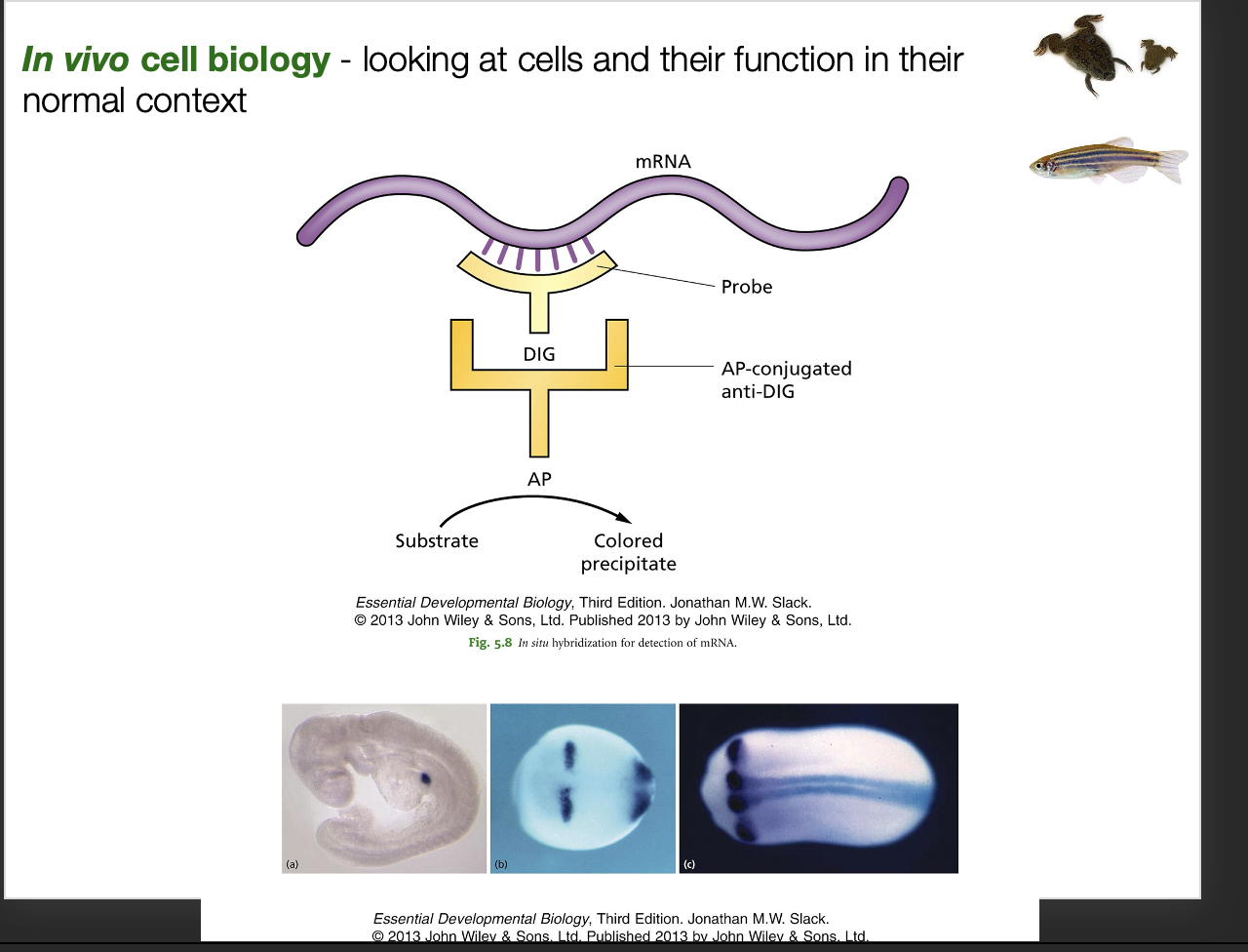
In vivo cell biology→ looking at cells and their function in their normal context Technique to find where and right time
mRNA→ in situ hybridisation
Probe mRNA expression in cells
in situ hybridisation
add a probe the attaches to mRNA
prob has label
secondary antibody can bind
has alkaline phosphates
akes precipiatte if the mRNA is there
ALTERNATIVE: use fluorescent
proteins (is protein in same place as gene?)→ antibody staining
Use first and second antibody
attach fluophore directly to the second
excite
Advantage of using fluorescent microscopy
label multiple things
organelles, proteins, mRNAs
All look at the same time→ just use different fluorphores and exite at different wavelength
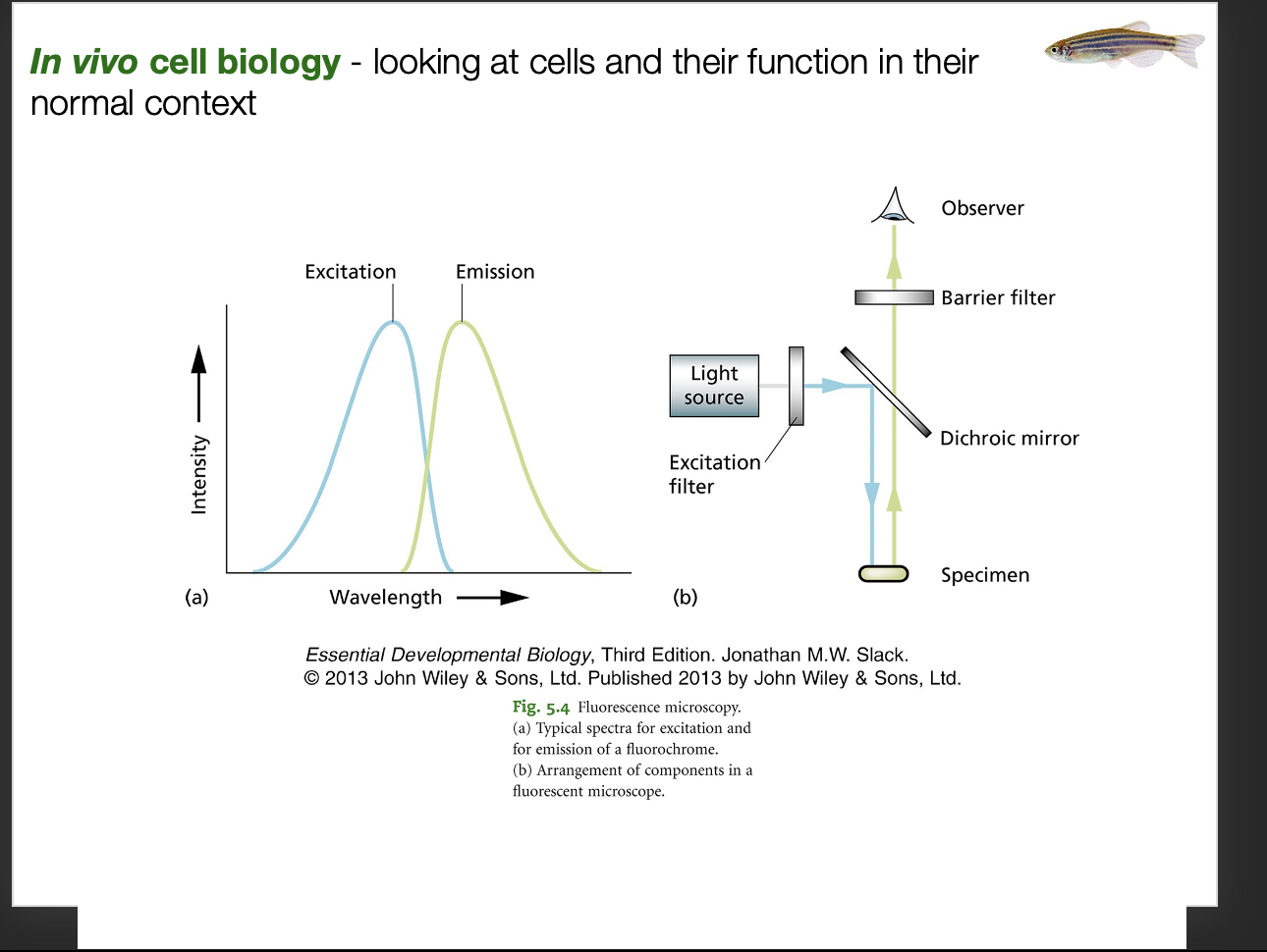
Problem with using widefeild
cells in vivo
will just blur the image
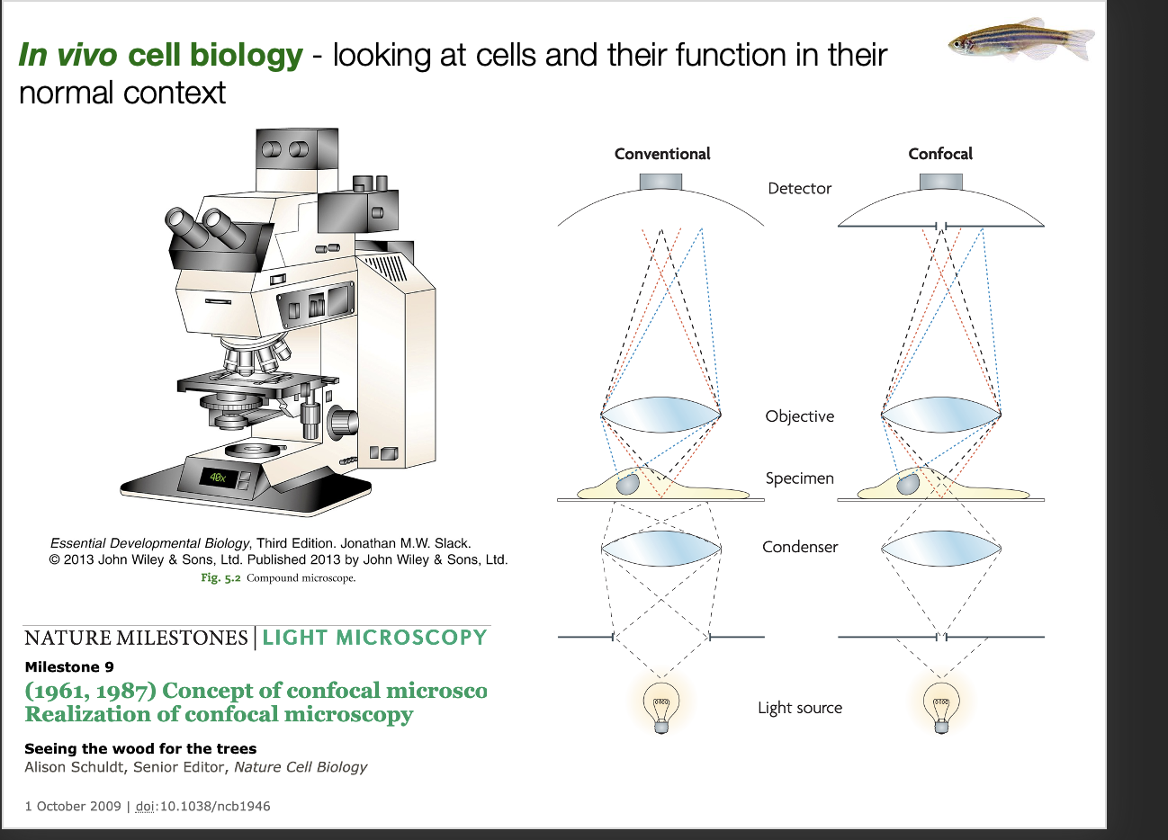
Solution
Confocal microscopy
One section of the embryo at a time
pin-hole
only collects light in one focal plane
can make Z-plane section
progeress this through
can form 3D image
Biochemistry→ ‘extracting and blotting’ why Xenopus is good for this
large size
lots of material can be extracted from single embryo
Why learn about gastrulation
Crucial moment → when body plan is established
Tech how cells both
Differentiate (cell fate decision)
morphogenesis (Generate shape change)
What happens in gastrulation
Image: with GFP (see how cell division in sync and then out of sync)
Ball of cells
cavity if formed
blastopore→ will become the anus
Folds in
closes
Forms ECTO and ENDO
Ecto ingression to form MESO inebtween the two
How does gastrulation become more complex
more cells
extra-embryonic tissue added (depends on the species)
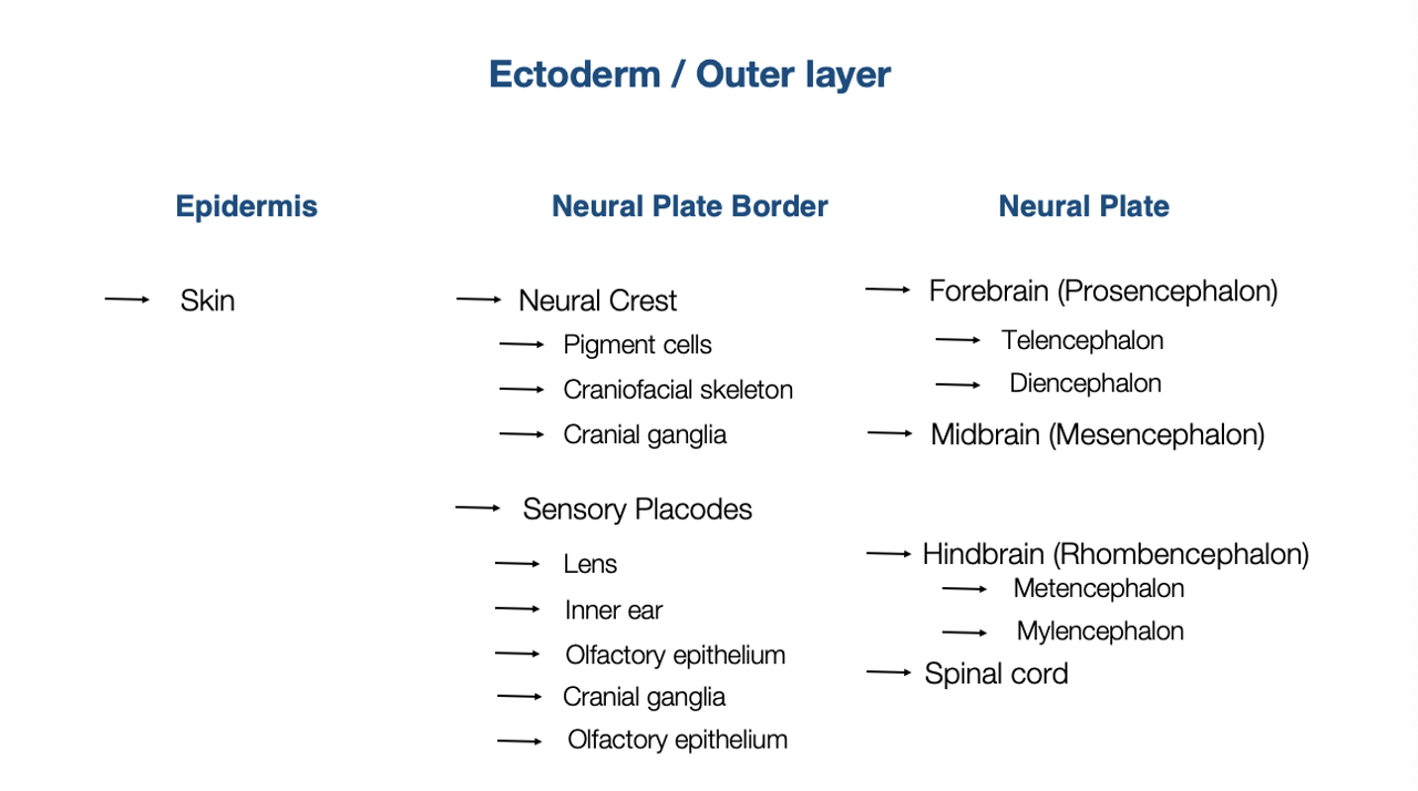
Cell types from Ectoderm
Neural plate border
Nerual crest
Pigment
craniofacial
cranio ganglia
Sensory placodes
lens, inner ear, olfactory
Skin
CNS
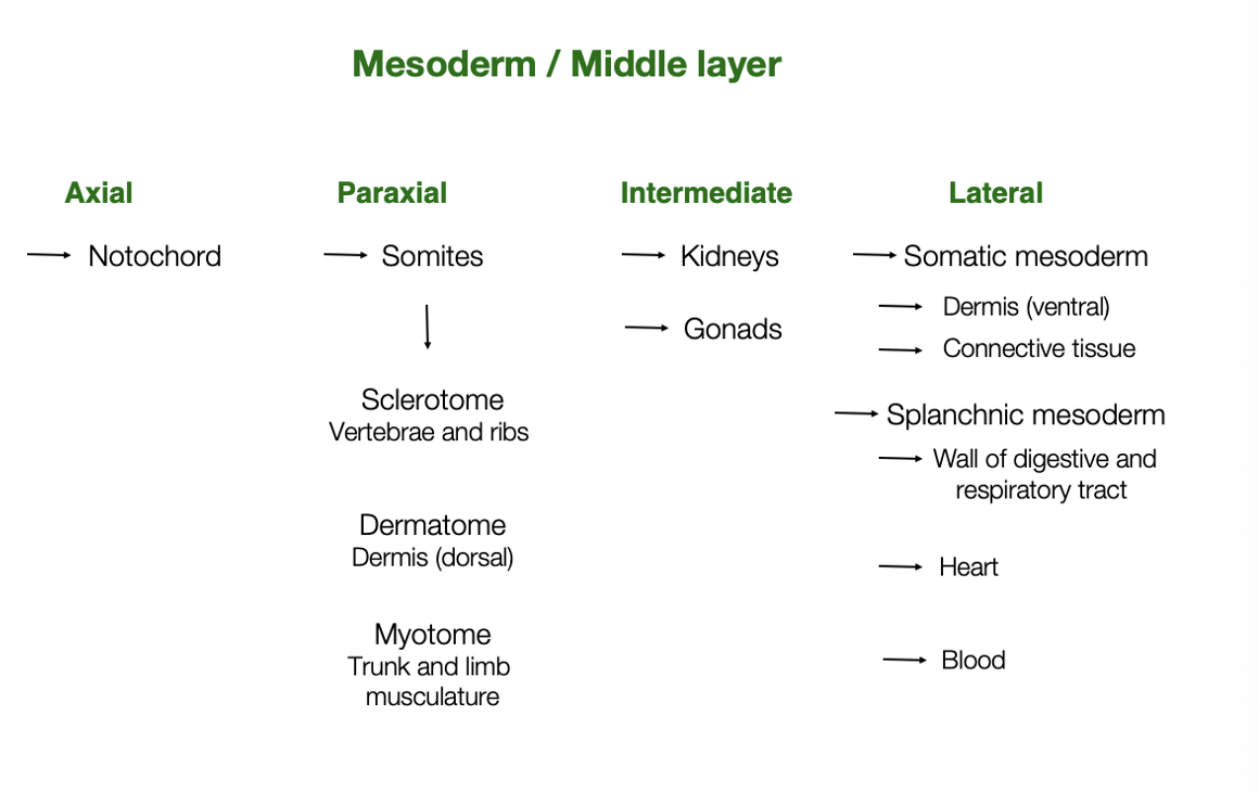
Cell types from Mesoderm
Axial
Notochord
At first→ embryogenic (reigitity)
Then→ Spinal column
Paraxial
somites formed in periodic manner
Basis of segmented body plan
Intermediate
lateral
Cells from the endoderm
Gut
Respiratory
endocrine

Further reading