BIMM 100 Final (COMEBACK BABYYY)
1/102
There's no tags or description
Looks like no tags are added yet.
Name | Mastery | Learn | Test | Matching | Spaced | Call with Kai |
|---|
No analytics yet
Send a link to your students to track their progress
103 Terms
Genes in cell are alike but differ
since not all genes are expressed
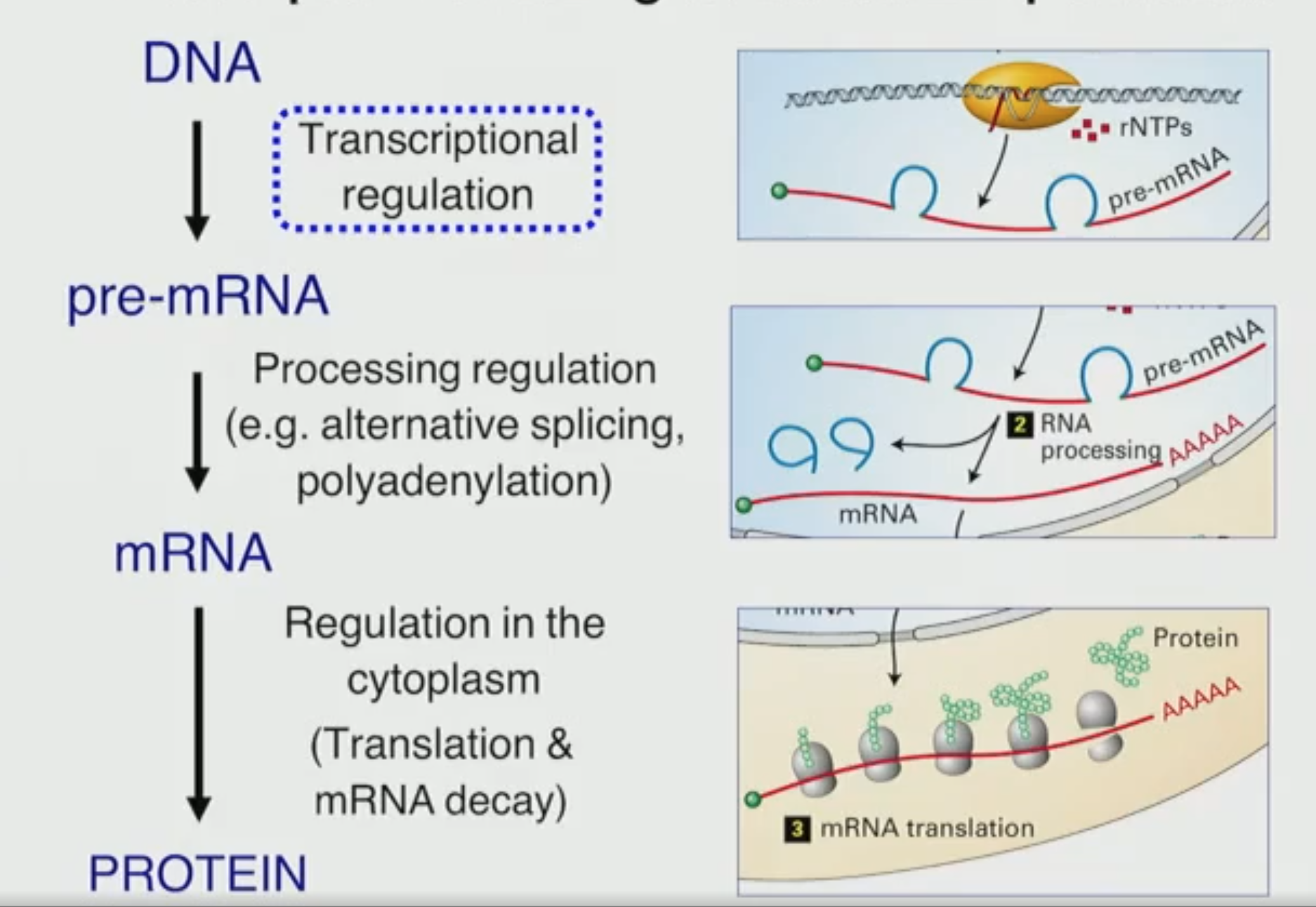
Gene expression is regulated at multiple levels
Different cell types
Same genes → different mRNAs → different proteins → different morphology functions
Same cell type under different conditions
Same genes → different mRNAs → different proteins → different cellular behavior (Ex: +hormone or -hormone)
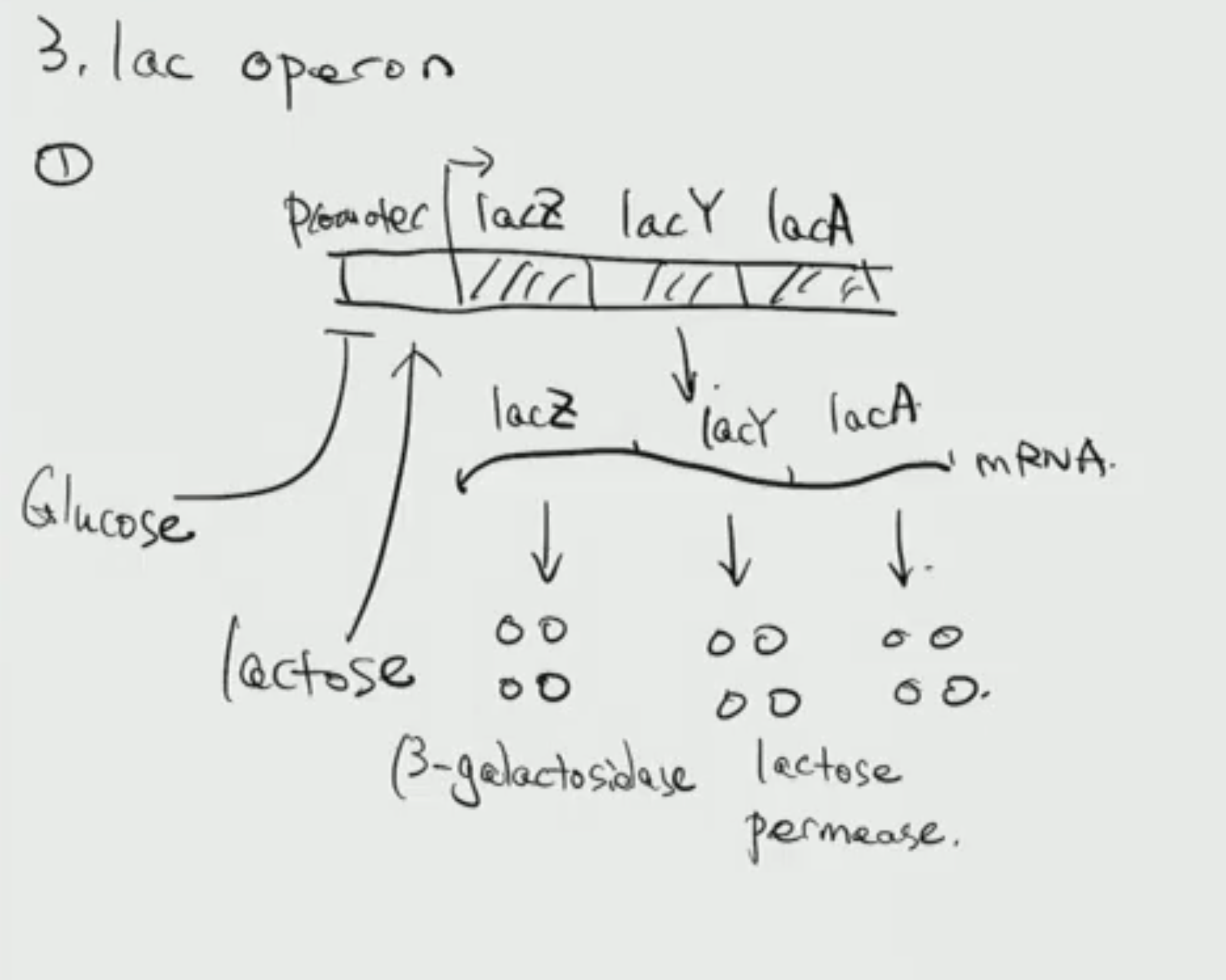
Gene organization in bacteria
One promoter regulates multiple genes (Operon - a cluster of genes under a single promoter)
During transcription will be transcribed into single mRNA (containing all genes)
During translation different parts of mRNA will be translated independently to independent proteins
How is pre-mRNA processed to mRNA in bacteria?
Recall: RNA Pol (ONLY one)
Does not undergo 5’ capping and 3’ poly A tail, instead ribosomes recognize site upstream of AUG (start codon) called ribosomal binding site (RBS) where Ribosome binds.
Operon consists of multiple genes how does Ribosome distinguish gene translation to produce separate proteins?
Find RBS at every individual gene found on Operon along with AUG (start codon) and UAA (any stop codon)
Sugar metabolism in bacteria
Consume glucose→glycolysis-energy ONLY until fully depleted, change gene expression (plateau until produce enzymes) to now digest Lactose ONLY
Lactose structure
Consists of Galactose-Glucose, Glucose→glycolysis→energy, and Galactose also digested/broken down, however need transporter lactose permease (brings Lactose into cell).
B-galactosidase used to break Lactose into Galactose-Glucose
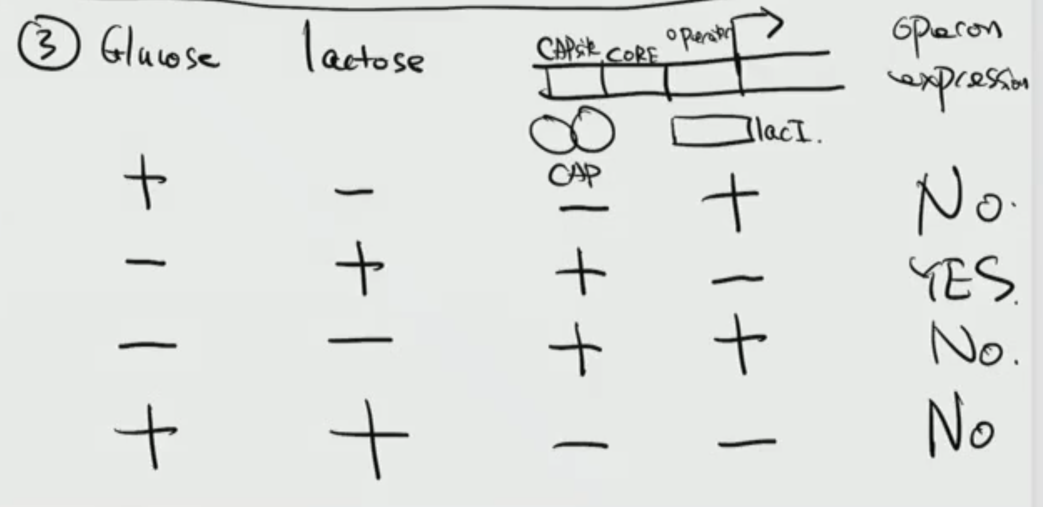
Lac Operon
Lac operon promoter elements
CORE element (functional element) recognized by Sigma factor (general TF) that will recruit RNA Pol to bind to CORE element, upstream of CORE element find CAPsite (activating functional element) bound by CAP activator (will promote binding of Sigma factor to CORE element) promote transcription
Downstream of CORE promoter find Operator (inhibiting element) bound by lacI (trans acting) which will repress binding of Sigma factor to Core element
Find that a gene constantly produces both lacI and CAP activator
How is CAP activator regulated?
Glucose acts to inhibit CAP activator, Glucose reduces cAMP production → cAMP important for CAP-DNA binding
High glucose/ low cAMP & low CAP binding
Low glucose/ high cAMP & high CAP binding
How is lacI regulated?
Lactose acts to inhibit lacI, Lactose binds to lacI causing conformational change preventing binding of Operator
Operon expression in different conditions
Experimental method for studying Gene regulation
Western blot: To detect specific proteins and relative levels
Extract total proteins (no charge), mix with SDS + heat causing protein to denature, run Gel electrophoresis, transfer Gel on top of membrane to transfer, incubate membrane with proteins nucleic acid with antibody that bind to protein of interest (be able to see band)
Band intensity represents protein level
Eukaryotic Gene regulation
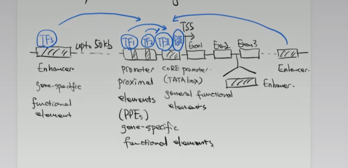
Whats the difference between Enhancers and PPEs
1.Can function over a long distance (found upstream, downstream, and inside introns), DNA is highly folded meaning the Enhancer could be found hundreds of nucleotides away but in reality is in proximity of CORE promoter
2.Position/Orientation of Enhancer does not matter still performs exact same function whereas position/orientation important for transcription direction in Promoter
Cis-regulatory elements
DNA seq that regulate gene transcription (anything found on DNA). Same in all cell types
Ex: Core promoter, PPEs, Enhancers, etc
How do cis-regulatory elements control gene expression?
Through TFs that bind to them
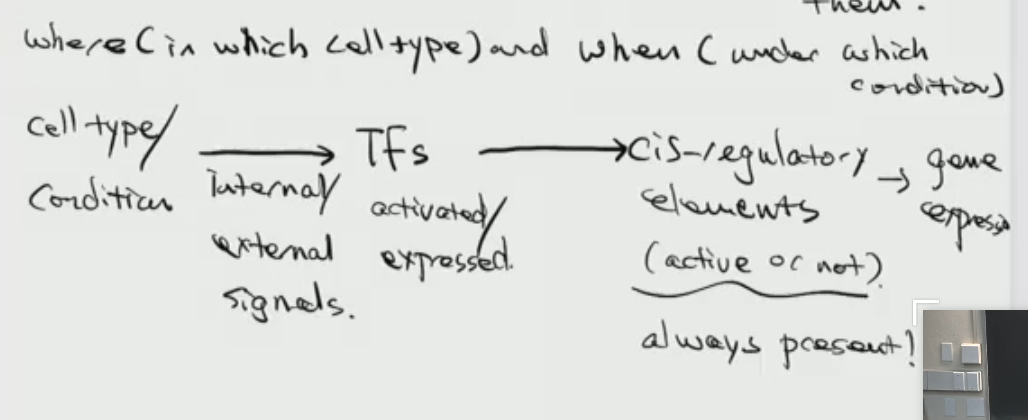
Trans regulatory elements
Transcription factors that bind to cis regulatory elements
Transcription factors are modular (functionally independent due to single protein containing multiple domains)
Find Activation/Repressor domain (regulates transcription) that is connected by Linker region to DNA binding domain (recognize specific DNA seq)
If remove Linker region and still have Activation/Repressor domain and DNA binding domain still observe transcriptional activity
Reacap Eukaryotic Gene regulation
All cells in an organism have the same DNA seq
But different genes are expressed depending on cell types , developmental stages, and environmental conditions
Controlled by cis regulatory elements and transcription factors bound to them
Cis regulatory elements: CORE promoters (general functional elements), PPEs and enhancers (both gene specific)
Trans regulatory elements
DNA binding domain components
Binding specificity - interaction with bases
Binding affinity - interactions with phosphate, sugars, or bases
How does activation domain promote transcription
TFII/Pol II recruitment to CORE promoter via mediator complex and/or activation domain itself
Chromatin regulation
Chromatin
DNA wrapped around histone (DNA more compact)
Function of Histones
protect dna and repress transcription
Histones are proteins (Proteins have N and C terminal)
Nucleosome
Repeated unit of chromatin, consists of 150bp DNA wrapped around 8 histones
Chromatin States (for a specific genomic region)
Heterochromatin (closed, inactive) - Chromatin modifiers allow transition between states but slow - Euchromatin (open, transcriptionally active) - Chromatin remodeling complex able to move position of nucleosome to free CORE promoter allowing recruitment of TFII/Pol II and FAST (w/ATP) - VERY ACTIVE Euchromatin state
Do all the genes have all three states?
No not all the genes have all three states
Chromatin modifiers and remodeling complex are recruited to specific DNA region by TFs or DNA binding proteins. Without unable to bind
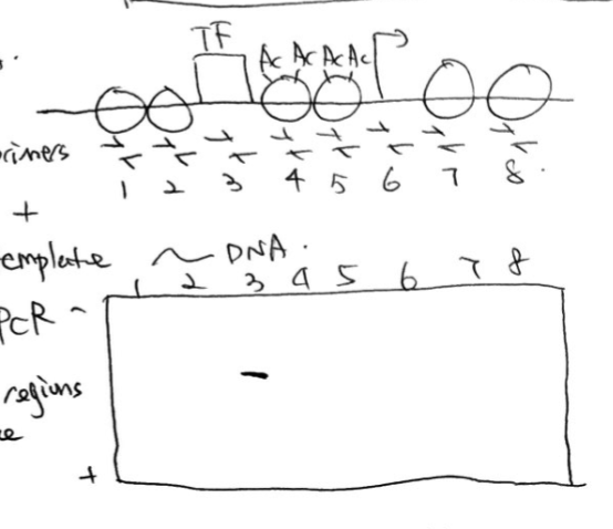
DNase Protection Assay
To determine the chromatin state of specific DNA region, use restriction site and DNase (cut any naked DNA not gene specific) to cute specific region, Euchromatin each Nucleosome is cut but in Heterochromatin state unable since closed, remove histones and restriction site digestion → find multiple pieces of DNA in Euchromatin but one long piece of DNA heterochromatin, use radioactive probe, run Southern blot gel (run DNA on gel rather than RNA), will notice different length fragments
Chromatin modifications and modifer
Histone N-terminal contain many Lysine (+ charged), modifications are chemical marks added on specific lysine on histones
Modifier: Enzymes that add or remove these chemical marks
Chromatin modifications
Histone acetylation/deacetylation
Histone methylation/demetyhlation
Histone Acetylation/Deacetylation
Histone acetylation (loss of + charge, results in less tight binding in DNA, opposite charges attract) and bromodomains (transcription activators) bind to acetylated histones, promote Euchromatin state
Enzyme called HATs
Histone Deacetylation, enzyme called HDAC
Find that for each lysine N terminal there is a pair of modifiers for Acetylation or Deacetylation
Histone methylation/demetyhlation
No effect on charge of Lysine, instead promotes the Heterochromatin or Euchromatin state depending on Lysine. Methyl binds to protein with chromodomains (activators/repressors)
Modifers: Histone methyltransferase or Histone Demethylase
How do you get Heterochromatin formation and spreading
Often forms across a large genomic region, DNA binding elements bind to TF (repressor) recruit modifier (methyl) which will trigger modification of neighboring histones, once modified will bind to proteins with chromodomain, will further modify those neighboring histones forming cascade chain rxn (Heterochromatin spreading)
How is Heterochromatin spreading regulated/stop?
Find boundary elements (DNA sequence that stops Heterochromatin spreading)
How do boundary elements work?
Generate nucleosome free region (if no nucleosome neighboring will stop cascade)
Recruit proteins that will repress Chromatin modifiers
Chromatin Immunoprecipitation (ChIP)
To determine whether and where a specific protein binds to a specific DNA region (both are known) interaction in the cell (different cell types and environmental conditions yield different results)
Fixation (Formaldehyde) stabilize protein DNA interactions in cell
Isolate chromatins from cell, sonication (shear DNA into pieces, leaving only DNA that is protected by proteins/histones)
Immunoprecipitation: use antibody with bead (specific to TF, protein of interest) to pull down the protein and its associated DNA. Add to soln and antibody + bead + protein of interest will be left only
Reverse crosslinking: Release DNA from protein
Analyze DNA through PCR or Sequencing
Chromatin Immunoprecipitation (ChIP) can also be used to detect which chromatin region is modified
Same as previous experiment besides STEP 3 need antibodies that bind to histones (acetylated or methylated)
ChIP-seq use sequencing instead of PCR if interested in
areas where TF are bound/ modified(acetylated or methylated) across whole genome
Transcription regulation (activation)
Transcription activator (trans acting) its DBD bind specific region, Activation domain could recruit TFII/Pol II or chromatin modifiers (acetylation/methylation) or recruit Nucleosome remodeling complex (DNA helicase, ATP)
Transcription regulation (repressor)
Transcription repressor (trans acting) its DBD bind specific region, RD will either prevent binding of TFII/Pol II recruitment or repress Chromatin modifiers
Environment dependent gene regulation
Find Transcription factor (RD-DBD-LBD) is confined in the cytoplasm unable to reach target gene due to inhibitor and lack of presence of ligand. When ligand is present it dissociates inhibitor from Transcription factor and induces translocation of receptor to the nucleus to intiiate an anti-inflammatory response
Environment dependent gene regulation What happens once TF translocates into nucleus
Initiates anti-inflammatory response when binds to inflammatory genes, RD will recruit chromatin modifiers (Histone deacetylase, HDAC) therefore repressing affect
Tissue specific gene regulation
Pax6 is an important gene for the development of certain tissues, RECALL present in every cell, however only expressed in pancreas, eyes, and brain.
Brain specific TF/Retina specific TF/Pancreas specific TF bind to their specific tissue enhancer to activate expression, no other TF can bind to these enhancers
Gene regulation post-transcriptionally (SAME GENE)
Alternative splicing (only certain Exons present, addition/removal of Exon before transcription yields different protein), Alternative polyadenylation (STOP site present at different areas yielding different protein based on where STOP is recognized), Alternative promoters
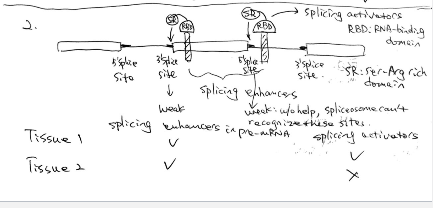
Alternative splicing mechanisms
In pre-mRNA find short RNA seq called Splicing Silencers, recognized by proteins called Splicing repressor (repress recognition of splice sites)
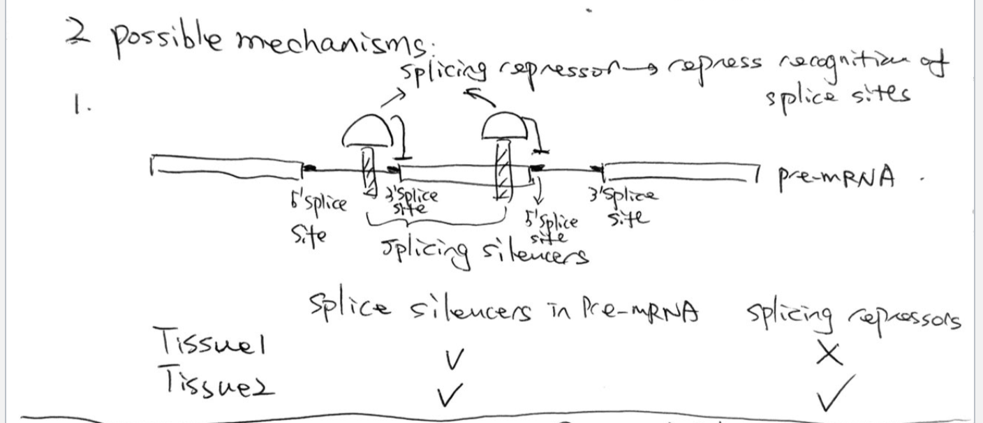
Both Tissue 1 and Tissue 2 express splice silencers since are apart of gene, however splicing repressors only present in Tissue 2 since only Exon 1 and Exon 3 are expressed. Due to presence of Splicing repressor in Tissue 2 Exon 2 is recognized apart of Intron in between Exon 1 and Exon 3.
In pre-mRNA find short RNA seq called Splicing enhancers, recognized by proteins composed of RBD connected to SR domain (Ser-Arg rich domain, similar to AD). These proteins are called Splicing activators, you find weak splice sites in gene (w/o help Spliceosomes can’t recognize these sites and a result cut out Exon along with Intron), however with Splicing activators they activate these splice sites therefore allowing Spliceosome to recognize Exon and not cut off.
DNase cuts fragments unless it has a protein or Heterochromatin state protecting DNA, whereas restriction enzymes cut at exact seqeunce
TFII is a trancsription factor that contains TATA binding protein, it binds directly to
Promoters of open/active genes tp help recruit RNA Pol II
Linker scanning experiment
Mutation analysis, purposely mutate areas located in promoter. If mutation drops trancsription below wt (mutation ruins trancsription thus activator element)
If mutation increases above wtf (mutation causes over expression, therefore find a repressing element)
EMSA (Gel shift)
Measures whether protein binds to piece of labeled DNA
Key Differences Between DNA and RNA
Structure: DNA is a double-stranded helix (like a twisted ladder). RNA is a single-stranded molecule, often shorter than DNA.
Sugar Type: DNA contains deoxyribose (missing hydroxyl in 2’ instead two H) sugar, while RNA contains ribose sugar (which has one more hydroxyl group, making it more reactive).
Location: DNA is found inside the nucleus and mitochondria. RNA is formed in the nucleus but moves to the cytoplasm and ribosomes.
Stability: DNA is more stable, protecting the genome, whereas RNA is highly reactive and easily broken down.
Bases are
Purines are Adenine and Guanine(6 and 5 membraned ring) and Pyrimidines are Thymine and Cytosine (Only 6 membrane ring)
Triphosphate
Provides energy for polymerization
Nucleotide = Nucleoside Tri-Phosphate (NTP)
DNA: dATP, ddGTP, dCTP, dTTP (dNTPs)
What are the 5’ end, 3’ end composed of
5’ end has a free phosphate group (start of sequence) and the 3’ end has a free hydroxyl group (end of sequence or phosphate group connecting nucleotides)
Phosphodiester bond
3’ → 5’
Be able to interpret and conclude results of Genetic material is transferable experiment
Inject smooth to mice and dies
Inject rough to mice and lives
Inject smooth (heat killed) to mice and lives
Inject smooth(heat killed) and rough to mice and dies (this is due to transfer of genetic material that creates protein coat to evade human immune system)
Be able to interpret and conclude results of Genetic material using bacteriophage being DNA Hershey and Chase experiment
Through bacteriophages (S35 contained protein coat and P32 contained DNA), phages S35 (sulfur exists in protein not DNA) and P32 (only DNA has phosphorous not protein), saw that phages inject DNA into bacteria rather than protein, confirming DNA as genetic material
Base pairs
A=T and G=C in DNA but [A+T] ≠ G+C
G can form three H bonds with C
A can form two H bonds with T
DNA forms double helix, base found inside and phosphate outside, strands are antiparallel
Does the same rule of A=U and G=C apply in RNA?
No since RNA is generally single stranded
Chemical features of DNA
Highly stable
Negatively charged (phosphate groups)
Denaturing (melting) and annealing (H-bonds)
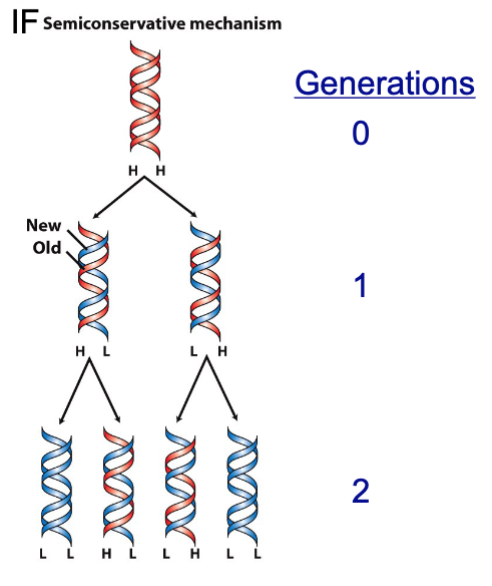
Is DNA replication conservative or semi-conservative, Meselson-Stahl experiment
Semiconservative: DNA helix split and each generates another strand
Conservative: DNA helix remains intact, but new synthesized DNA made
Found that DNA is semiconservative due to using parental strands synthesized in 15N and after replication introduce 14N, through Ultra-Centrifugation can separate strands due to weight.
Found that all newly synthesized DNA is 14N not 15N
What is required for DNA polymerization in vitro?
DNA synthesis is unidrectional
DNA polymerase, dNTPS, DNA single strand template and Primer (cannot start from scratch without these two)
DNA sequencing using chain terminators
ddNTPS (missing both OH groups unable to attack) cause visualization of multiple fragments, dNTP 3’ attacks phosphate of 5’ nucleotide
What role does the 3’ hydroxyl group play in addition to DNA polymerase
3’ OH group attacks alpha phosphate group and the diphosphate group leaves, underwent hydrolysis releasing energy, forming sugar bond
Energy: Triphosphate on nucleotides
Nucleic acid synthesis always 5’ to 3’ due to hydroxyl group attacking triphosphate, the 5’ end does not have hydroxyl group unable to attach new nucleotides
DNA synthesis in vivo (in cells)
DNA replication determined to be bi-directional in nature by treating with low radioactivity (thus nucleotides added appear) and after high radioactivity (thus newest nucleotides added are all highly radioactive) showed that nucleotides are being added both sides
What troubles are encountered due to bi-directional replication
Replication fork: Leading strand DNA synthesize and fork movement are parallel, however Lagging strand DNA synthesize and fork movement are antiparallel (requires multiple RNA primers). Nuclease removes multiple RNA primers along lagging strand. Different DNA polymerase synthesizes in gaps (however it can’t connect these DNA fragments), require Ligase (uses ATP) to connect phosphate sugar bond OH and Triphosphate
Each end has a leading/lagging strand
DNA polymerase functions
5’→3’ nucleic acid addition activity and 3’→5’ exonuclease activity (remove nucleotide if wrong)
Types of Nuclease
Endonuclease breaks phospho-sugar bond in middle of DNA (regenerate hydroxyl and phosphate) this is called a nick and is fixed by ligase
Exonuclease breaks phospho-sugar bond at end of DNA (3’ or 5’)
Ligase
Connect phospho-sugar bond between fragments on lagging strand
Gap in DNA described as
Missing nucleotides, this is created by endonuclease + exonuclease. Require DNA polymerase and Ligase to fill gap
Helicase
Breaks H bonds between base pairs
General strategy of repair of DNA single strand damage or replication error
DNA damage/error recognized
Nuclease removes nucleotides
Gap filled (DNA Pol + Ligase)
Promoter
Not apart of coding sequence rather recruit transcription factors and RNA polymerase
if no promoter → no gene product
5’ UTR (Untranslated region)
Occurs in nucleus as mRNA molecules is being syntehsized by RNA Pol II. Transcribed into mRNA
Coding region
Region that is transcribed into mRNA, only region translated into amino acid sequence to protein
3’ UTR
Occurs in nucleus as mRNA molecules is being syntehsized by RNA Pol II. Transcribed into mRNA
Molecular cloning: Transforming and obtaining bacterial clone library
Plasmid vector + dna of interest, insert dna into vector, mix plasmid with cell along with heat, cell will digest vector, plate w/amp, bacteria containing plasmid will only survive
Restriction enzyme
cut DNA at specific sequences (a specialized endonuclease)
Vector
Plasmid or viral DNA that can replicate in a desired organism
Each bacterial cell will take one single piece of DNA
What do we need in a plasmid/vector
Origin of replication (without wont be able to replicate
Selectable marker: antibiotic resistance gene (AmpR)
Gene of interest
Sticky ends
Generated by restriction enzyme, short single stranded DNA overhangs, can automatically form H bonds to complementary fragments (anneal: form H bonds)
During transformation of plasmid and bacteria, one bacterial cell can only take up one plasmid, seperated by applying amp and only those with ampR gene survive and produce colonies
If there is no origin of replication even if has ampR gene, cell will not recognize plasmid, and no replication of DNA. Will not see any colonies (only few amounts) under Amp
Gel electrophoresis to separate DNA molecules based on size
Molecules move through pores in gel (agrose), DNA is negatively charged so goes from - → +. Smaller fragments able to travel faster rate than larger fragments
Genome sequencing
Genomic DNA, use a restriction enzyme to cut specific sequences of DNA, from there you ligate the fragments with vectors (w/ origin of replication and ampR gene), put on dish and watch colonies grow (put amp) to kill off any colonies not resistant to amp and divide into separate test tubes, from there you align and get whole genome seq. Are able to determine order by sequencing genome again but with different restriction enzymes and if see overlap are able to confirm fragment order
Steps of PCR
Require two primers (since double stranded DNA)
Denature DNA by heating up to 95*C (single strand)
Annealing of primers by cooling 50-72*C
Elongation/Extension of primers (polymerase + dNTPs makes new DNA) 72*C
Design Primers
Forward and Reverse and are just complementary sequences
Single digest (one singular enzyme, found on plasmid)
Opens plasmid into one long piece (only see one band)
Double digest (multiple enzymes same/different)
distance between two enzyme sites