DH228: Oral Pathology Picture
1/75
There's no tags or description
Looks like no tags are added yet.
Name | Mastery | Learn | Test | Matching | Spaced | Call with Kai |
|---|
No analytics yet
Send a link to your students to track their progress
76 Terms
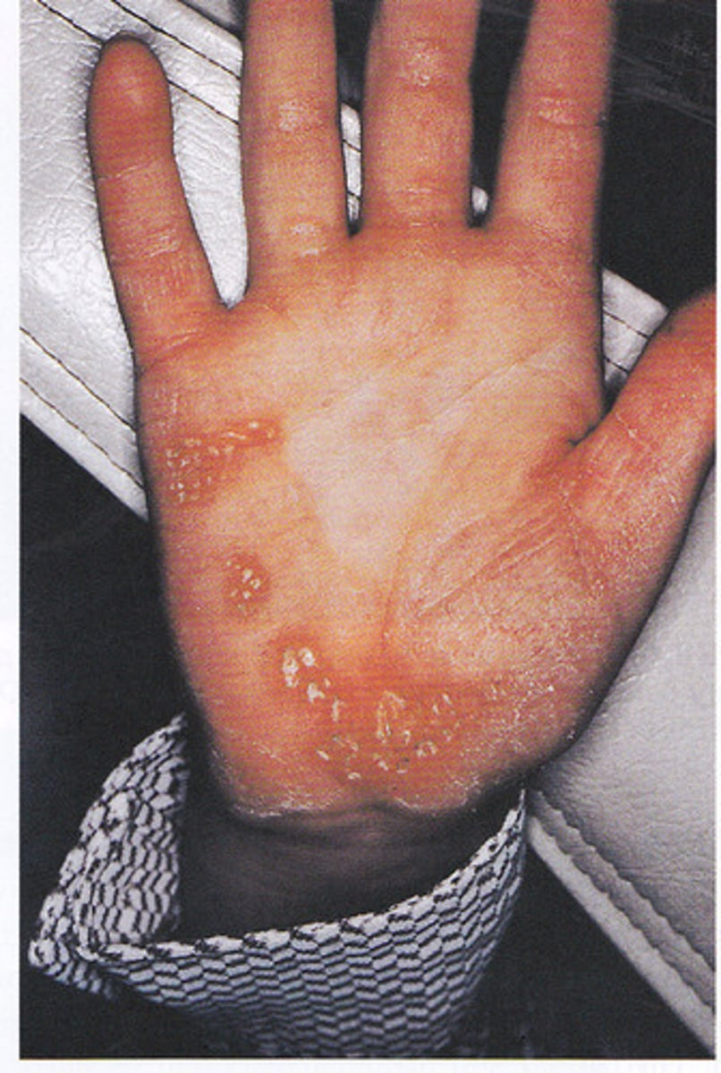
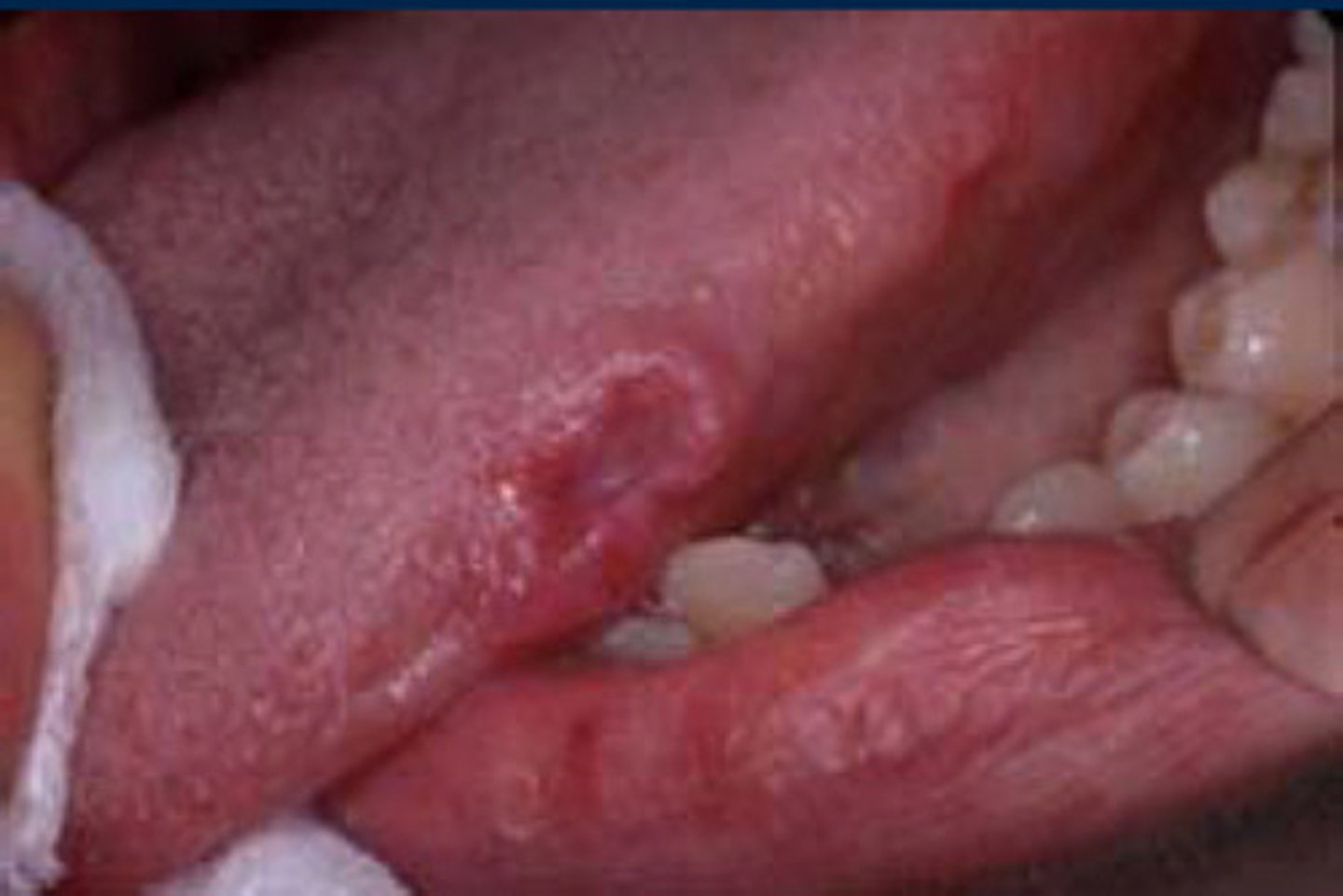
Aspirin Burn
Topical application is a common misuse of this product; The tissue becomes necrotic & white; Surface may slough off, leaving a painful ulcer; Usually heals in 7-21 days
Your patient is complaining of a toothache in the LL posterior. During the EIOE, you notice a white to gray patch on the left buccal mucosa.
•What do you suspect is the cause of this lesion?
What question would you ask the patient?
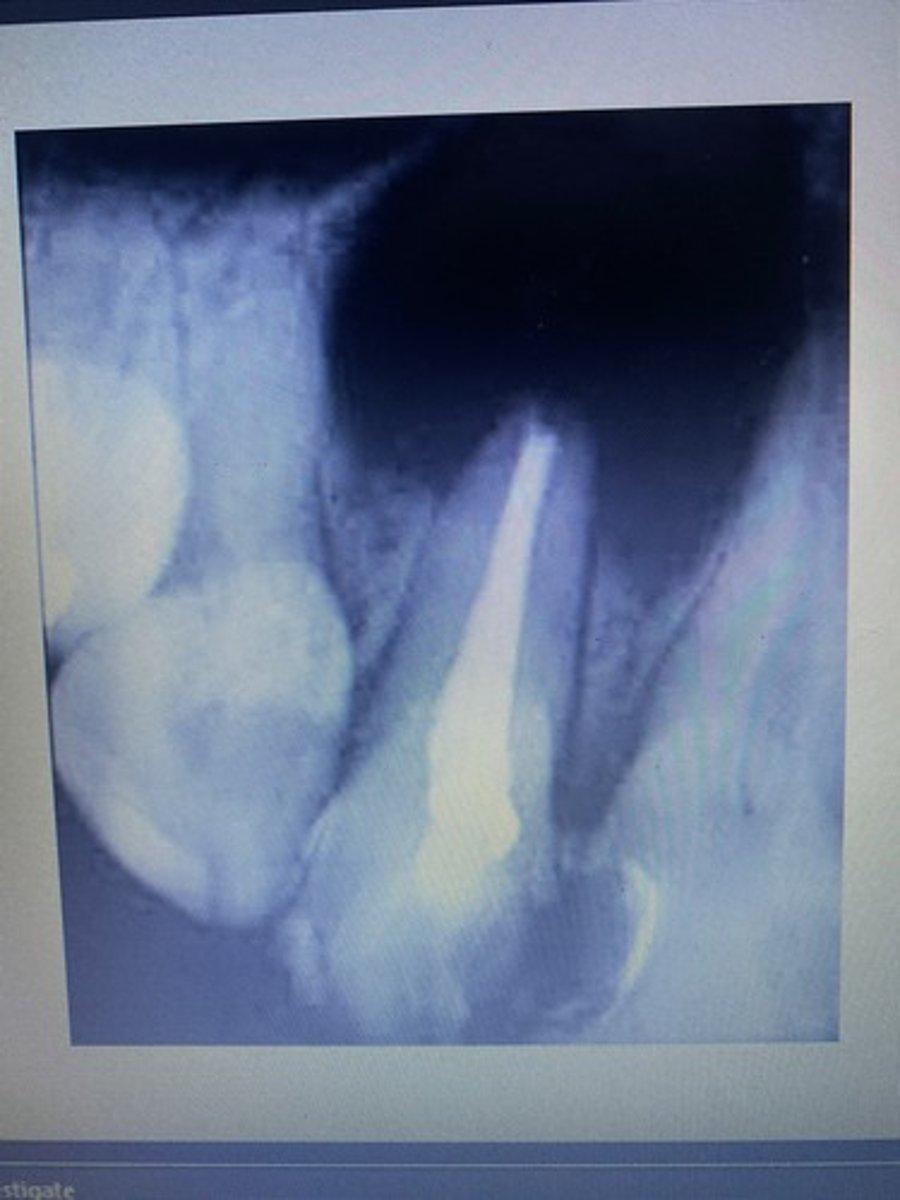
Radicular cyst
Name this condition (Most common cyst observed in the oral cavity, caused by pulpa inflammation and often a side effect of a long lasting or recurrent periapical infection)
kaposi sarcoma
-diagnosis is made via biopsy
-no effective treatment exists
The most common intraoral locations for this lesion are the gingiva and palate. When diagnosed, this vascular lesion meets the criteria for the diagnosis of acquired immune deficiency syndrome (AIDS). What is the name of this vascular lesion?
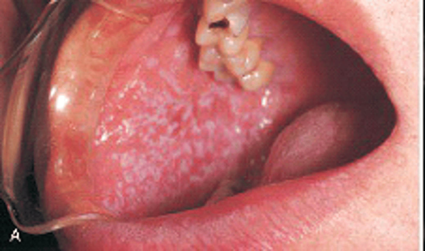
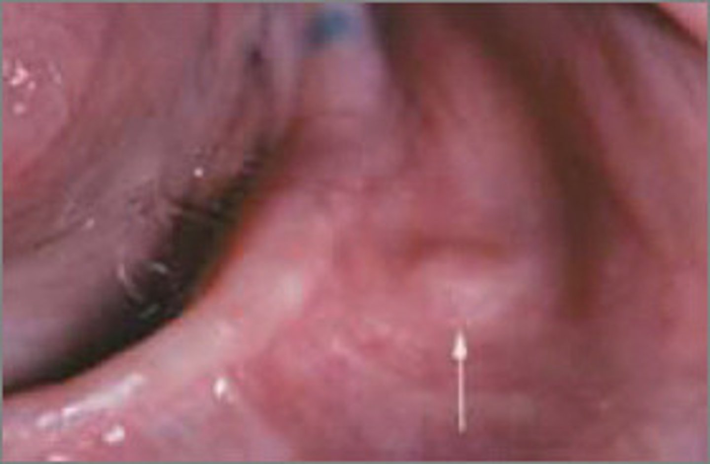
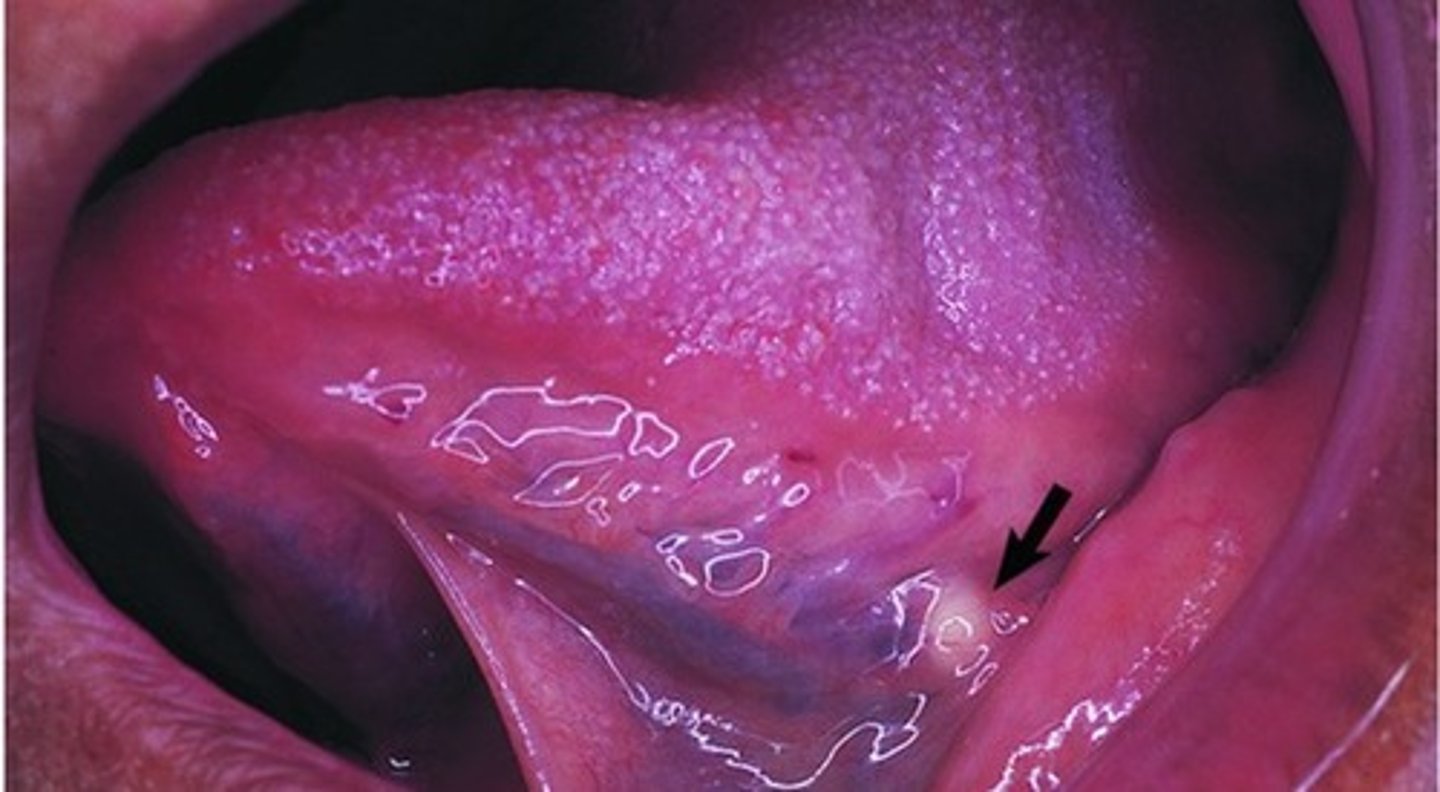
erythema migrans (geographic tongue)
the lesions by the arrows on the dorsal of the tongue are characterized by diffuse areas devoid of filiform papillae that form on the dorsal and lateral borders of the tongue are called
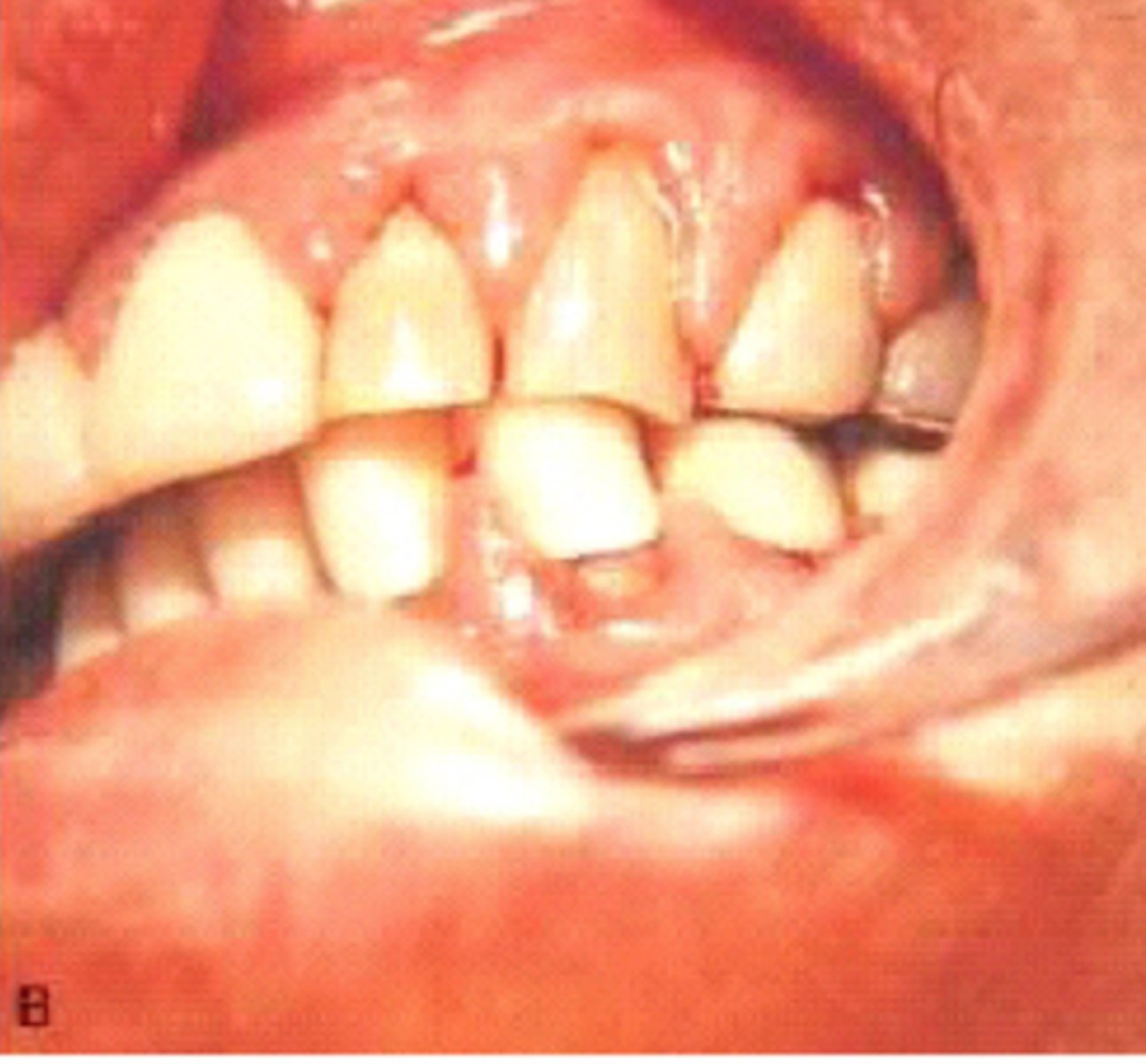
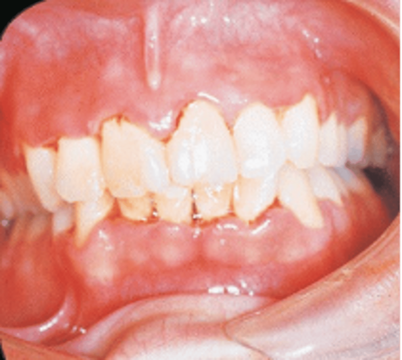
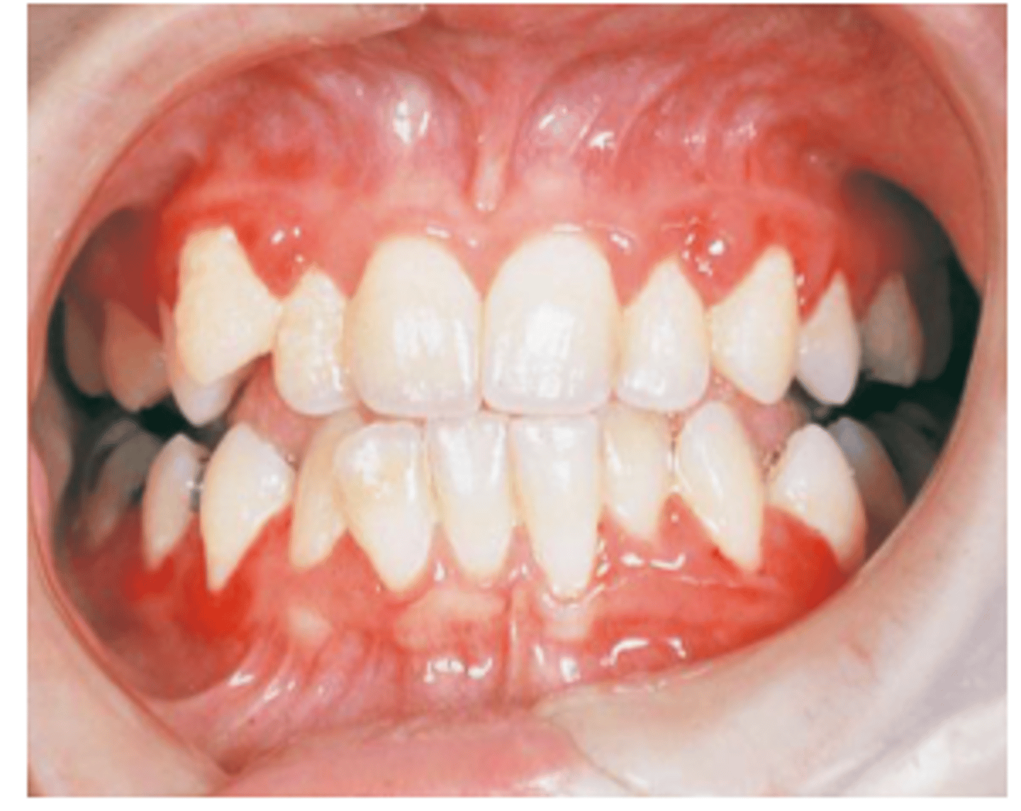
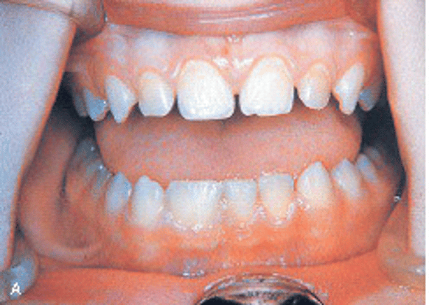
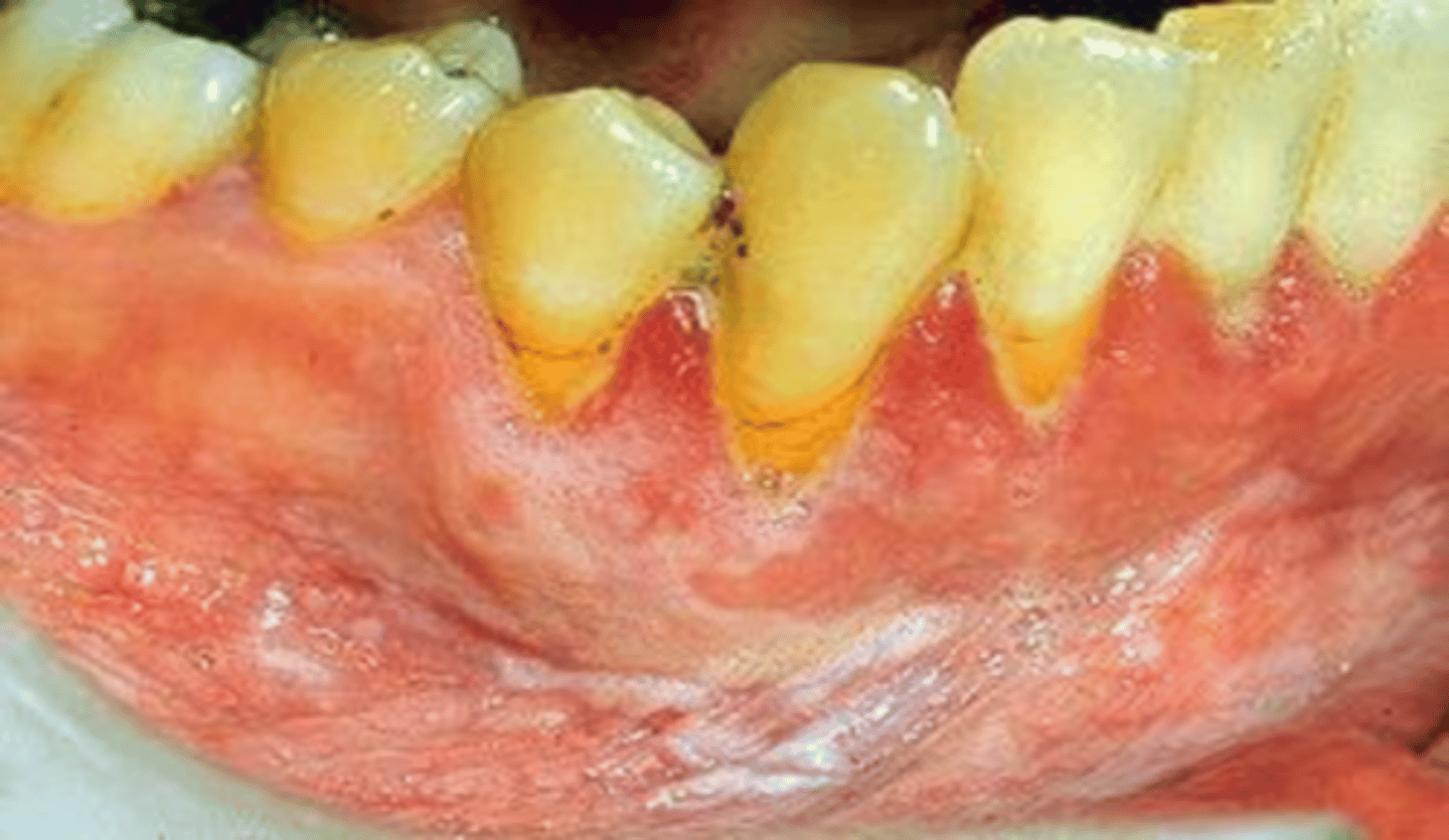
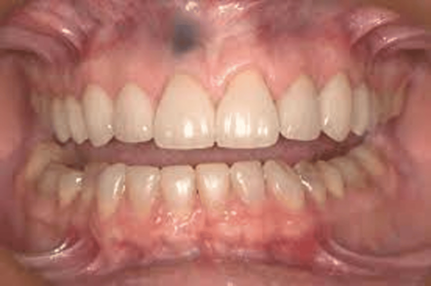
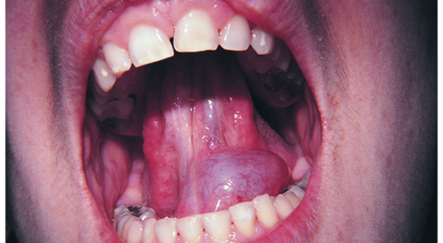
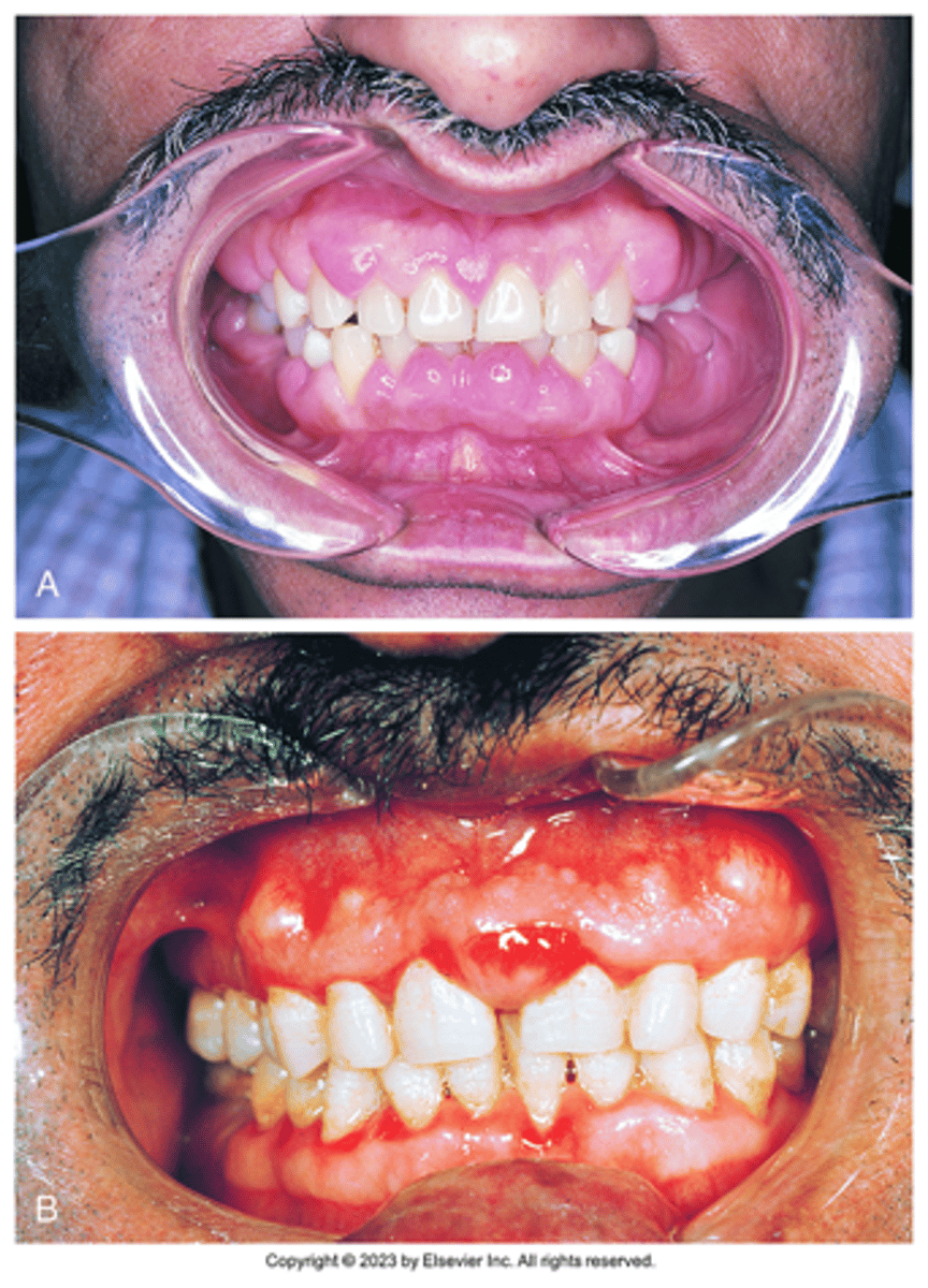
calcium channel blockers.
the common drug is nifedipine
What class of drugs can cause gingival enlargement as seen in the image below?
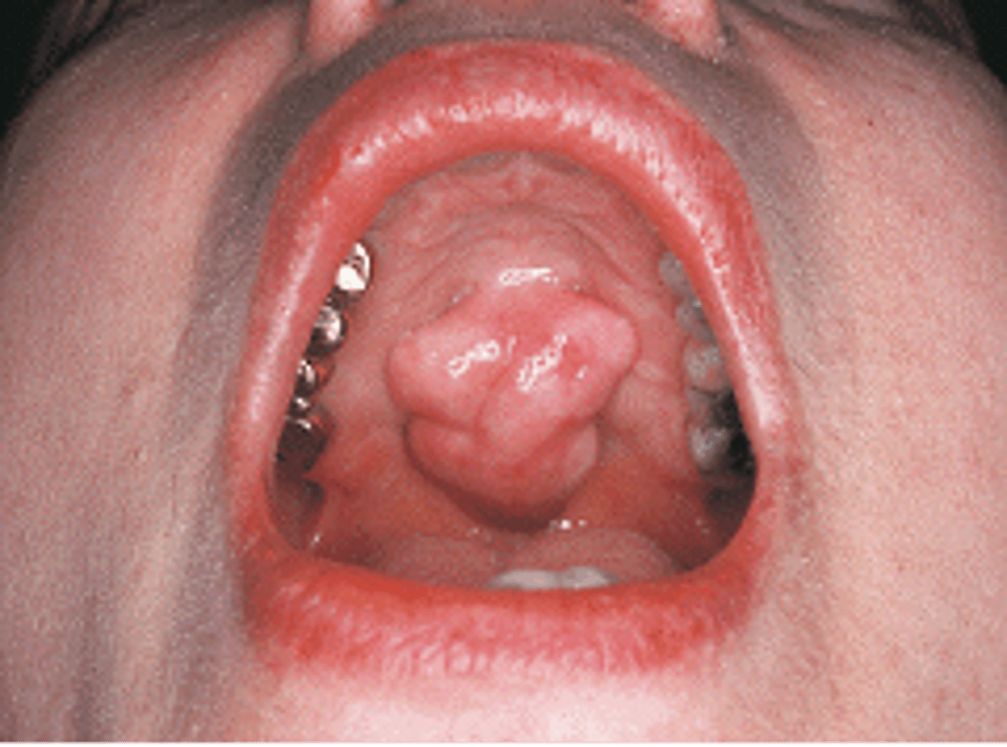
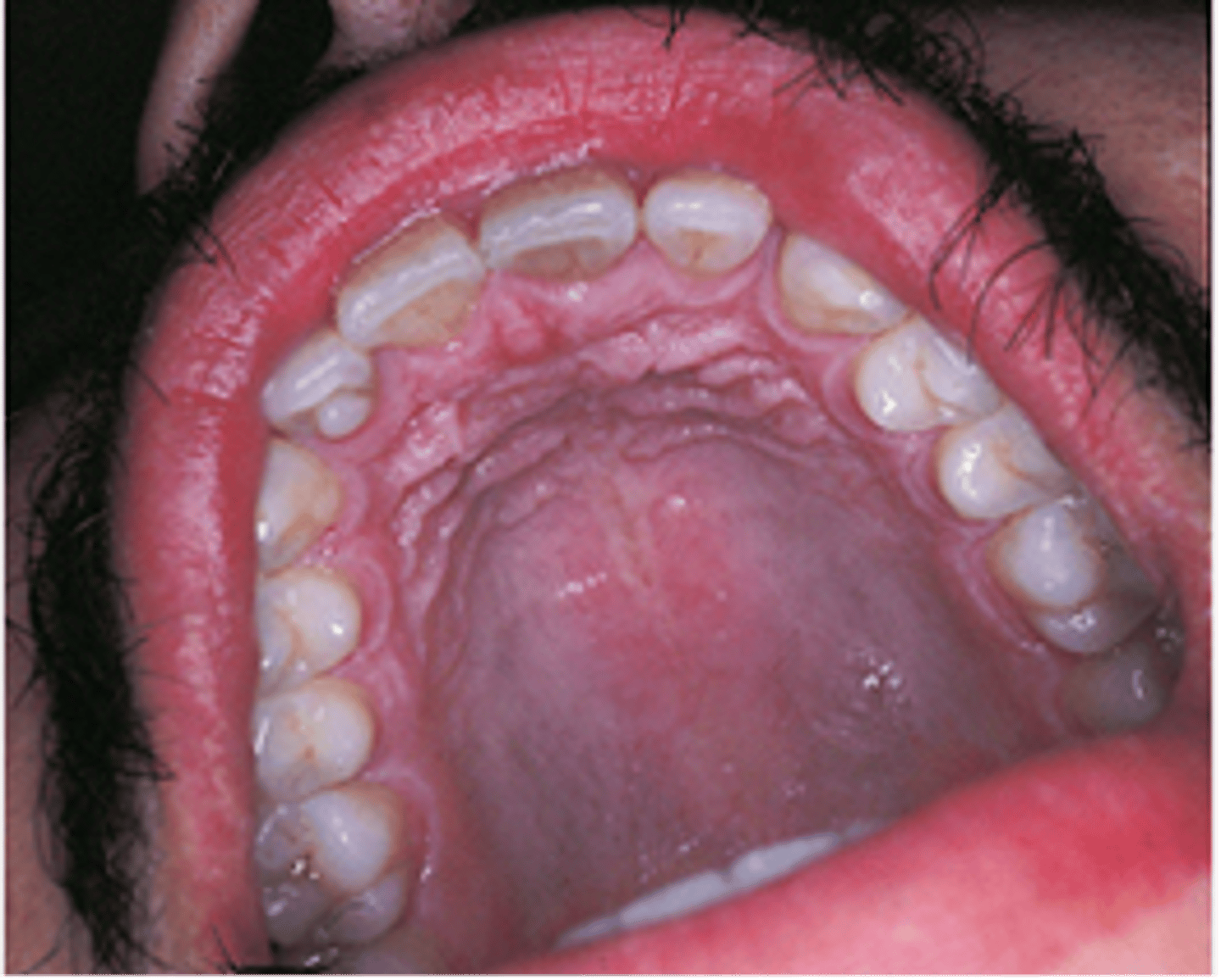
Torus Palatinus (AKA palatine torus)
This bony hard, radiopaque structure in the midline of the hard palate is diagnosed through clinical diagnosis. It is genetic and inherited in an autosomal dominant manner...

osteonecrosis
staged based on size, location and symptoms. -bisphosphonate therapy
iron deficiency anemia
-tongue is devoid of filiform papillae
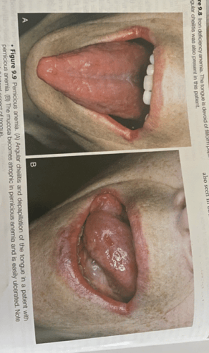
pernicious anemia
-Vitamin B12 deficiency that is caused by a deficiency of intrinsic factors a substance secreted by the parietal cells of the stomach
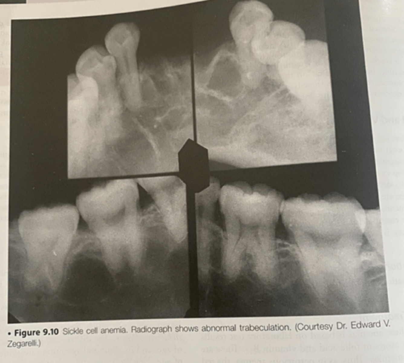
sickle cell anemia
-abnormal trabeculation
-most common inherited disorder of red blood cells
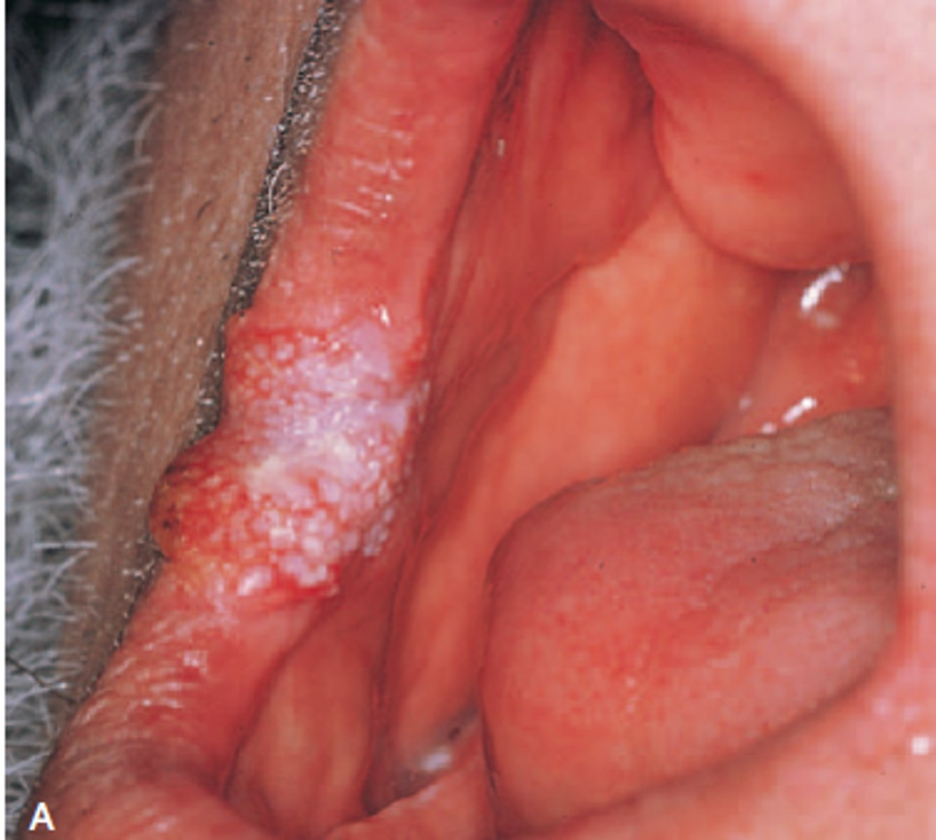
Verrucous Carcinoma
-commissure and anterior buccal mucosa
-is a specific type of squamous cell carcinoma that is separated from other squamous cell carcinoma because it has a much better prognosis
-appears as a slow growing exophytic tumor with a pebbly white and red surface
-most cases occurs in men older than 55 years of age and involves the vestibule and buccal mucosa
ameloblastomas B
-show multilocular radiolucencies in the molar areas if the mandible
-is benign, slow growing but locally aggressive epithelial odontogenic tumor that may arise in either the maxilla or the mandible
-it is a non encapsulated tumor that infiltrates into surrounding tissue and can cause extensive destruction
Benign Cementoblastoma
-shows a well circumscribed radiopaque mass surrounded by a radiolucent halo and attached to the root of a mandibular first molar
-pain is a frequent symptom
-occurs in young adults and most occur in patients under 30
paget disease
-enlargement of the maxilla with spaces between the teeth
-radiograph demonstrating irregular opacification that is also referred to as a cotton wool appearance
Calcifying Odontogenic Cyst
-composed of odontogenic epithelium that contains "ghost cells"
-also referred to as gorlin cyst
-most commonly seen individuals younger than 40 years
-lesions occur equally in the maxilla and mandible, most often in the incisor and cuspid area
-present as well defined unilocular or multilocular radiolucency
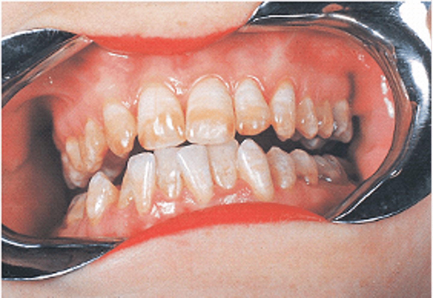
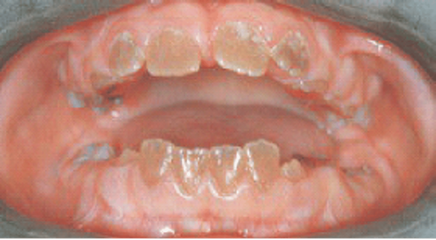
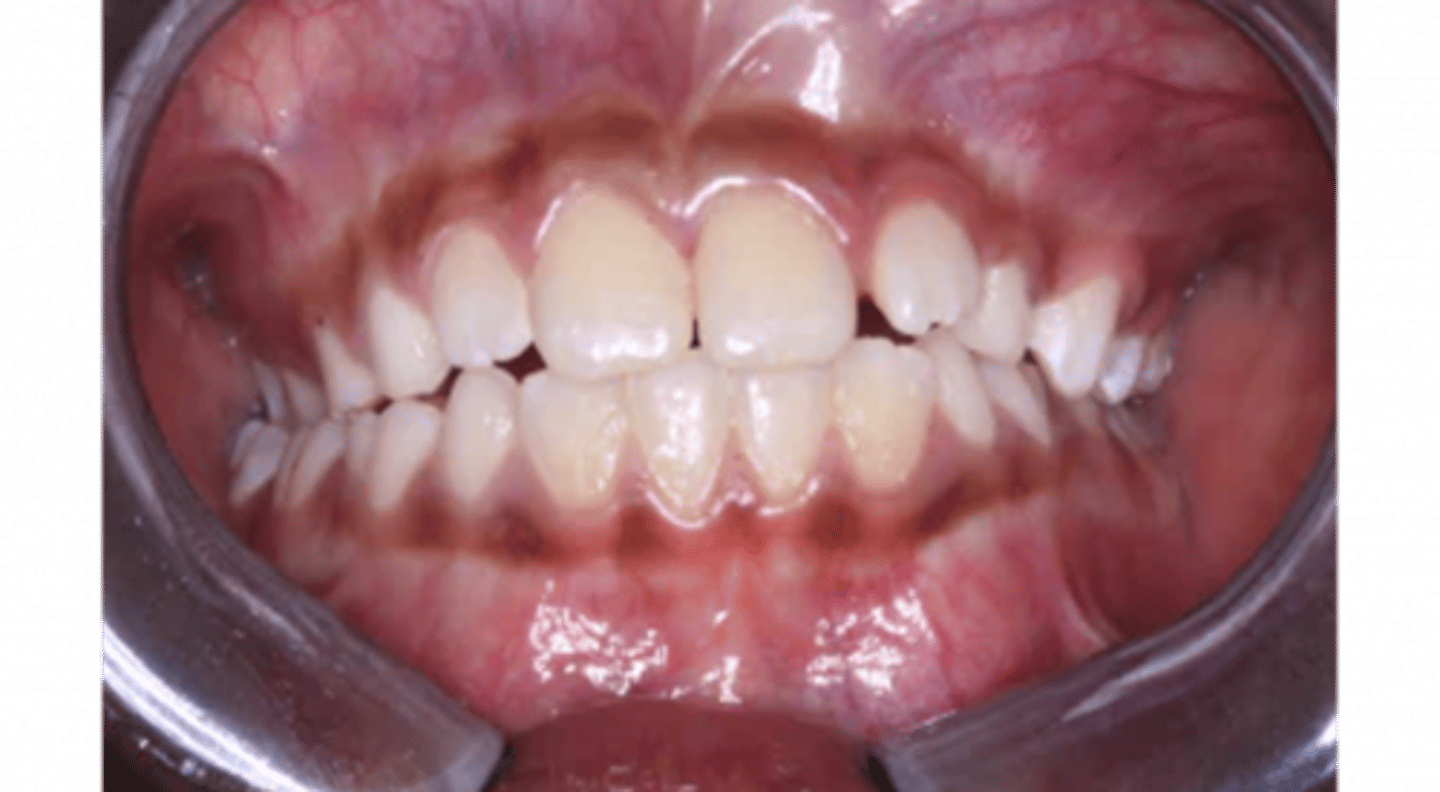
Mottled enamel- fluorosis
Discoloration of enamel from high fluoride intake
The patient in this image is healthy with no local or systemic infection or disease. The patient's teeth are caries free, as are all of the teeth of all of the patients who exhibit this defect. What is this condition?
Lichen Planus
Benign, chronic disease that affects the skin and oral mucosa
-White lines= Wickham Striae
The classic appearance of lichen planus affecting the oral mucosa is a pattern of interconnecting slender white lines referred to as?
Abfraction
loss of tooth surface in the cervical area, caused by tooth grinding compression forces
What are these wedge-shaped defects at the cervicofacial surfaces of these teeth called?
Kaposi sarcoma (KS)
Cancer caused by the human herpes virus 8 (HHV-8) that mainly affects the skin and mucous membranes but may also cause extensive visceral organ involvement; also called malignant neoplasm of soft tissue
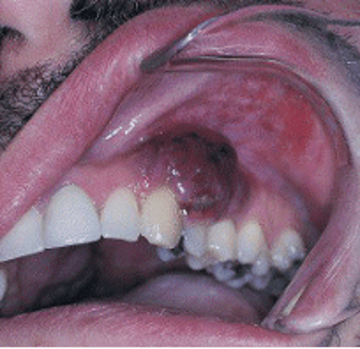
Gingival Enlargement by a calcium channel Blocker
Seen by a patient taking nifedipine (Ca Channel blocker)
Concrescence
Union of teeth by cementum
What is it called when two or more teeth are joined by cementum?
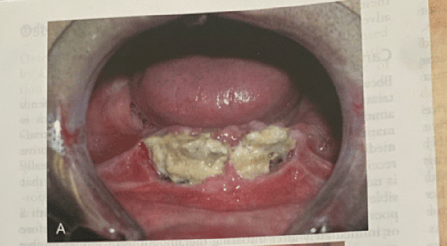
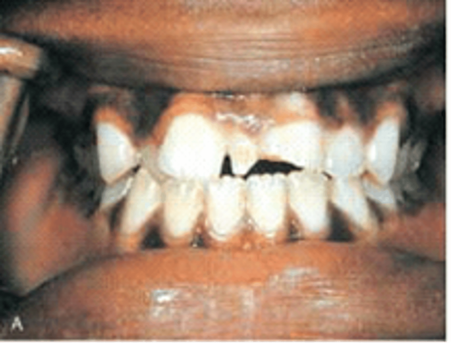
NUG (Necrotizing Ulcerative Gingivits)
Fusiform bacillus and Spirochete cause this, cratered papilla, and white tissue because of cell death
This patient presents with painful, erythematous gingiva and the interdental papillae appear as punched-out, necrotic, cratered areas. Sloughing of necrotic tissue presents as a pseudomembrane over the tissues. What condition do you suspect?
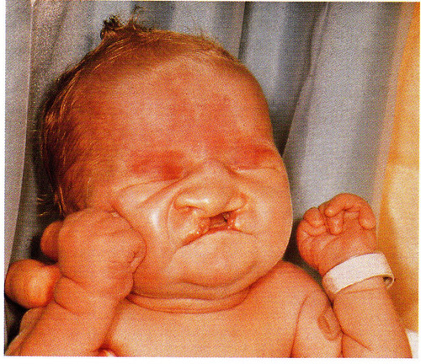
Trisomy 13 (Patau Syndrome)
-abnormalities in multiple organs
turner syndrome
-webbing of the neck
-caused by patient has only one X chromosome or monosomy X
Papillon-Lefevre Syndrome
-lesion of the hand remain reddish white scaly, thick areas of hyperkeratinization
-autosomal-recessive inheritance
Cyclic Neutropenia
-by decrease in the number of circulating neutrophilic leukocytes, white blood cells help fight infection and remodel the body.
-autosomal dominant
cleidocranial dysplasia
-multiple extracted supernumerary teeth rom the patient with cleidocranial dysplasia
-results in cranium developing into a mushroom shape because the fontanelles remain open
-the premaxilla is generally underdeveloped
cherubism
-bilateral facial swelling that appears when the patient is between 1.5 and 4 years of age
-soap bubble appearance
Gardner syndrome
-panoramic radiograph of patient with gardner syndrome shows multiple osteomas and odontomas
-also known as familial adenomatous polyposis
-benign bone growths
-teeth can exhibit hypercementosis and fail to erupt
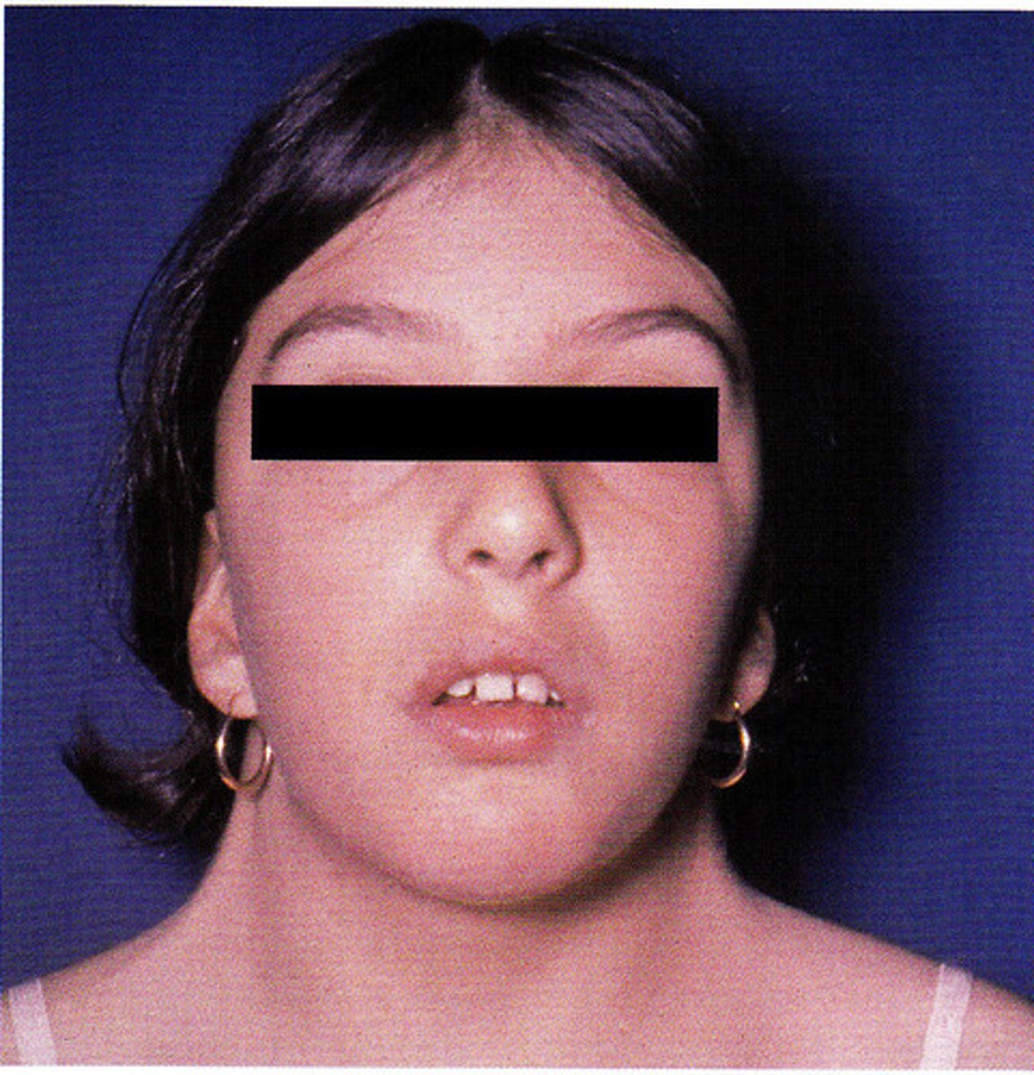
Mandibulofacial Dysostosis
-markedly high-arched palate and malpositioned teeth in a patient with mandibulofacial dysostosis
-also known as treacher collins syndrome
-results in a dysmorphic face, including downward sloping of the palpebral fissures.
-mouth appears fishlike
Peutz-Jeghers syndrome
-multiple small to medium sized pigmented macules on the labial mucosa of a patient with peutz-jeghers syndrome
-also known as hereditary intestinal polyposis syndrome
-pigmentation occurs around the eyes, nose and mouth
-associated with gastrointestinal polyposis
dens invaginants aka dens in dente
-tooth within a tooth
The most common location for this developmental anomaly is the maxillary lateral. What is this anomaly called?
Dilaceration
an abnormal bend or curve, as in the root of a toot
The sharp bend or curve in the root as seen in the image is called?
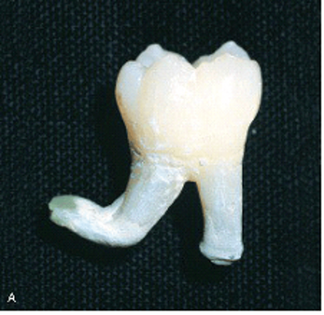
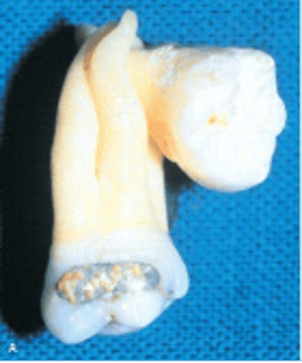
Fusion
Two roots, one crown on MND lateral incisors (in this picture- usually two roots, two heads, 2 pulps)
When the large tooth in the mandibular arch is counted as one, then there's one less tooth than normal in the mandibular dentition.What condition do you suspect?
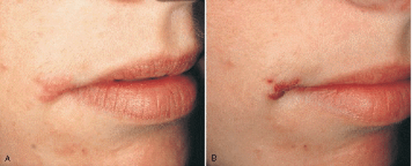
Angular Cheilitis
-inflammation in corners of the mouth
-sore, swollen, cracked
This lesion in the commissure is most likely caused by Candida albicans or a nutritional deficiency.
Which condition do you suspect?
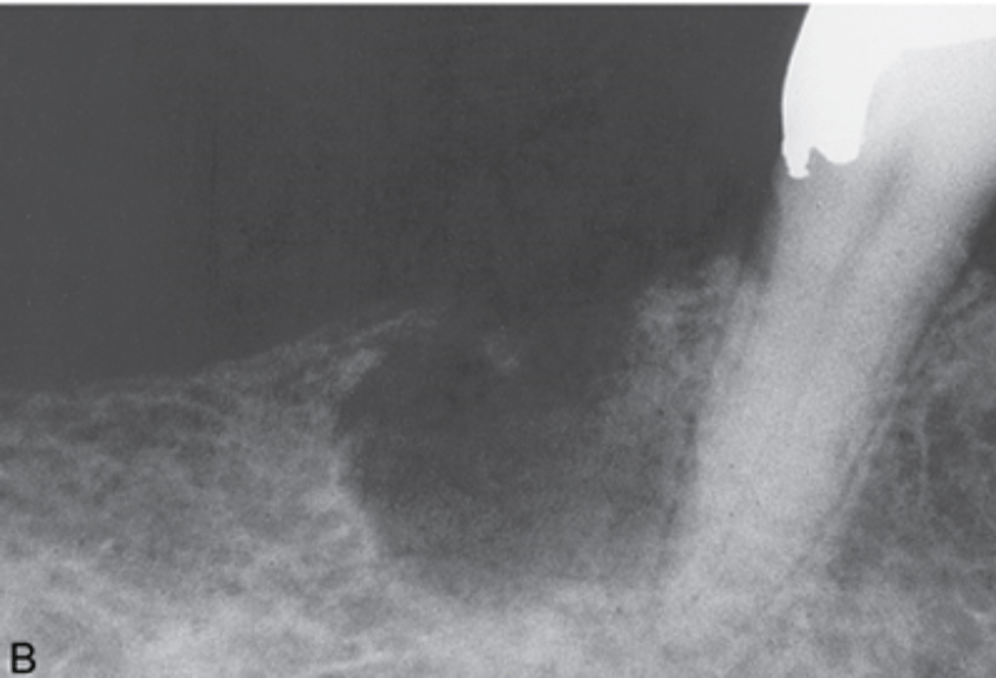
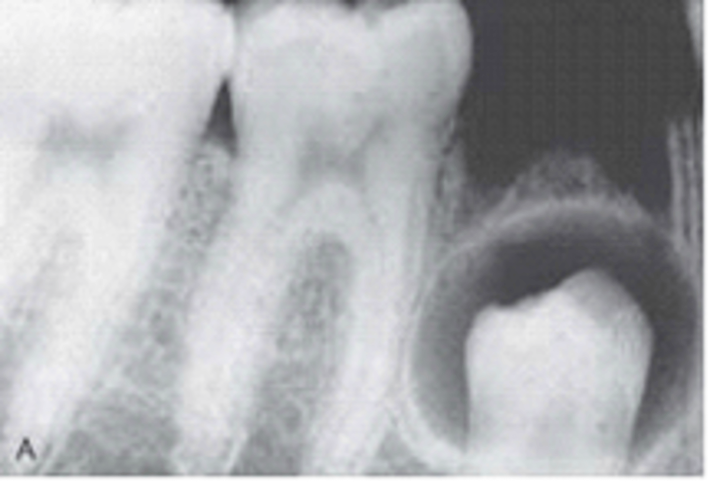
Lateral periodontal cyst
-True cyst (epithelial lining)
-Well circumscribed Unilocular or Multilocular lucency in the lateral periodontal membrane of adults between roots of adjacent, erupted teeth
-most found in mand premolar region
-associated tooth is vital
This radiolucent lesion located interproximal to the mandibular lateral incisor and canine is most likely which of the following?
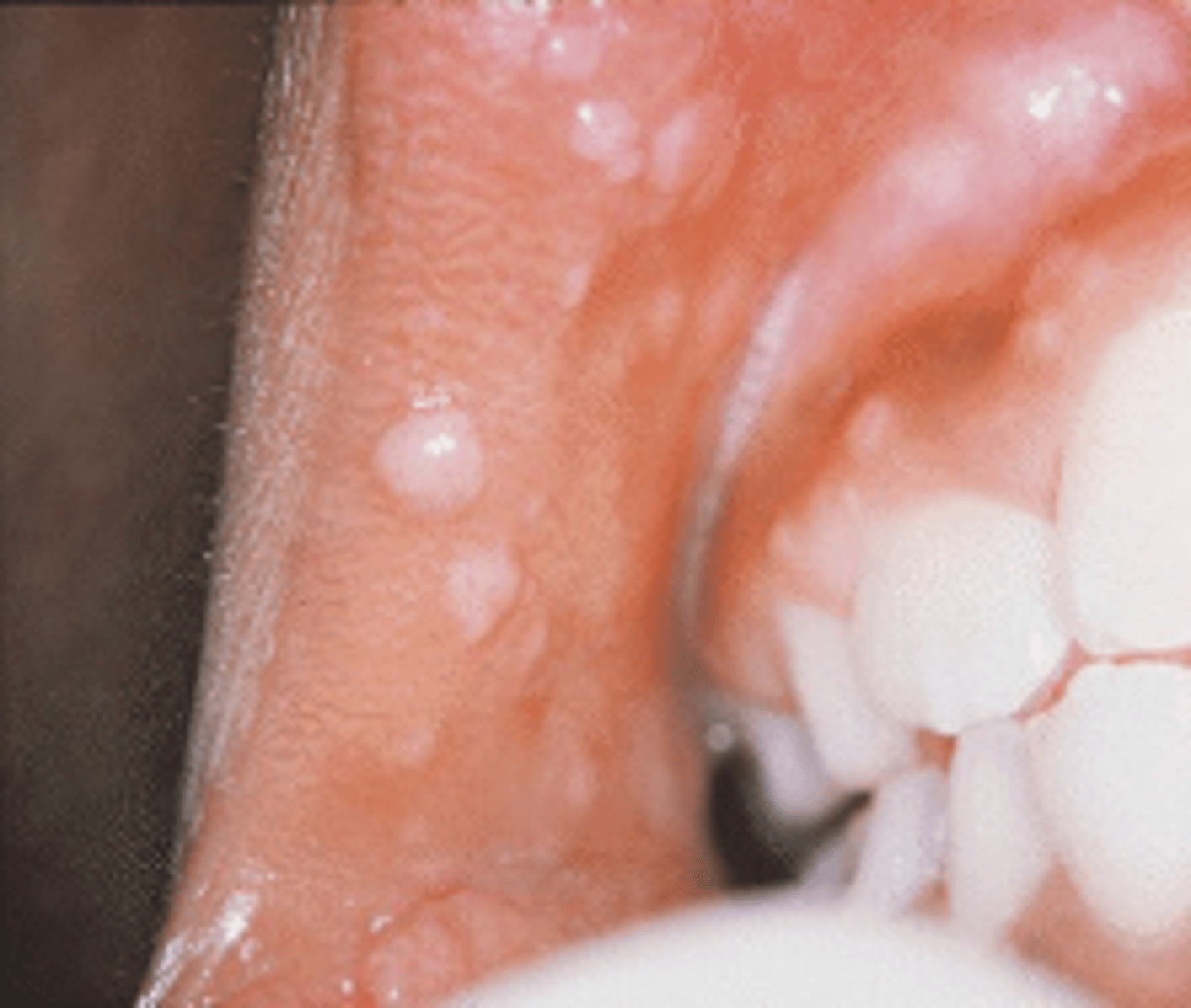
Heck Disease / human papillomavirus (HPV)
is a cutaneous condition characterized by white to pinkish papules that occur diffusely in the oral cavity. It is caused by the human papilloma virus types 13 and 32.
This condition is referred to as Heck disease or multifocal epithelial hyperplasia, is most common in children, and infrequently seen in patients with HIV. What is the etiologic agent for this condition?
Ankyloglossia (tongue tie)
Significant adhesion of the tongue to the floor of the mouth caused by a short lingual frenum is called?
developmental anomaly characterized by a shortened lingual frenum that limits movement of the tongue.
Can cause speech problems, periodontal defects, and problems with breast feeding.
More common in males
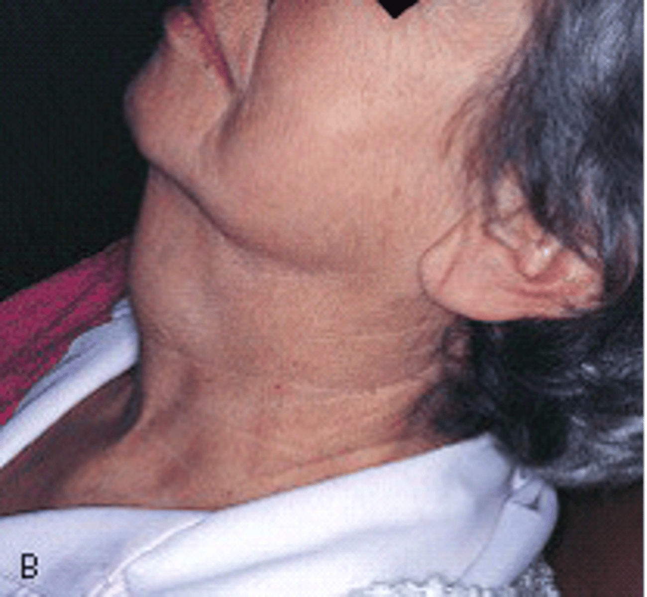
Thyroglossal duct cyst
a palpable cystic midline mass in the neck due to incomplete closure of the thyroglossal duct.
The enlargement on the midline of this patient's neck has been present for months and is slowly increasing in size. It is soft and painless.Histologic examination shows this to be an epithelium-lined sac filled with clear, yellow fluid. What type of cyst is it?
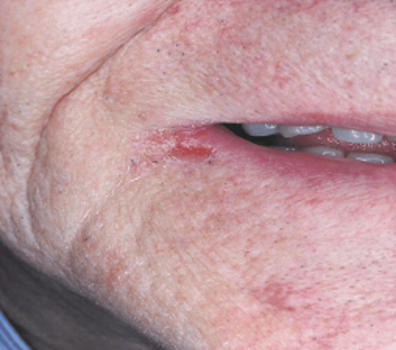
Herpes Labialis
blister-like sores on the lips and adjacent facial tissue that are caused by the oral herpes simplex virus type 1 (HSV-1); also known as cold sores or fever blisters
The cluster of vesicles seen at this pt's vermillion border is commonly called a fever blister or cold sore. What is your DHDx?
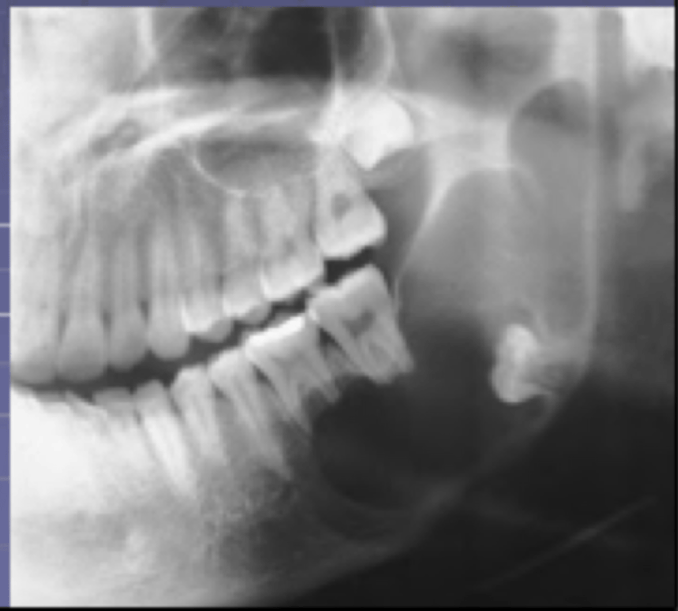
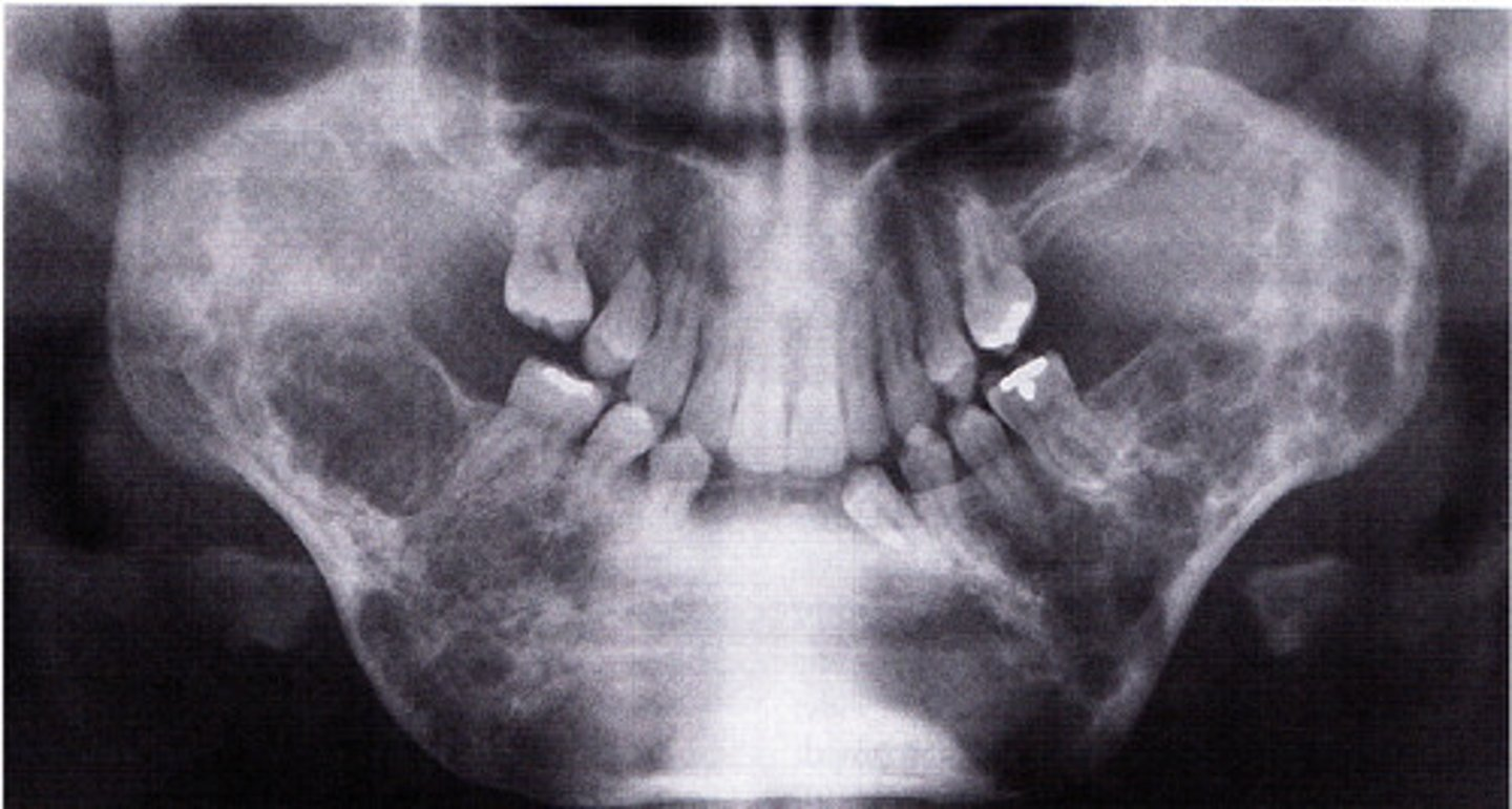
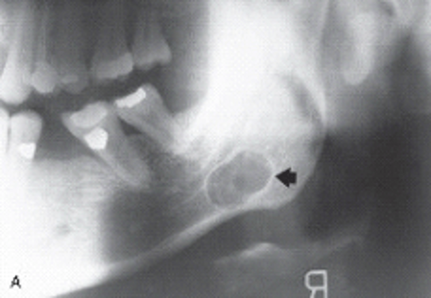
Dentigerous cyst
A cyst that forms around the crown of an unerupted or developing tooth. Also called a follicular cyst. Second most common cyst.
This unilocular radiolucency extending CEJ to CEJ around the crown of this unerupted tooth, is most likely a ?
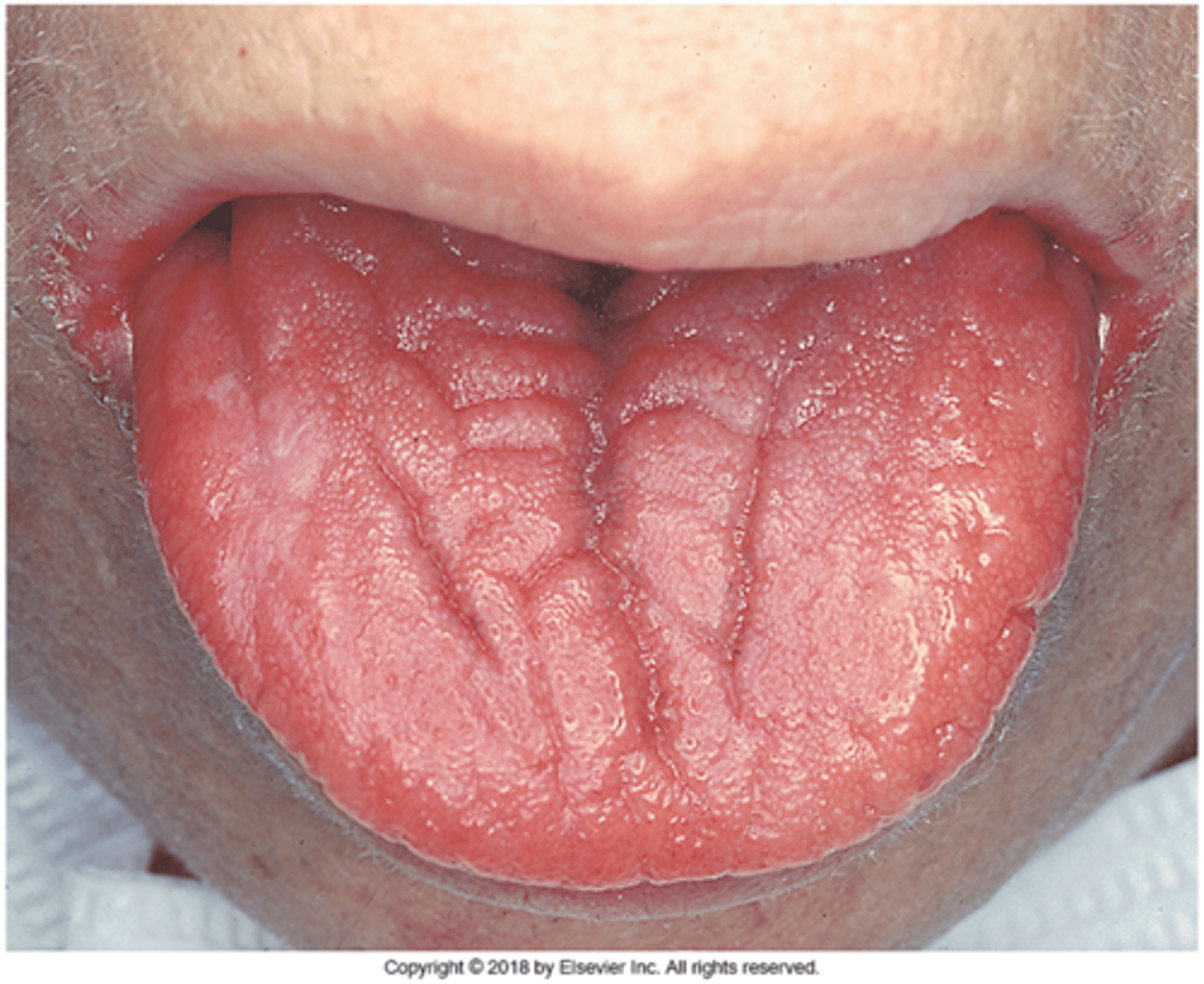
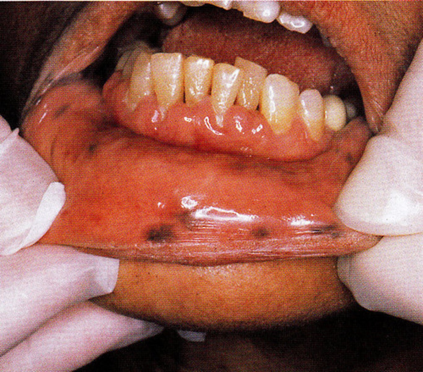
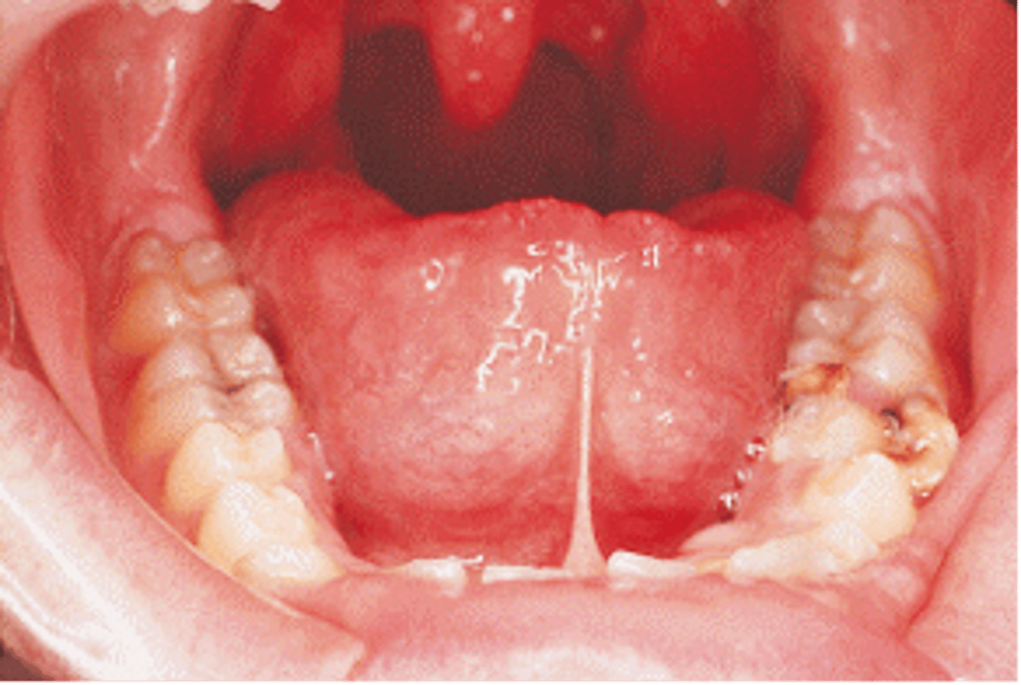
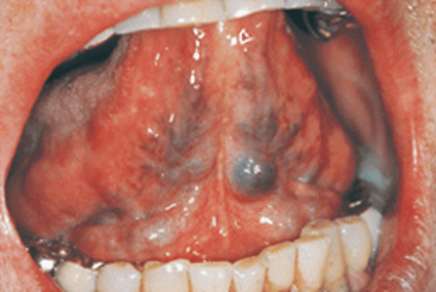
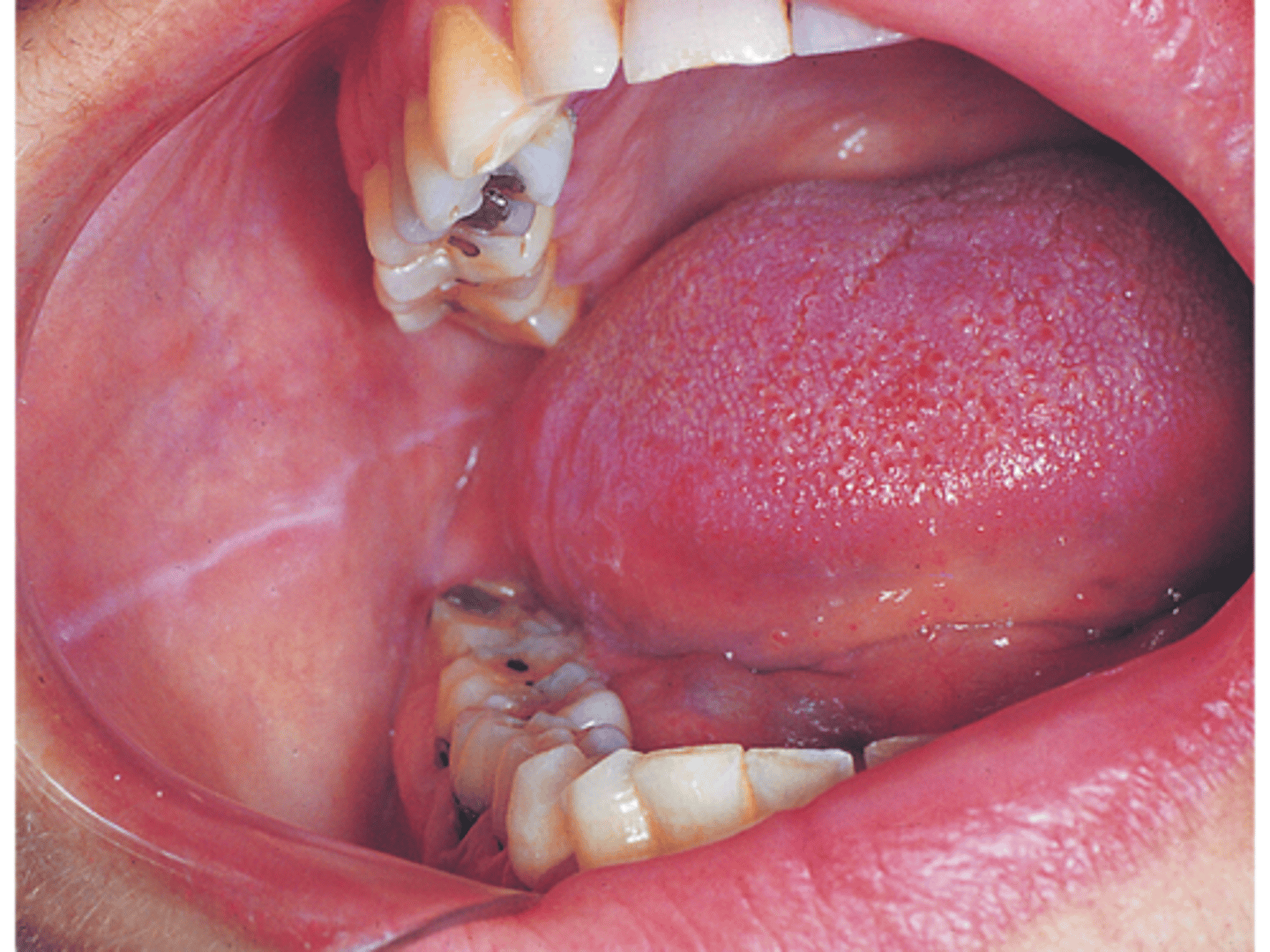
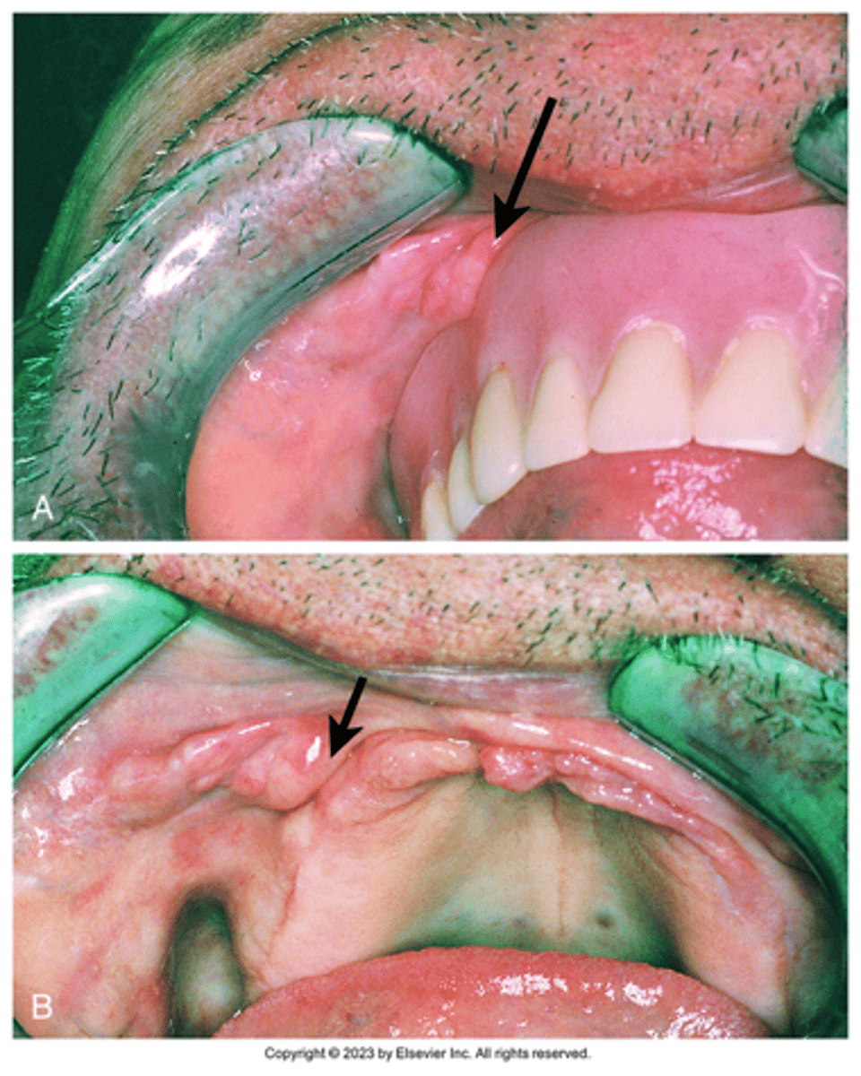
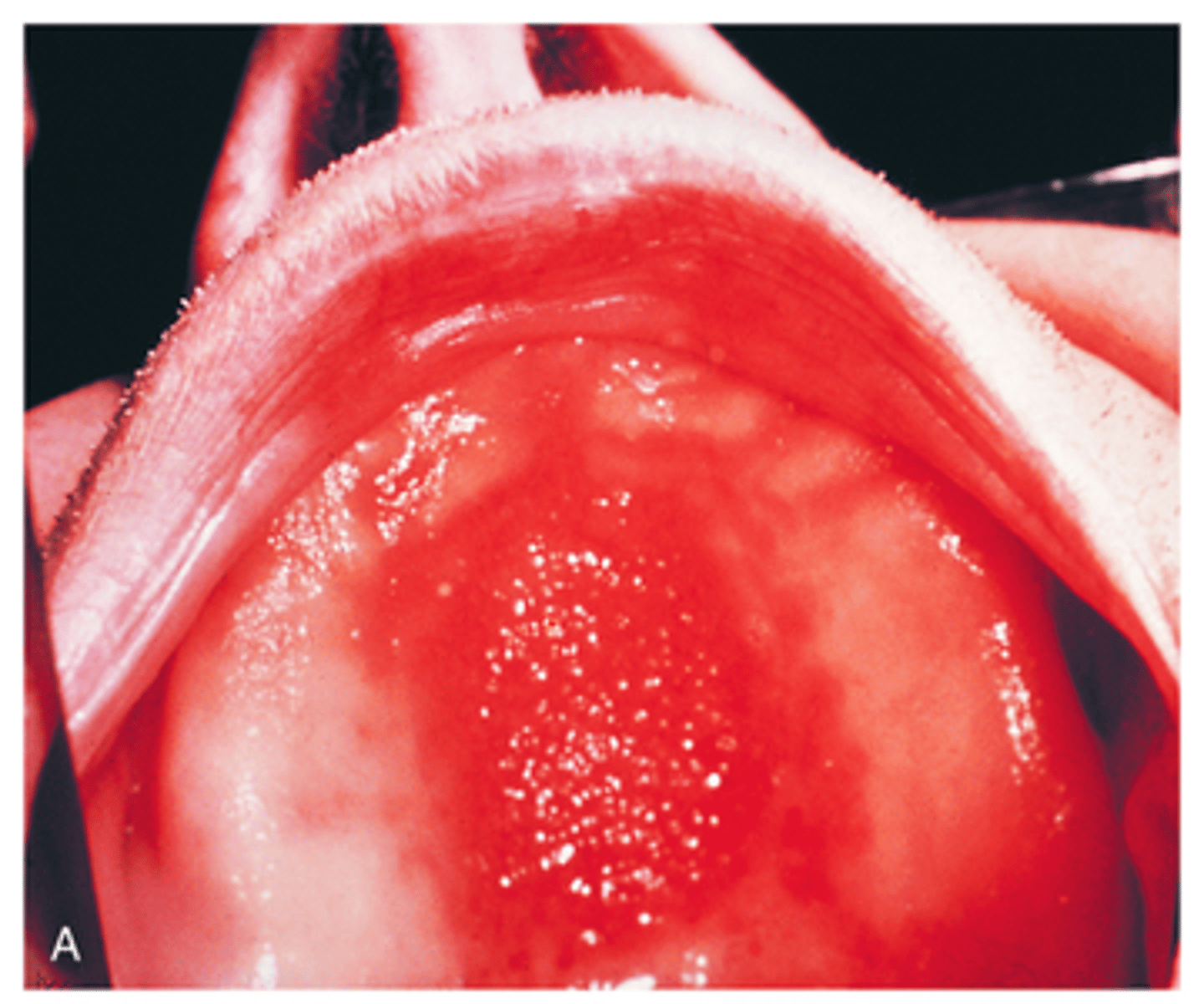
Lingual Varicosities
- Normal
- May be very pronounced
- Protruding veins on ventral surface of the tongue
what are the reddish-purple clusters on the ventral surface of the tongue that are variants of normal and diagnosed via clinical exam called
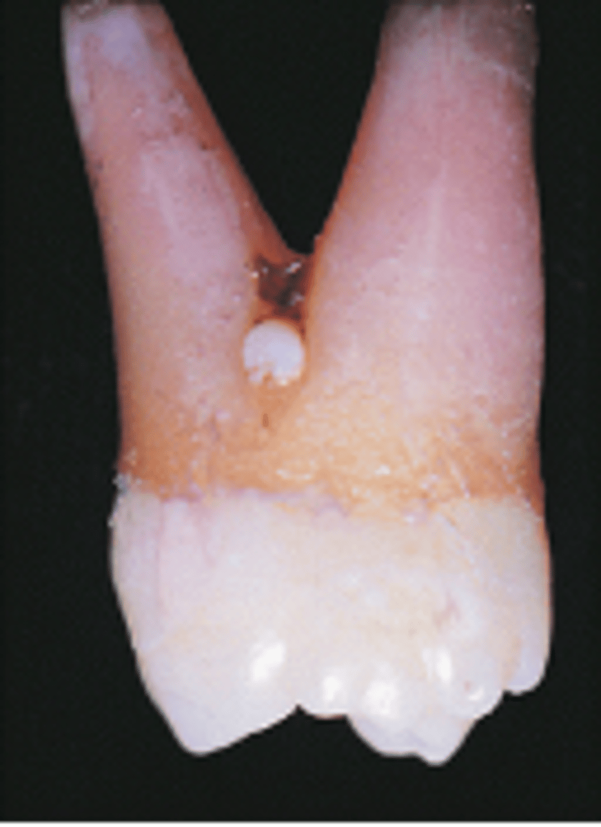
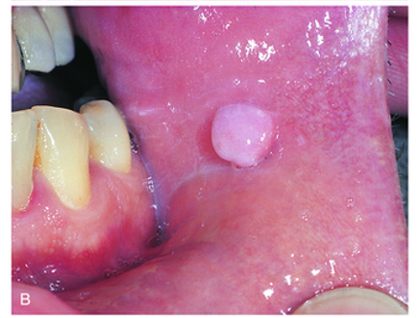
Enamel Pearl
-Ectopic enamel
-Tooth enamel on the root surface (usually in the furcation)
What is the white, spherical enamel projection located at the furca of the tooth in the image called?
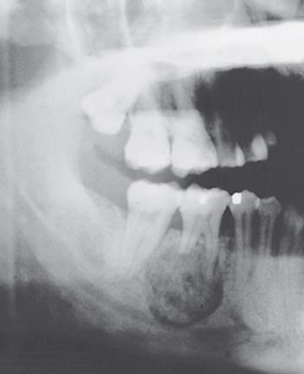
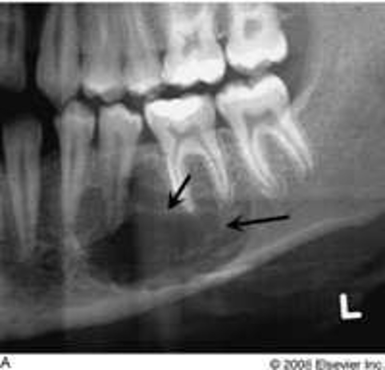
Stafne bone cyst / defect
Radiolucency in posterior mandible below mandibular canal due to lingual concavity of jaw
This pseudocyst seen is surrounded by salivary gland tissue.
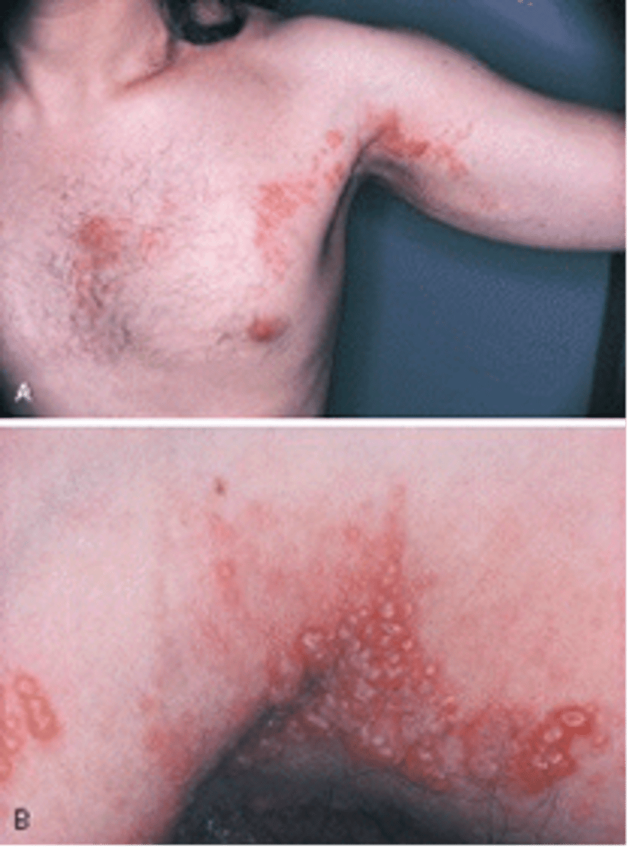
shingles (herpes zoster)
viral infection producing the eruption of highly painful vesicles that may follow a nerve path
The unilateral eruption of vesicles along the distribution of a sensory nerve seen in this adult patient is called?
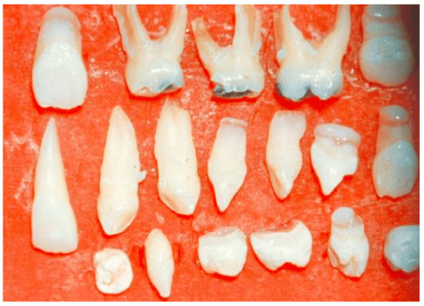
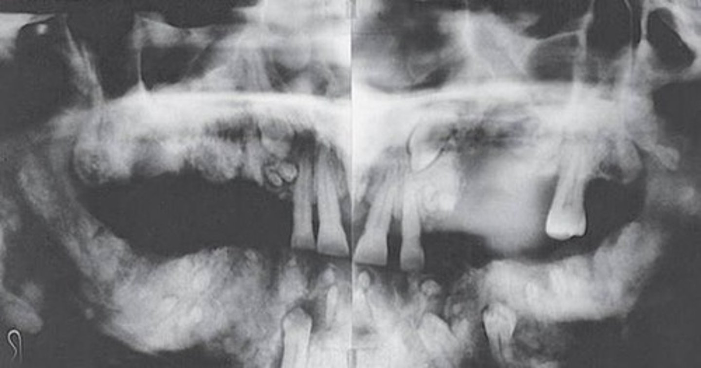
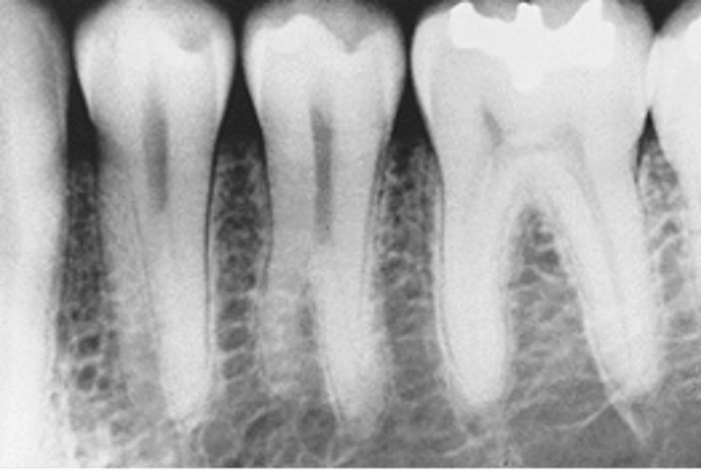
supernumerary roots on the mandibular premolar
Location: maxillary and mandibular 3rd molars, premolars, and cuspids
*visible extra roots
What is the developmental anomaly seen in this radiographic image?
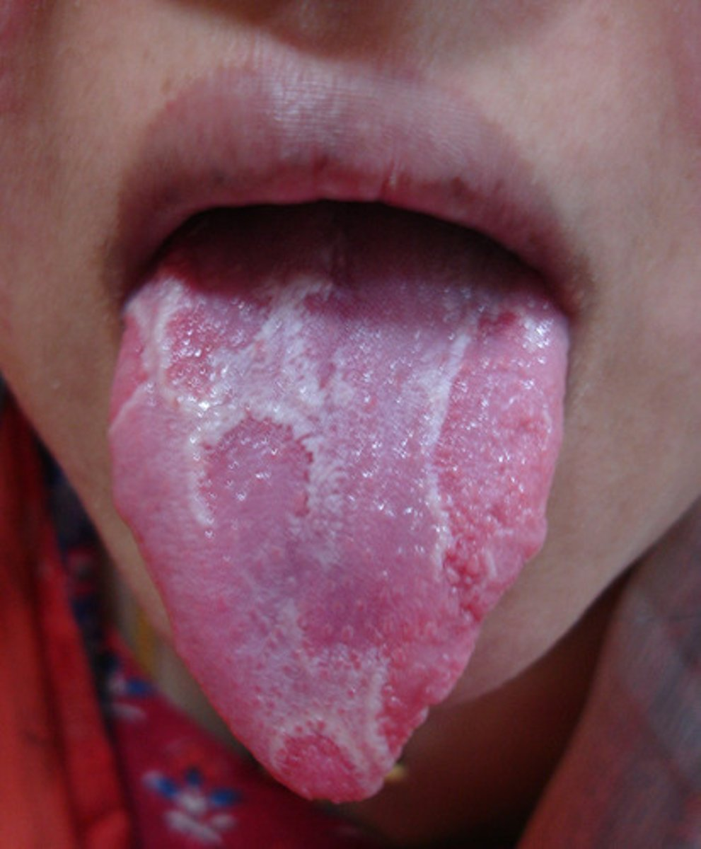
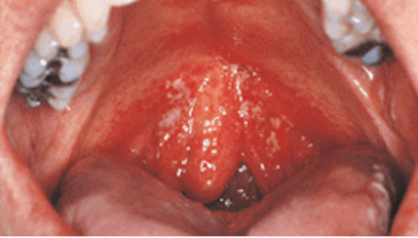
Candidiasis (Thrush)
Thick, white, raised patches in the mouth
This condition most commonly appears in patients with HIV. What is the clinical diagnosis?
condyloma acuminatum
lesion that appears as a result of human papilloma virus; on the skin, lesions appear as cauliflower-like warts, and on mucous membranes, they have a flat appearance; also known as venereal or genital warts
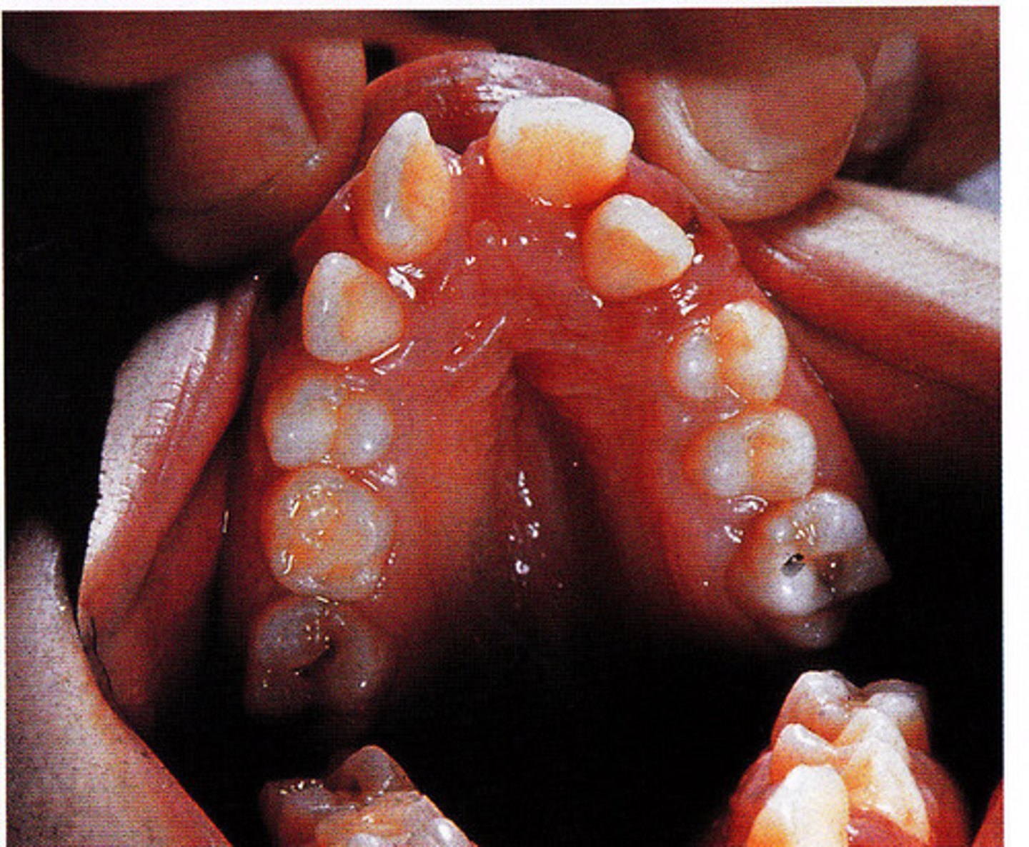
Localized Juvenile Spongiotic Gingivitis
Distinct subtype of gingivitis that does not respond to local plaque control. Appears as papillary or velvety, red nodule that bleeds easily
This flat, red, velvety lesion involving the maxillary anterior gingiva in an 8-year-old male resolved entirely after topical corticosteroid therapy. What is your DHDx?
Mesiodens
Supernumerary tooth between two permanent maxillary central incisors
The supernumerary tooth seen between teeth #8 & 9 is called?
Palatal Lobule tori
Tori on the palate with defined lobules
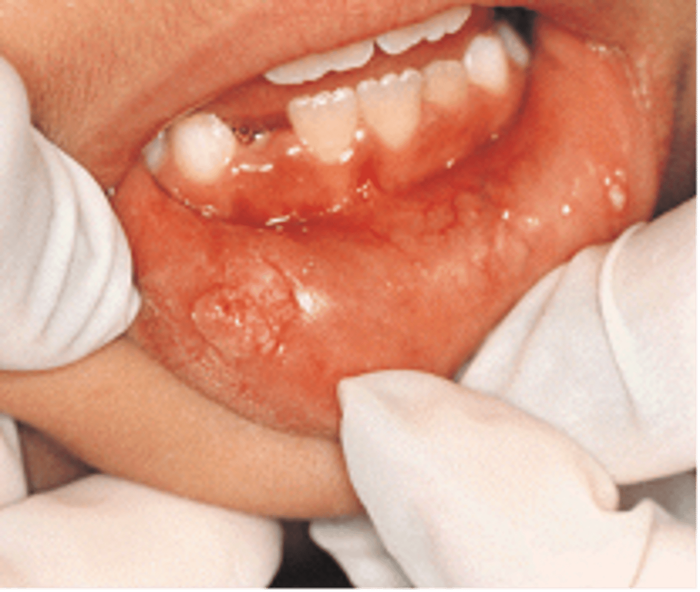
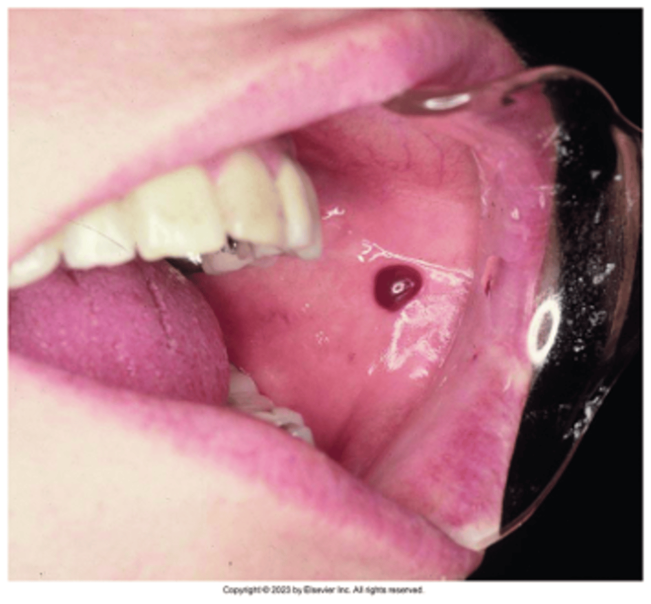
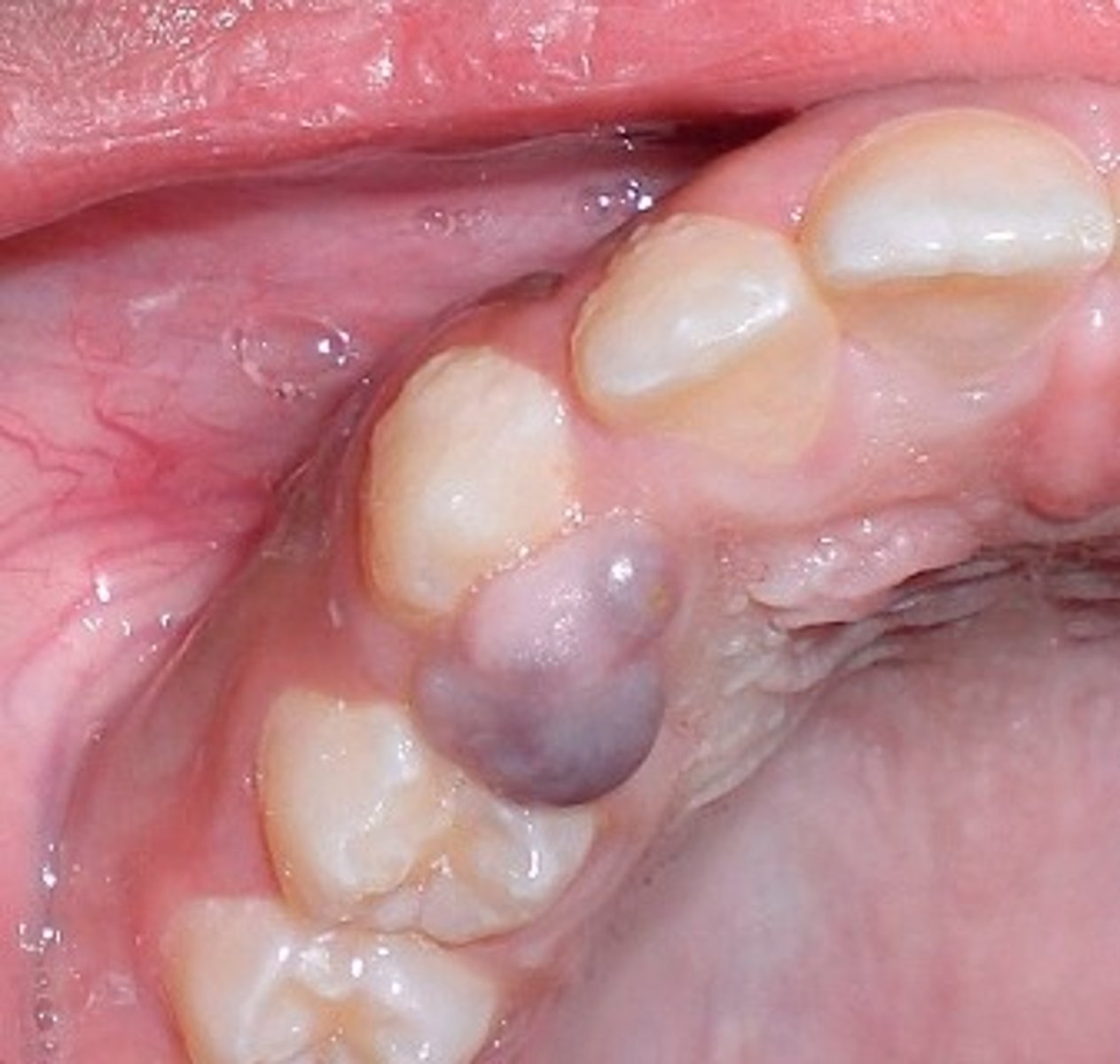
Hematoma
Accumulation of blood within tissues as a result of trauma; Appears as a red to purple to bluish-gray mass; Frequently seen on labial or buccal mucosa
Traumatic Ulcer
Cheek, lip, or tongue biting; Denture irritation; Mucosal injury; Overzealous brushing; Usually heals within 7-14 days unless the trauma persists; May require a biopsy
Frictional Keratosis
Form of hyperkeratosis; Chronic rubbing or friction against an oral mucosal surface; resembles a callus on skin; appears opaque white
Hairy Leukoplakia
a white rough patch that arises on the LATERAL tongue. Usually seen in immunocompromised and is due to EBV induced squamous cell hyperplasia. NOT pre-malignant.
Isolation of the Eppstein-Barr virus is the most reliable method for diagnosis of this lesion on the lateral border of the tongue. What is this lesion called, and what systemic disease is it most often associated with?

dentinogenesis imperfecta
incomplete or improper development of dentin tissue, between enamel and cementum
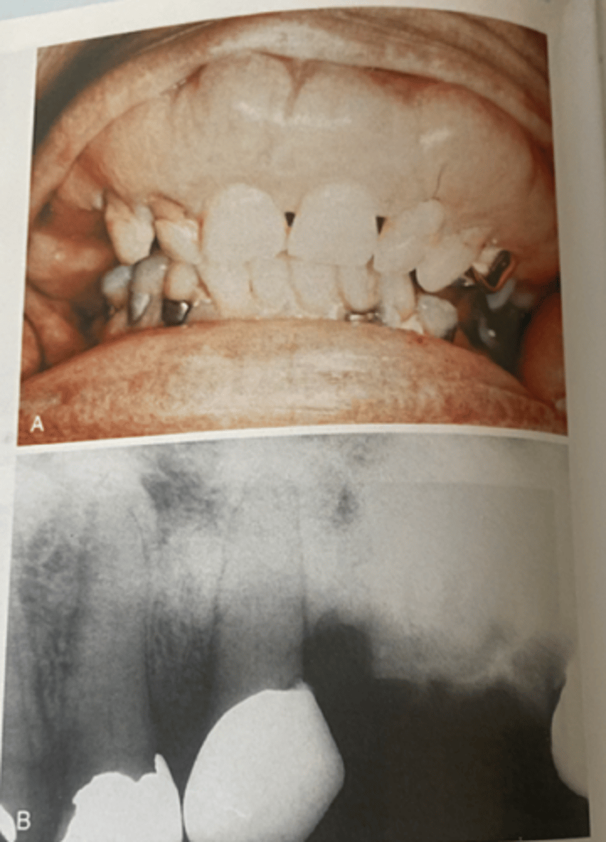
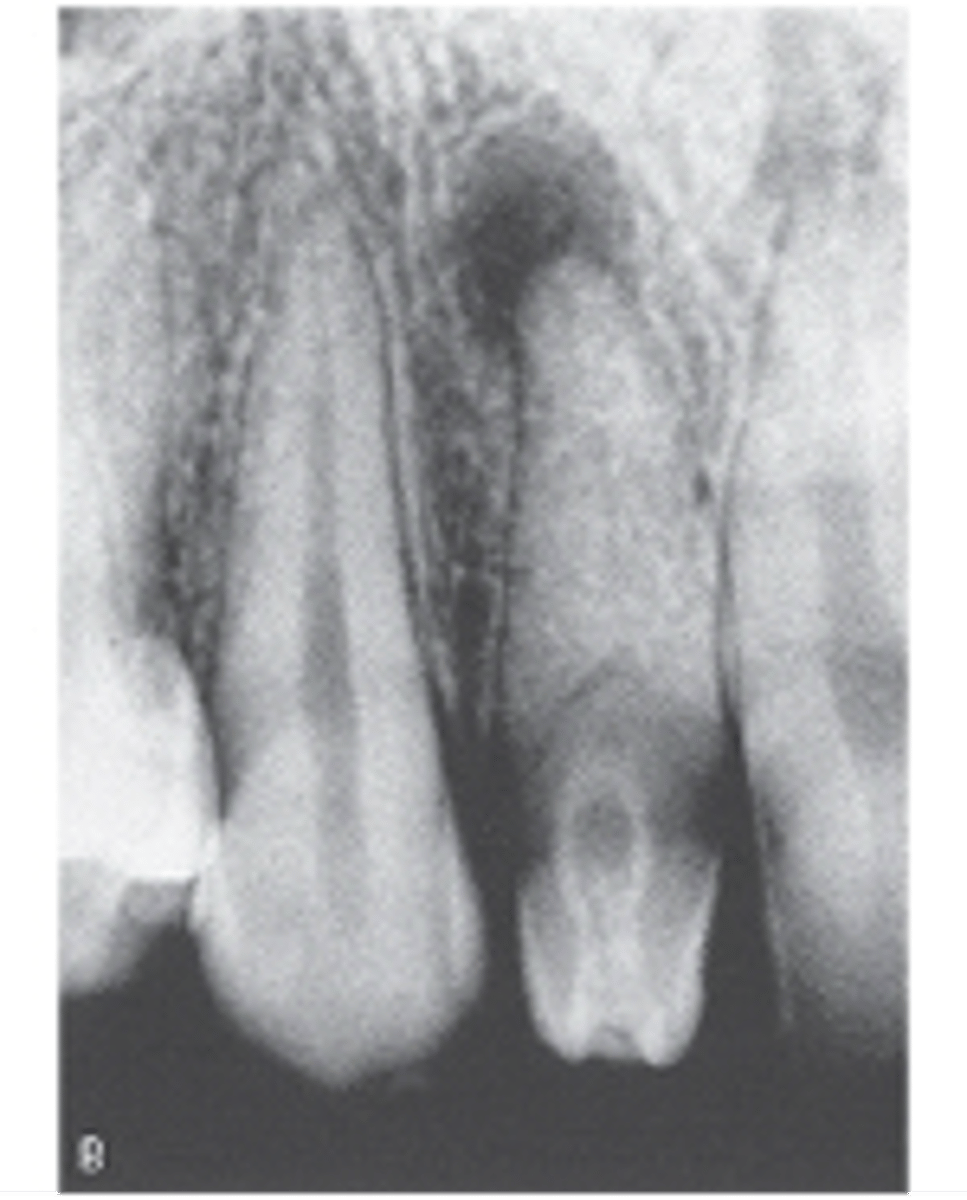
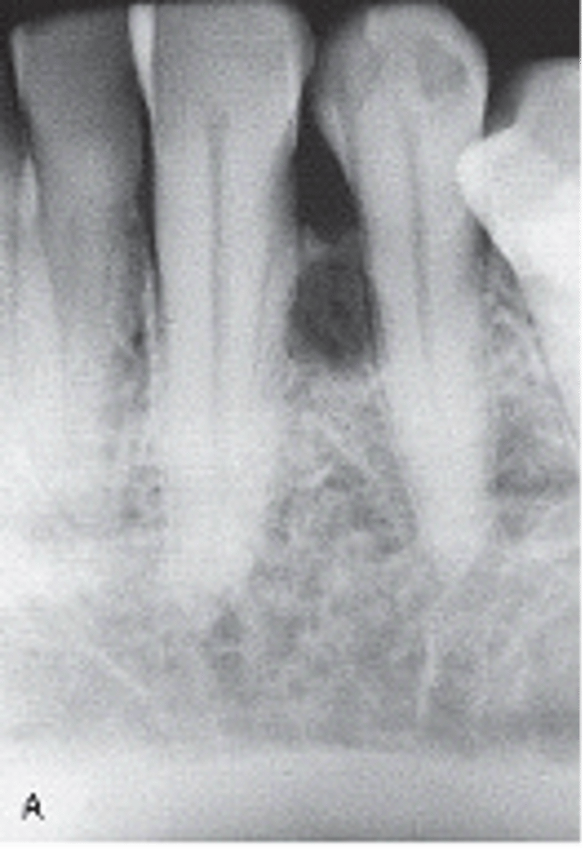
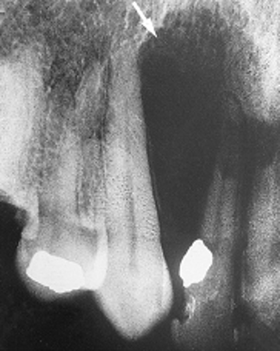
Globulomaxillary cyst
well-defined, pear-shaped radiolucency found between the roots of the maxillary lateral incisor and canine
The pear-shaped radiolucency seen in this radiographic image was historically referred to as what type of cyst?
talon cusp
Additional cusp on the surface of an anterior tooth that projects at least half the distance from the CEJ to the incisal edge; usually lingual
The accessory cusp located in the cingulum of the maxillary right lateral permanent incisor is called
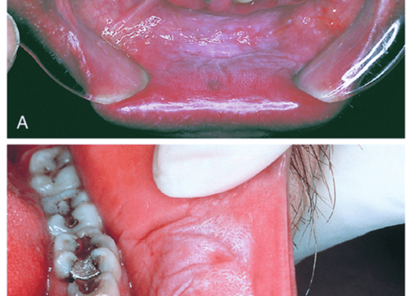
Linea Alba
White, raised line most commonly on the buccal mucosa at the occlusal plane; No treatment necessary
Nicotine Stomatitis
Benign lesion typically associated with pipe and/or cigar smoking; May also occur with cigarette smoking
This benign palatal lesion is typically associated with heavy, long-term pipe and cigar smoking and is caused by heat on the palatal mucosa.
Smokeless Tobacco Keratosis (epithelial dysplasia)
White lesion located where chewing tobacco is placed, most often in the mucobuccal fold
Traumatic Neuroma (peripheral nerve damage/painful)
Lesion caused by injury to a peripheral nerve; Painful, ranging from pain on palpation to severe, intractable pain
Amalgam Tattoo (focal argyrosis)
Flat, bluish-gray lesion of the oral mucosa, caused by the introduction of amalgam into tissue; Must be differentiated from malignant melanoma
Melanosis / melanin pigmentation
Normal physiologic pigmentation of oral mucosa
what is the coloration of the mandibular gingiva that is considered a variant of normal called
Solar/Actinic Cheilitis (sun exposure/vermillion border of lips)
Degeneration of the tissue of the lips, caused by exposure to the sun; Smoking & alcohol use increase risk of squamous cell carcinoma

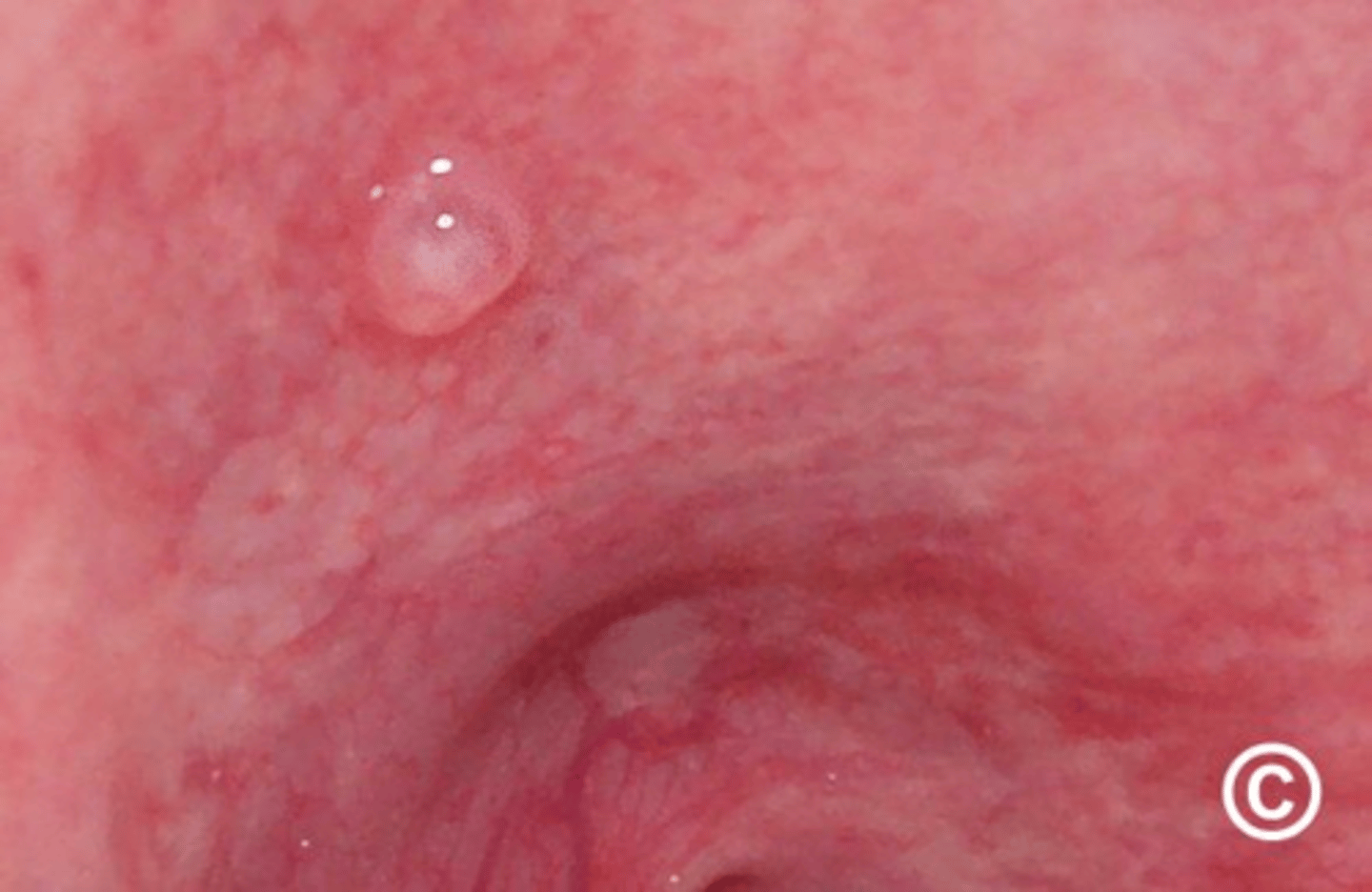
Mucocele (damage to salivary duct/small/lower lip)
Lesion formed when salivary gland duct is severed & the mucous salivary gland secretion spills into the adjacent connective tissue; Most often seen on lower lip; NOT A TRUE CYST because it is NOT lined with epithelium
Ranula (blocked duct/sialolith/large/unilateral)
Unilateral mucocele-like lesion that forms on the floor of the mouth; Associated with the ducts of submandibular & sublingual glands
Sialolith (salivary stone/ seen on x-ray)
Salivary gland stone; May be found in minor & major salivary glands; Formed by precipitation of calcium salts around a central core; May often be seen on radiographs
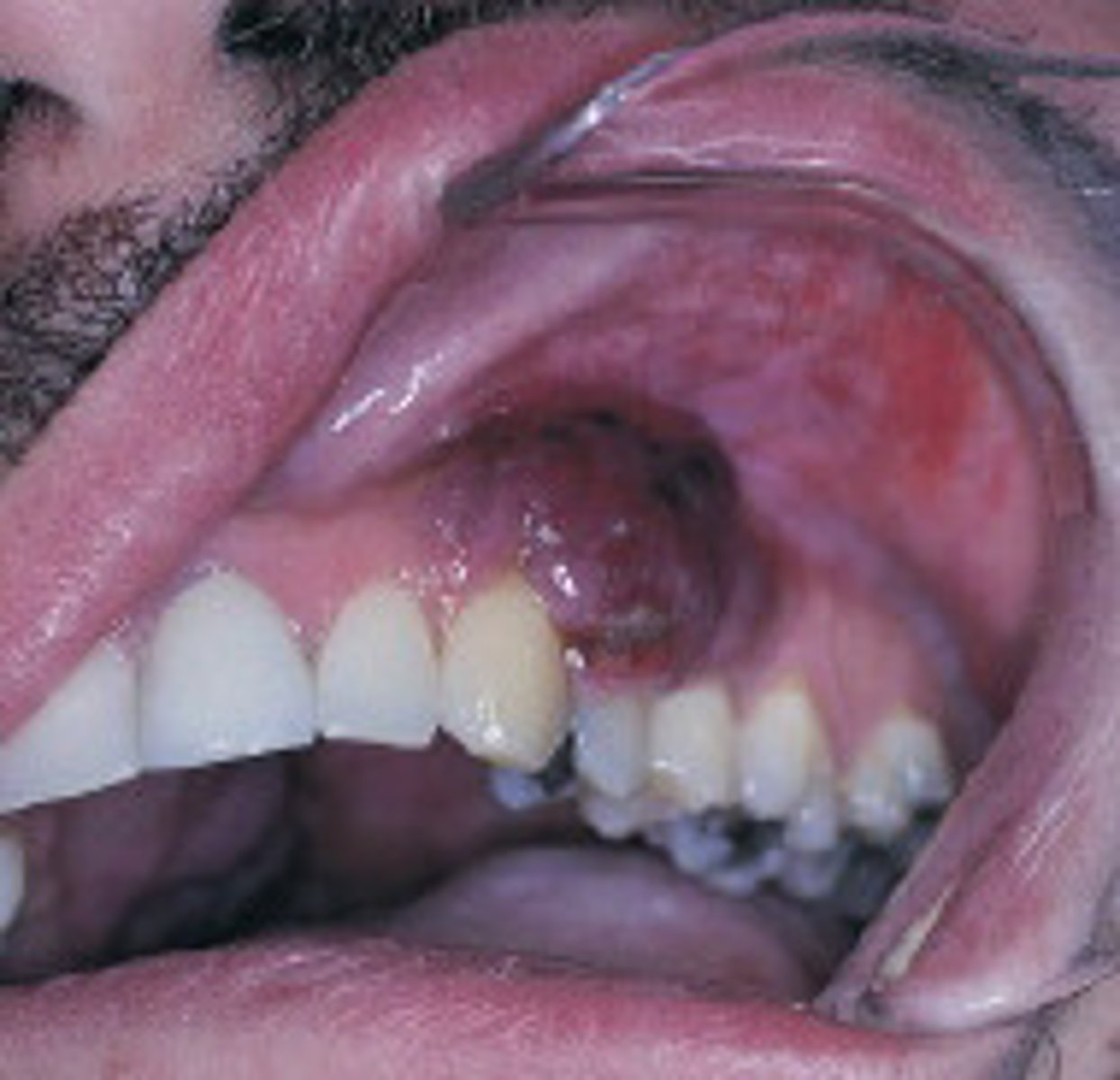
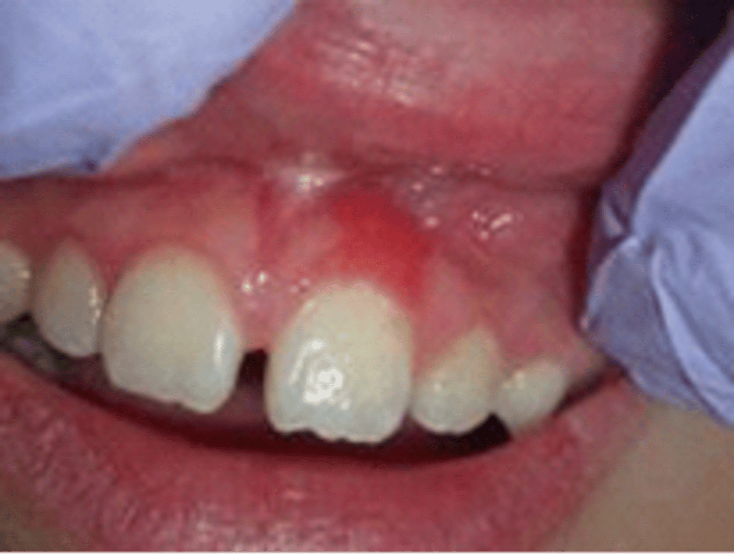
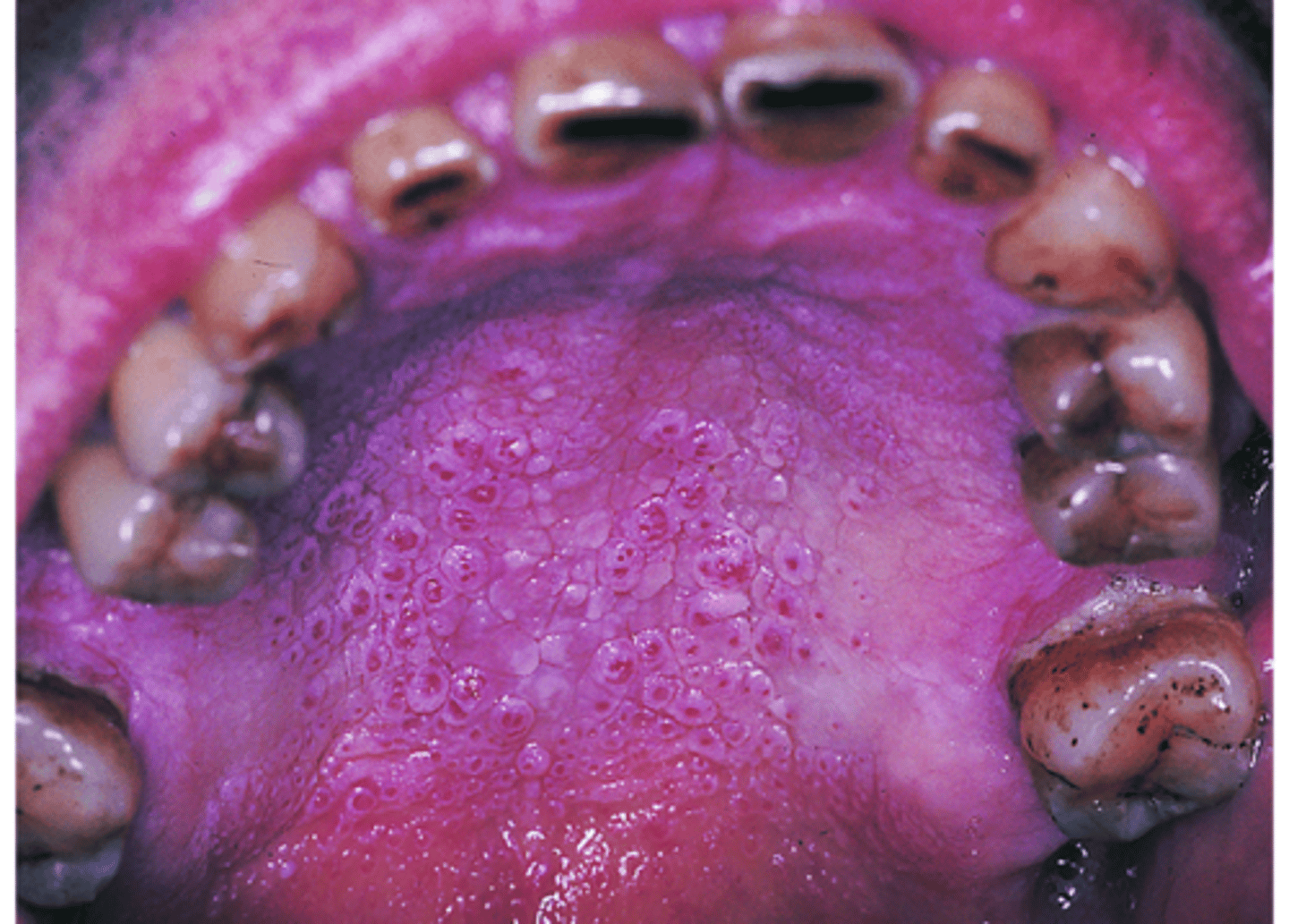
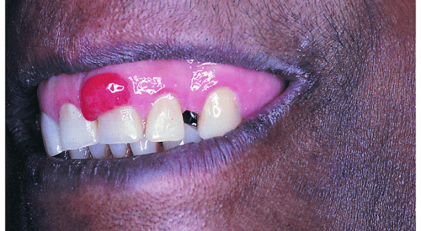
Pyogenic Granuloma (pregnancy tumor)
Most commonly observed on the gingiva; Most common in teenagers & young adults; If seen in pregnant female, it is called a pregnancy tumor; May be caused by hormonal changes & increased response to plaque
Peripheral Giant Cell Granuloma (in soft tissue- seen clinically)
Appearance resembles pyogenic granuloma; Reactive lesion; Contains many multinucleated giant cells, well-vascularized connective tissues, RBCs, & chronic inflammatory cells
Central Giant Cell Granuloma
Within bone; Can be seen radiographically
-may occur on the gingiva or alveolar ridge or in the maxilla or mandible
-divergence of the root of adjacent teeth is a common feature
Irritation Fibroma (traumatic fibroma)
Most common mass on the gingiva; Caused by trauma: Broad-based, usually small lesion, less than 1 cm in diameter
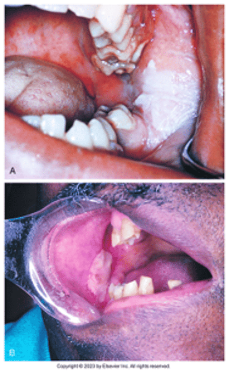
Denture-Induced Fibrous Hyperplasia (epulis fissuratum)
Caused by ill-fitting denture; Usually seen in vestibule due to denture flange
These elongated folds of tissue composed of dense, fibrous connective tissue form as a result of an ill-fitting full or partial denture. What is your DHDx?
Papillary Hyperplasia of the Palate (denture stomatitis) candida albicans
Associated with ill-fitting maxillary denture/partial; Denture-induced hyperplasia; "Cobblestone" appearance
This granular, erythematous papillary surface of the palatal vault is called? What was it caused by?
Gingival Hyperplasia (overgrowth/drug induced)
Gingival enlargement; Increase in the bulk of free & attached gingiva, especially the interdental papillae
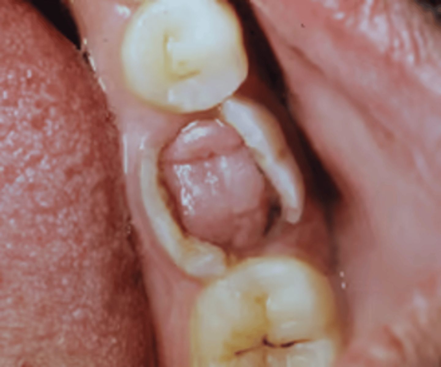
Chronic Hyperplastic Pulpitis
Excessive proliferation of chronically inflamed dental pulp tissue; Occurs in teeth with large, open carious lesions often in primary & permanent molars; Usually asymptomatic