6.1-6.3
1/105
Earn XP
Description and Tags
(Added 6.4-6.12 -> on 1/27/26)
Name | Mastery | Learn | Test | Matching | Spaced | Call with Kai |
|---|
No analytics yet
Send a link to your students to track their progress
106 Terms
What does the skeleton do?
they support and protect the organs/inside body
What are the 5 primary functions of Bones?
Support, Storage, Blood cell Production, Protection, Movement
(Support, Store, Produce, Protect, Move)
What does it mean that a Bone’s function is “to storage” ?
calcium salts of a bone = a mineral reserve which maintains normal concentrations or CALCIUM and PHOSPHATE IONS in body fluid
***yellow marrow = stress lipids
How is bone classified?
Shape
Internal Structure (Spongy vs Compact)
Bone is also called what Tissue?
OSSEOUS TISSUE
What is part of the Skeletal System
Bones/Joints
Connective tissue that stabilize/connect→Cartilage, Ligaments
Why does bone have it’s texture
Deposition of calcium salt in MATRIX
What dominates bones
Calcium Phosphate
Different percentages of what makes up a bone’s weight
Calcium phosphate = 2/3
Collagen Fibers = 1/3 (density)
osteocytes.other cells = 2% of bone mass
Macroscopic features include (other than the 4 shapes)
2 types of bone tissue
Periosteum
Cellular Endosteum
Tendon
Ligaments
What are the 4 shapes of bone
Short - Carpals, Tarsals
Long - Femur, Humerus
Irregular - Vertebrae, Some Skull bones
Flat - Scapula, Ribs, Sternum, Some Skull bones
Short bones
short and roughly equal dimensions
Flat Bones
thin and relatively broad
Irregular Bones
Complex shape/Doesn’t fit with the rest (it’s giving emo boy)
Long Bones
… long :)
Features of a Long Bone
Diaphysis (“Central shaft”)
Epiphysis (Proximal and distal)
Articular cartilage
Spongy Bone
Compact Bone
Marrow cavity
Bone Marrow
Endosteum
Periosteum
Epiphyseal Line
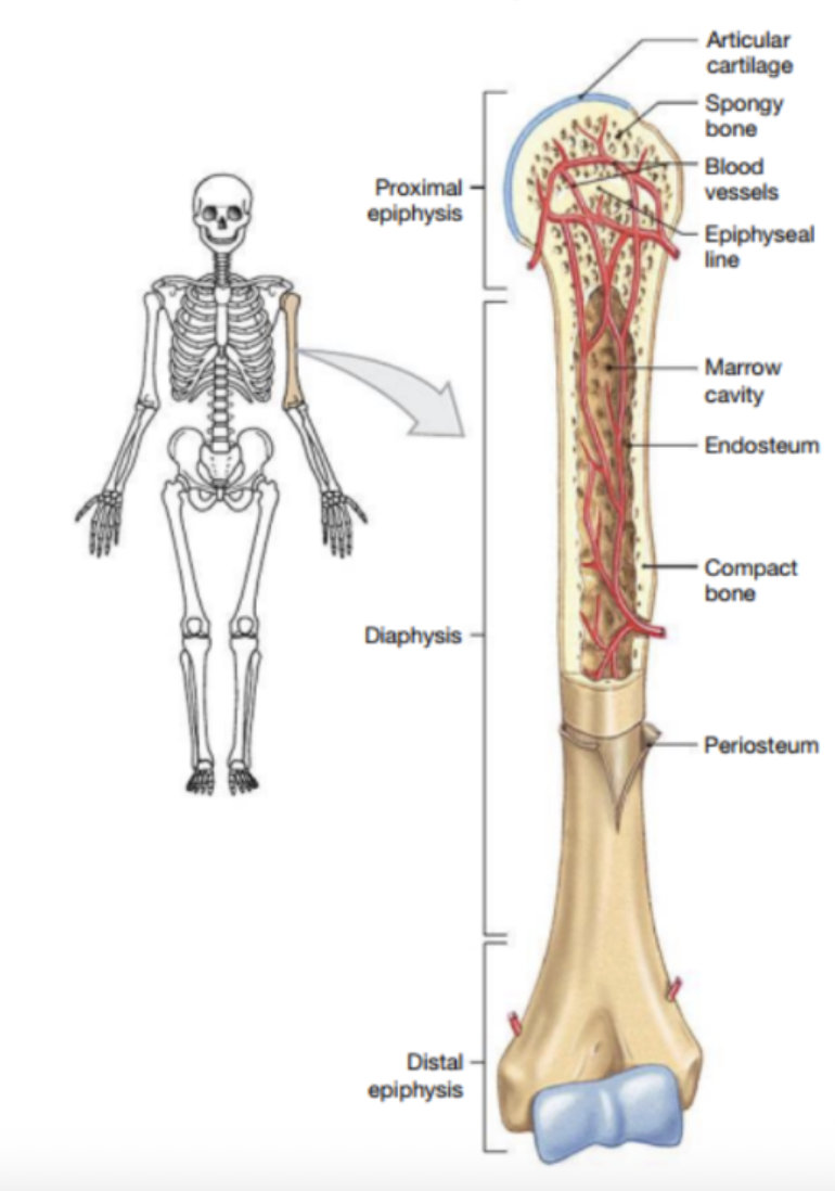
Diaphysis
Aka “Central Shaft” -> its Marrow Cavity is filled with Bone Marrow
Epiphysis (Proximal and Distal)
the wider portions at each end (covered by articular cartilage)
What is cellular Endosteum?
Covers spongy bone of marrow cavity and over inner surfaces
***Active role in bone growth and repair/remodeling
Lines the inside
Articular Cartilage
Cartilage Covering Joint Ends (Usually in long bones)
Periosteum is what?
made of fibers and tendons
provides route for supplies
Helps bone repair and growth
Outside layer of bone
Spongy Bone
AKA “Cancellous Bone” : has projections of bone separated by spaces
What are Epiphysis covered by?
Articular cartilage
Each Epiphisis articulates an adjacent bone at a joint
Compact Bone
Densely Packed (forms the Diaphysis)
What does the Marrow cavity do?
house the bone marrow : a soft fatty tissue
Has epiphysis = expended portions at the ends
—-
Red Marrow: Hematopoietic tissue (“hematopoiesis) that produces red blood cells, white blood cells, and platelets.
Yellow Marrow: Composed mostly of fat cells, it serves as an energy reserve and can convert to red marrow if needed.
Hallow Part inside of Diaphysis and usually Contains Spongy Bone
filled with bone marrow
Epiphyseal Line
Marks where Cartilage is Replaced (burial Site for Epiphyseal plate of the bone)
How to spell the long bone part which is the “wider portions at the end”
Epiphysis or Epiphyseal
Central shaft surrounds what?
Central marrow cavity of long bones
Why immature bone growth?
problems with junctions between epiphysis and diaphysis
2 types of Bone tissue
Compact (Dense) vs Sponge (cancellous)
What the the 3 cells in the Bones?
Osteocytes
OsteoBlasts
OsteoClasts
Osteocytes are what?
in both bone tissue (and are mature bone cells)
Inside Lacunae (small pockets)
part of Canaliculi
Osteon
Haversian System (Basic Functional Unit of Compact Bone)
Osteocytes arranged Concentric Layers around the Haversian Canal (circular formation)
How are nutrients/waste of osteocytes diffused?
through extracellular fluids surrounding the call and cytoplasmic extensions
How do Osteocytes maintain bone structure?
recycles calcium salts in bone matrix and assists repairs
Lacunae vs Lamellae
Lacunae = between sheets of calcified matrix (the gap)
Lamellae = calcified matrix
What are canalicui?
Small channels in the matrix
Connect lacunae to blood vessels
Contains cytoplasmic extensions of osteocytes
Trabeculae
Lamellae Rod formation
(“3 strands is not easily broken…” bible verse)
OsteoCLASTS
Bigguh cells (50 or more nuclei)
Secrete acids and enzymes
“Osteolysis”/Resorption Release store minerals
DISSOLVES bony matrix
regulates calcium and phosphate concentrations
OsteoBLASTS
CREATES new bone and matrix (“ossification”)
promotes deposition of calcium salts in organic matrix
surrounded by calcified matris = becomes a osteoclast (like jedi becoming sith)
Compact Bone
Has Osteon (“Haversian System”).. basic functional unit
Osteocytes arrange in a CONCENTRIC pattern/in concentric layers around a central canal/”Haversian canal'“ (Big circle, circle, circle)
Has 1/more vessels
Lamellae = cylindrical = parallel to long axis of central canal
Perforating canals = passage ways which links vessels of central canals with periosteum and marrow cavity
Covers every bone except joint capsules (articular cartilage go there)
** Covers area with limited direction stress … Stronk
Ex: Limb Bones
Spongy Bone
No osteon
Lamallae = rods/plates formation; branching, open networks
The trabecular netowrk = supports/protects cells of red bone marrow
Canaliculi from LACUNAE = at the end of the bone, exposed surface of trabeculae
Nutrients/waste diffuses between marrow and osteocytes
LIGHTER than compact bones (muscles move bone easier)
Covers interior of bone
Stress from many directions : weakuh (not heavy stress)
Growth of bone stars from when a embryo is
6 weeks after fertilization
skeleton = all cartilage
Bone = keeps growing till usually 25
Ossification is what?
Process of replacing other tissues with bone (Making new bone)
Calcification is what?
Deposition/accumulation of calcium Salts
usually during ossification (can happen in non-bone tissue)
2 types of ossification are what?
Intramembranous and Endochondridal
Intramembranous Ossification is what?
bone develops in sheets/membranes of connective tissue
usually in deep dermis layers
* Looks like sponge but changes later
Steps for Intramembranous Ossification are what?
OSTEOBLASTS differentiate in ebryonic/fetal fibrous connective tissue
Matrix is secrete by stem cells (stem cells become calcified) … Osteoblasts differentiate from connective tissue stem cells
1 and 2 happen ins the ossification center
new bone grows outward ; osteoblasts get trapped in calcified matric and turn to osteocytes (NOT CLASTS)
Blood vessels grow ; supply osteocytes and also get trapped in the bone too
the intarmembranous bone looks spongy but remodeling around vessels = osteons are created (ONLY SEEN IN COMPOUND BONE)
Example of Intramembranous Ossification processes bones
Flat bones of skull, mandible, and clavicles
Endochondia Ossification is what?
forms in hyaline cartilage and covers it up laterz (more common than intra.)
cartilage into true bone
ONLY FORMS IN HYALINE CARTILAGE
Steps of Endochondia Ossification are what?
Chondrocytes (in cartilage) enlarge; surrounding matrix calcifies
Chondrocytes die, matrix slows its nutrient diffusion
Bone forms thin layer around shaft area; blood vessels invade perichondrium
Cells in inner layer differentiate into OSTEOBLASTS (starts making bone matrix)
Blood vessels invades inner region of cartilage
Migrating fibroblasts differentiate into OSTEOBLASTS
new osteoblasts form spongy bone in center shaft in. a PRIMARY OSSIFICATION CENTER
Bone enlarges; OSTEOCLASTS breakdown spongy bone = makes a marrow cavity
Cartilage dont fill bone bcauz epiphysisal cartilage/”plates” on the ends are growing/enlarging
Bone length increases
** osteoclasts break, and osteoblasts create (ying and yang motion)
Center of Epiphysis starts calcifying; blood vessels and osteoblasts fo into the Epiphysis
** Make a SECONDARY ossification center; Epiphysis is filled with spongy
** cap of of cartilage is exposed to joint cavities = “ARTICULAR CARTILAGE”
SHAFT Bone and Epiphysis = separated by cartilage
Osteoblasts produce more than the epiphysis cartilage expands = cartilage narrows till diaapears
END OF GROWTH = “epiphyseal closure”
**Time it is done varies by person, and bone type (plus sex hormones too)
sex hormones = speed up growth —- puberty (some boys say their bones/GROWING hurt)
this is why
IN adults = marks of former locations of epiphysial cartilage is marked by a line (like the berlin wall)
Appositional growth is what?
Diameter of bone increases
Why does Appositional growth happens?
Cells of periosteum become OSTEOBLASTS and more bony matrix (outer)
Osteoclasts erode inner surface; marrow cavity enlarges
One must decrease so the other increases
What are the requirements for regular bone health?
Calcium (salts)
Phosphorus
Vitamin A, C
Vitamin D 3
HORMONES
Phosphate and calcium help with what?
reliable source for minerals; during prenatal/embryo… baby absorbs calcium and could absorb mass of mom (done through absorbing minerals in mom’s blood stream)
Viamin D3
Calcium metabolism role
Goes into liver and kindey to turn into CALCITROL
Calcitrol = hormones; stimulates absorption of calcium and phosphate ions
Vitamin A/C
Essential for bone growth and maintenance
Vitamin C deficiency = scurvy; reduced osteoblast # ; brittle/weak bones
Rickets
Flexible bones
- as a result of lack of Vitamin D 3
soft/bending of bones (usually during kids years it shows)
BOW LEGGED STANCE… bros couldn’t win against gravity frfr (too soft)
What are the 3 things that bone growth and development need to be in balance?
Mineral supply : Especially calcium salts
Vitamins
D3 : helps calcium metabolism
Deficiency in D3 = RICKETS
A and C : supports osteoblasts functions
Growth, sex, thyroid, and calcium-balancing HORMONES
Two items that maintain the bone MATRIX are…
Osteoblasts
OsteoClasts
What is the Turn over rate for Bone?
1/5 is remodeled every year (20%)
Influenced by…
Age
Mechanical Stress
Hormonal Balance
Is every part of the bone remodeled? Explain
Yes however, the amount of time it takes differs from…
person to person
bone type
location of the bone
What allows bones the ability to adapt to new stresses:?
Remodeling
If bone is heavily stressed, what happens to it?
Could Fracture/break
What happens if bone isn’t subjected ot daily stresses?
Becomes weaker and brittle
What does regular exercise do for bones?
Maintains Normal Bone Structure and strength
What happens to a bone when someone wears a cast (and now removes it)
lose up to 1/3 of bone mass (work out to strengthen it)
What is the Most Abundant Mineral in the Human Body?
Calcium (salts)
Explain what happens in the body to certain physiological processes if calcium ion concentrations increase and if they decrease.
Increase : Muscle and neurons shut down/become unresponsive
Above 5% -> unusual
30% -> unresponsive
Decrease : Become energetic/excited… they convulse…
35% -> convulse
50% -> death
How can calcium levels in body fluids become elevated?
Parathyroid Hormone (PTH) ; Parathyroid Glands
Calcitrol ; Kindeys
How can calcium levels in body fluids become lowered?
Calcitonin : THYROID GLANDS
What are 3 main causes of bone fractures mentioned in the text?
Weak bones
Overstress/ repetitive stress
Injuries/Trauma/Falls
In order for bones to heal on their own, what do they need?
Blood vessels/Supply remains
Cellular Parts of the Endosteum & Periosteum are intact
How long can the Healing of Bones take?
4 months to a year
Explain the 4 steps in the repair of a fracture.
Blood clot : Fracture Hematoma
Closes off blood supply
KIlls osteocytes
Results in dead bones on either side of the fracture
Cells of Periosteum and Endosteum go to fracture
Makes an external and internal Callus
** external callus = develops hyaline cartilage
Osteoblasts replace cartilage with spongy bone
Spongy bone is replaced by compact bone
Leave a slightly thicker bone patch at the fracture site
Explain what happens to bones as we age.
Weakens and gets more brittle
Why is osteoporosis more common in older women than in older men?
Their sex hormone keeps being produced till 60 (androgens)
Where do elevations and projections form on bones and what are they used for?
Form where ligaments and tendons attach or where adjacent bone articulate at joints
Depressions, grooves, and Openings
Shows where blood vessels and nerves run along and/or penetrate bone
Process
Any Projection or bump
Trochanter
A large, rough projection
Tuberosity
A smaller, rough Projection
Tubercle
A small, rounded projection
Spine
a pointed process
Head
The expanded articular end of an Epiphysis, separated form the shaft by a neck
Neck
a narrow connection between Epiphysis and Diaphysis
Condyle (median and Lateral)
A smooth, rounded articular process
Trochlea
A smooth Grooved articular process shaped like a pulley
Crest
A prominent ridge
Foramen
A rounded passageway for blood vessels/nerves
Canal
A duct or Channel
Fissure
An Elongated cleft/slit
Sinus
A chamber within a bone, normally filled with mucus
What are the two Skeletal Divisions and their subdivisions?
Appendicular (126)
Limbs
32 Upper (each limb)
31 Lower (each limb)
Axial (80)
Skull (22)
7 Associated (includes 1 Hyoid Bone)
8 Cranial Bones
14 Facial Bones
Thoracic Cage (25)
24 ribs
1 Sternum
Vertebral Column (26)
Partial Fracture
A break that doesn’t go through the bone completely
Complete Fracture
A break that goes Completely through the Bone
Open Fracture
AKA Compound fracture
Fracture goes through the skin and can be seen externally
Closed Fracture
AKA simple fracture
Fracture does not pierce though the skin (all internal)
Non-Displaced Fracture
two broken ends of the bones are still lined up (not moved)
Displaced Fracture
two broken ends of the bones are not lined up (moved out of place and it needs to be set)