A&P Chapter 6: Bone Tissue
 Call Kai
 Learn
Learn Practice Test
 Spaced Repetition
 Match
Match Flashcards
 Knowt Play
1/95

There's no tags or description
Looks like no tags are added yet.
Name | Mastery | Learn | Test | Matching | Spaced | Call with Kai |
|---|
No analytics yet
Send a link to your students to track their progress
96 Terms
What is Bone?
bone aka osseous tissue is a hard, dense connective tissue that forms most of the skeleton.
support structure for the body
in areas where bones move (ribcage/joints), cartilage provides flexibility and smooth surfaces for movement
skeletal system is composed of bones and cartilage
What are the five functions of the skeletal system?
supports the body
facilitates movement
protects internal organs
produces blood cells
stores/releases minerals and fats
[Functions of Skeletal System]
Supports the body
provides structural support for entire body
individual bones/groups of bones provide framework for the attachment of soft tissues/organs
[Functions of Skeletal System]
Facilitates Movement (and leverage)
facilitate movement by serving as points of attachment for your muscles
many bones function as levers that can change magnitude/direction of forces generated by skeletal muscles
bones act as levers and joints serve as fulcrums (pivot point)
movement produced range from delicate motions to strong changes in position
[Functions of Skeletal System]
Protection (internal organs)
delicate tissues/organs are surrounded by skeletal elements
ribs protect the heart/lungs
skull encloses the brain
vertebrae shield the spinal cord
pelvis cradles urinary/reproductive organs
[Functions of Skeletal System]
Stores/Releases Minerals and Fats
bone matrix acts as reservoir for minerals (calcium and phosphorus)
they are incorporated into bone tissue but can be released back into bloodstream to maintain homeostasis
calcium is most abundant mineral = muscle contractions and controlling flow of ions and nerve impulses
humans have 1-2kg of calcium and 98% of it is in bones
yellow marrow: contains adipose tissue
triglycerides stored in adipocytes can serve as source of energy
[Functions of Skeletal System]
Blood Cell Production
hematopoiesis (aka blood cell production): red blood cells, white blood cells, and platelets are produced in the red bone marrow
this fills the internal cavities of bones
Axial skeleton
(the human body has 206 bones)
axial skeleton: 80 bones; consists of the bones of the skull, hyoid, sternum, rib cage, vertebral column, sacrum, and coccyx
Appendicular Skeleton
(human body has 206 bones)
appendicular skeleton: 126 bones; includes bones of the limbs and pectoral/pelvic girdles that attach the limbs to the axial skeleton
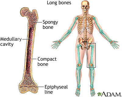
Long Bones
cylindrical in shape (longer rather than wide)
located in the arm, forearm, thigh, lower leg, palms, soles, fingers, toes
femur: largest/heaviest bone in body
ex: humerus, tibia/fibia, radius, ulna, clavicle, phalanges
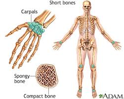
Short bones
cube-like in shape
bones of the wrist (carpals) and bones of the ankles (tarsals)
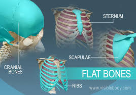
Flat bones
thin, often curved
form the roof of the skull, sternum, ribs, and scapulae
provide protection from underlying soft tissues + surface for attachment
they are made of spongy bone (diploe) covered by compact bone
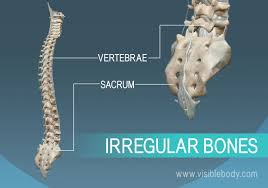
Irregular Bones
complex shapes with short, flat, notched, or ridged surfaces
don’t fit into any other category
ex: spinal vertebrae, bones of pelvis, some skull bones (mandible)
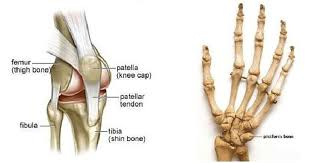
Sesamoid bones
generally small, flat, and shaped like a sesame seed
develop inside tendons and most commonly located near joints
ex: joints of knees, hands, feet
sesamoid patellae/kneecaps
amount can vary
[Anatomy of a Long Bone]
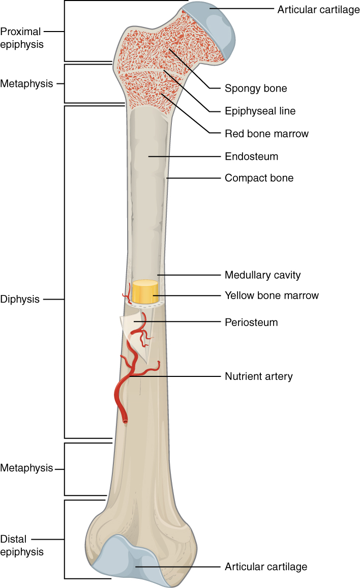
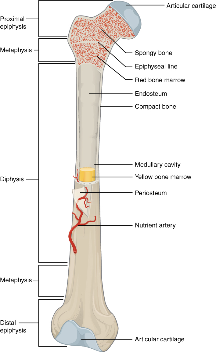
Diaphysis
long tubular shaft that forms the axis (middle section)
walls of shaft are made of compact bone
[Anatomy of a Long Bone]
Epiphyses
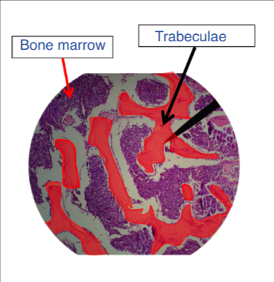
ends of the bones composed of spongy bone or trabecular bone
consists of open network of struts/plates (trabeculae) that resemble latticework
red bone marrow fills the space
proximal epiphyses: ends closest to origin of attachment
distal epiphyses: ends furthest from origin of attachment
articular cartilage: covers portions of the epiphysis that articulates with other bones (hyaline cartilage that reduces friction/shock absorber)
[Anatomy of a Long Bone]
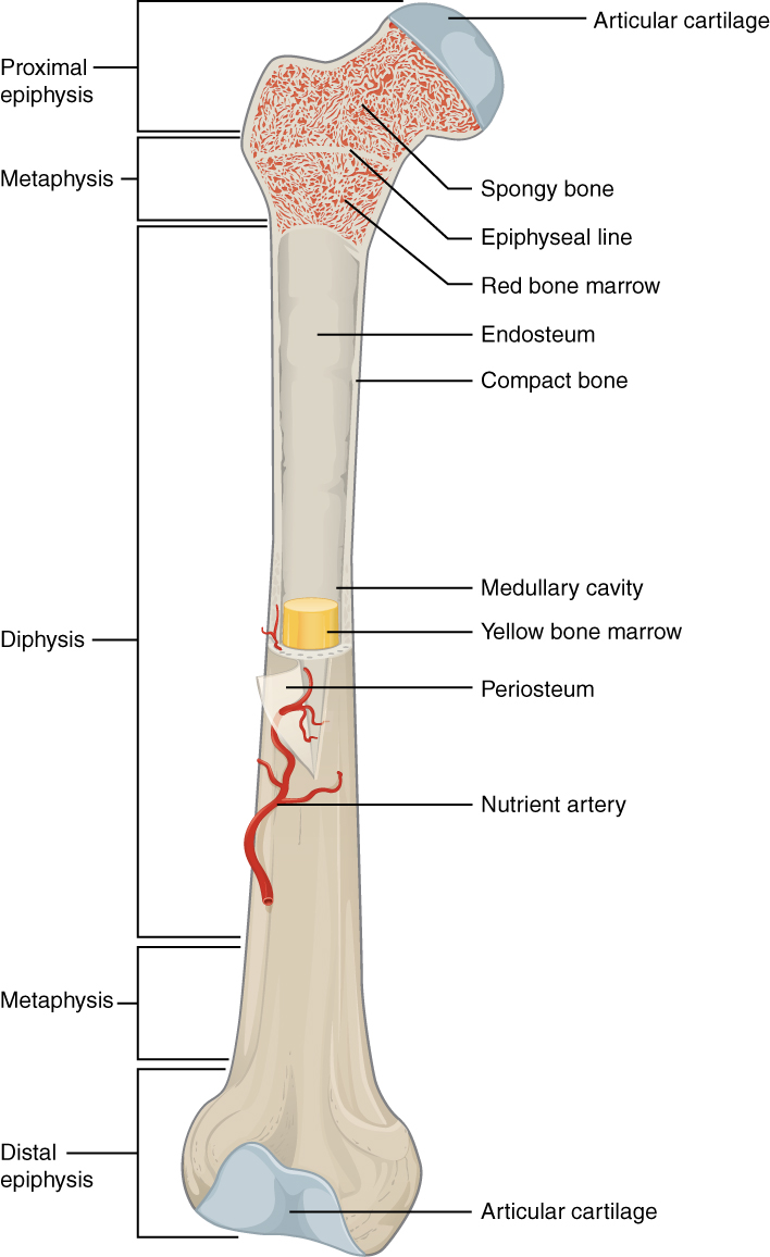
Metaphysis
narrow zone that connects the diaphysis to the epiphyses
epiphyseal plate: aka growth plate; thin layer of hyaline cartilage that helps growth in length of the bone
when the bone stops growing in early adulthood, cartilage is replaced by osseous tissue + epiphyseal plate becomes epiphyseal line
[Anatomy of a Long Bone]
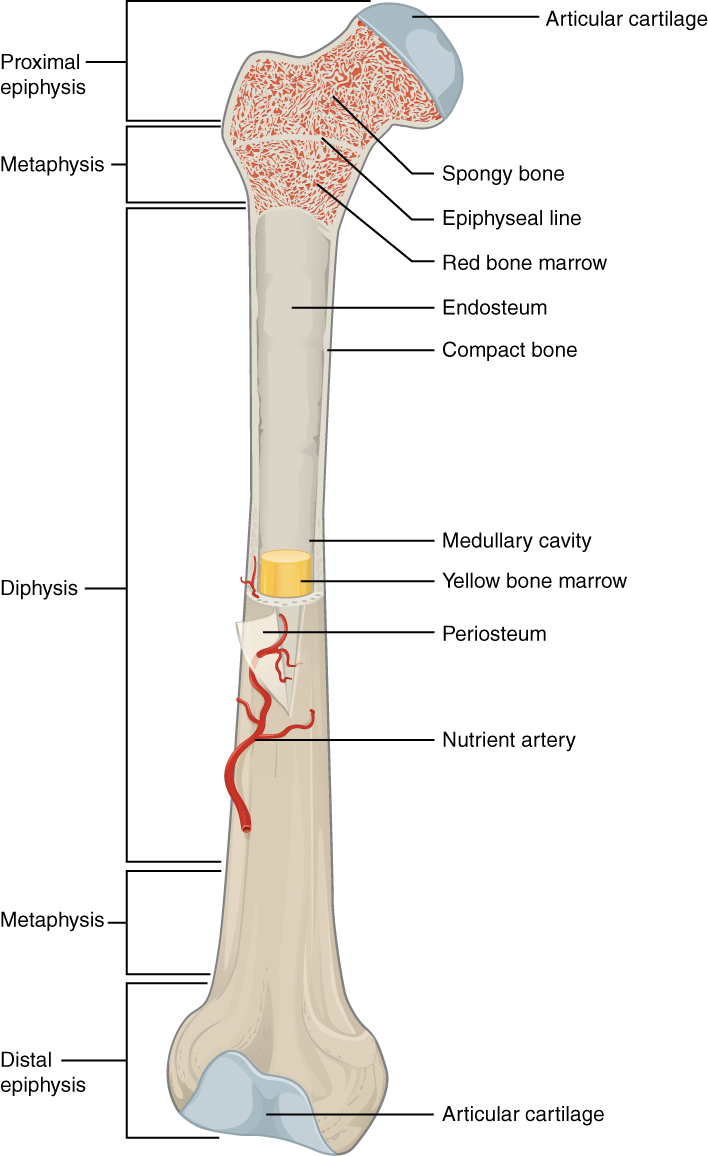
Medullary Cavity
within shaft of a long bone is cavity where bone marrow is located
in childhood: medullary cavity is filled with red bone marrow
important for hematopoiesis
as we age: fat accumulates within red marrow transforming it to yellow bone marrow
stores fat as important energy source
[Anatomy of a Long Bone]
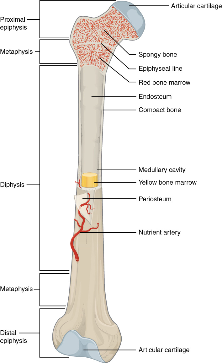
Membranes - Periosteum
outermost covering of bone made of dense irregular tissue + collagen fiber
contains blood vessels, nerves, and lymphatic vessels = nourish compact bone
tendons/ligaments attach to bones at periosteum
covers entire outer surface except wehre epiphyses meet other bones
[Anatomy of a Long Bone]
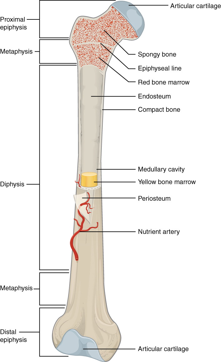
Membranes - Endosteum
internal membrane of bone made of connective tissue
where bone growth, repair, and modeling occurs
lines the canals that pass through bone to supply blood/nerves to the bone
[Bone Markings - Depressions/Openings]
What are depressions?
they are depressions or openings that allow blood vessels and nerves to pass
[Bone Markings - Depressions/Openings]
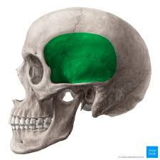
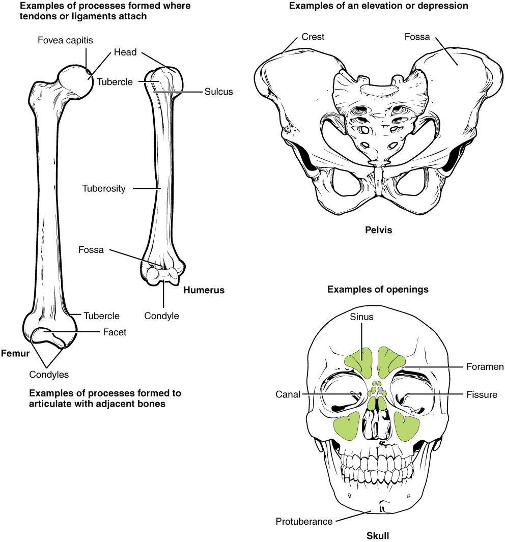
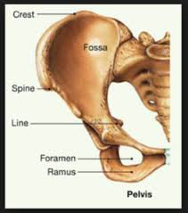
Fossa
shallow depression or recess in surface of a bone
[Bone Markings - Depressions/Openings]
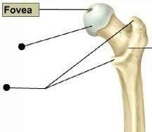
Fovea
small pit
ex: fovea capitus
[Bone Markings - Depressions/Openings]
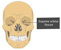
Fissure
narrow, slit-like opening or elongated cleft/gap
[Bone Markings - Depressions/Openings]
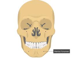
Foramen
round or oval opening through the bone
[Bone Markings - Depressions/Openings]
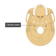
Canal or Meatus
large passageway through the bone
[Bone Markings - Depressions/Openings]
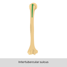
Sulcus or Groove
furrow or narrow trough in a bone
[Bone Markings - Depressions/Openings]
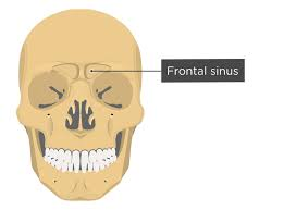
Sinus
chamber within a bone filled with air and lined with a mucous membrane
[Bone Markings - Depressions/Openings]
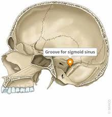
Sulcus
shallow groove
ex: sigmoid sulcus
[Bone Markings - Projections]
What are projections?
they are an area of a bone that projects above the surface and serves as sites for muscle/ligament attachment
[Bone Markings - Projections]
Tuberosity
large, round/rough projection that may cover a broad area
[Bone Markings - Projections]
Crest
narrow ridge of bone; normally prominent
[Bone Markings - Projections]
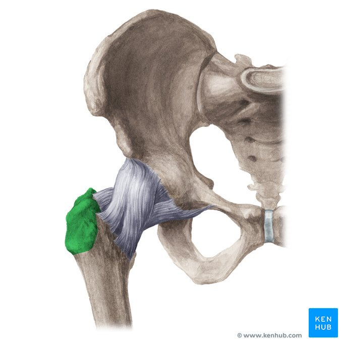
Trochanter
very large, irregularly shaped projection
[Bone Markings - Projections]
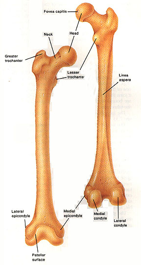
Epicondyle
raised area above a condyle
[Bone Markings - Projections]
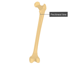
Line
narrow ridges of bone, less prominent then a crest
[Bone Markings - Projections]
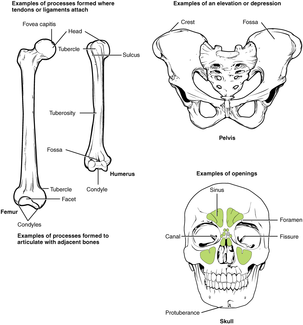
Tubercle
small, rounded projection
[Bone Markings - Projections]
Spine
sharp, slender, often pointed process
[Bone Markings - Projections]
Protuberance
protruding area such as a chin
[Bone Markings - Projections]
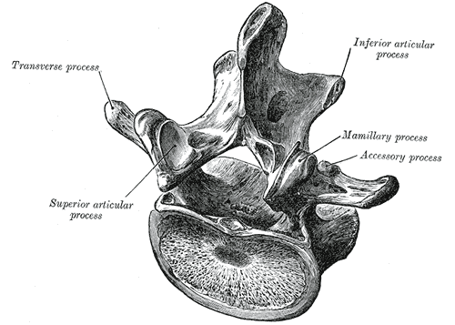
Process
prominent feature that stands out
ex: transverse process
[Bone Markings - Articulations]
What are articulations?
They are projections that form joints
[Bone Markings - Articulations]
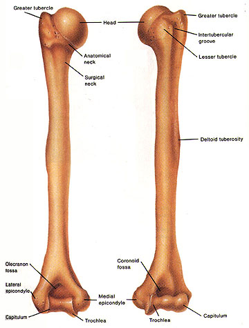
Head
expanded proximal end of a bone carried on a nar
[Bone Markings - Articulations]
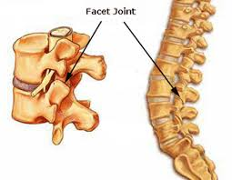
Facet
smooth, flat articular surface
[Bone Markings - Articulations]
Condyle
smooth, rounded articular surface
[Bone Markings - Articulations]
Ramus
arm-like bar of a bone
[Bone Tissue]
Organic Osteoid
roughly 1/3 weight of the bone is contributed by collagen fibers
strong/flexible, but can be bent if compressed
[Bone Tissue]
Inorganic Hydroxyapatites
mineral salts account for 2/3 weight of bone
calcium phosphate interacts with calcium hydroxide = hydroxyapatite
incorporates other salts to form a strong/flexible material
resistant to shattering
[Bone Cells]
Osteogenic Cells
mesenchymal cells located within the periosteum and endosteum
stem cells divide to produce daughter cells that differentiate into osteoblasts + important in formation of osteocytes
[Bone Cells]
Osteoblasts
immature bone cells located on the surface of bone
produce new bone matrix in process called osteogenesis/ossification
make and release proteins in matrix
before calcium salts are deposited, it’s called the osteoid
osteocytes develop from osteoblasts that are trapped in a lacuna
incapable of mitosis
[Bone Cells]
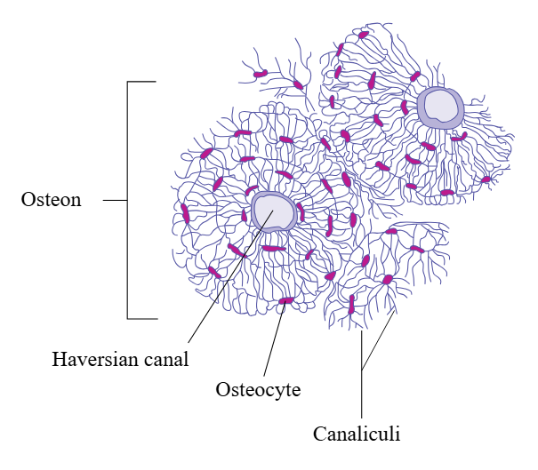
Osteocytes
mature bone cells that maintain protein/mineral content of surrounding matrix
secrete enzymes that dissolve the adjacent matrix + release minerals into circulation
rebuild matrix = deposition of mineral crystals
repair damaged bones
communication + receive nutrients via canaliculi channels
incapable of mitosis
[Bone Cells]
Osteoclasts
bone digesting cells that remove/recycle bone matrix
have 50+ nuclei
originate from white blood cells, not osteogenic cells
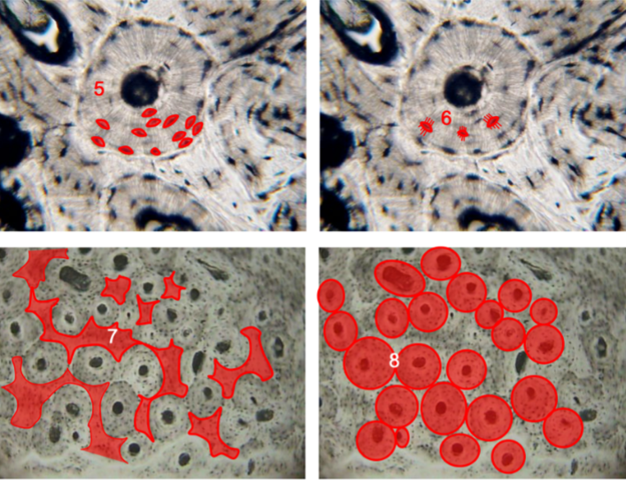
osteolysis: acids/enzymes are secreted by osteoclasts and release stored minerals; important in bone remodeling
[Compact Bone]
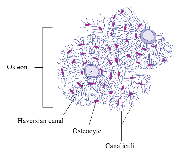
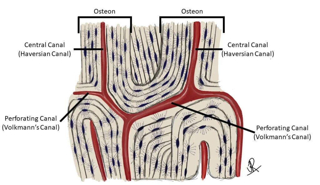
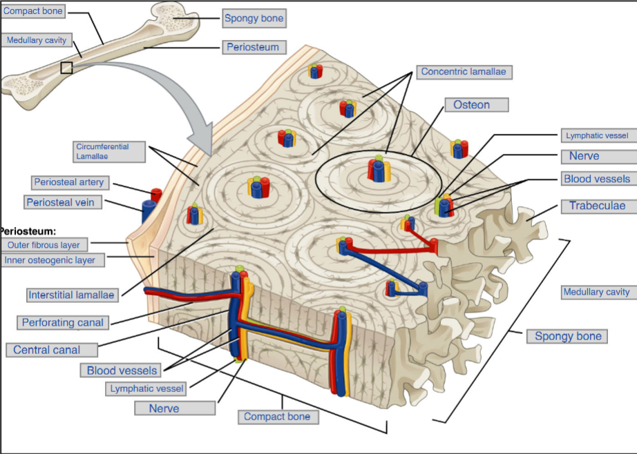
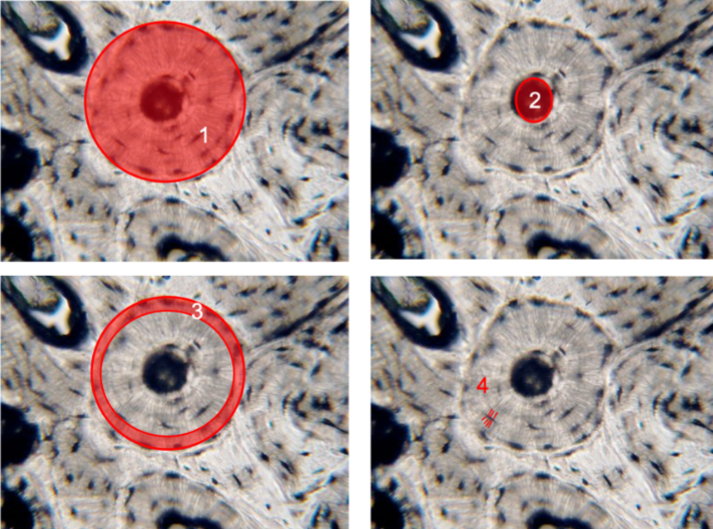
Osteon
basic structural/functional unit if bone
consists of bone cells organized around a central canal and separated by concentric lamellae
[Compact Bone]
Central Canal/Haversian Canal
runs parallel to the axis of bone
located in the middle of each osteon
possesses an artery and vein, lymph vessel, and nerve
[Compact Bone]
Perforating Canals
passageways that extend perpendicular to the axis of bone
connect the central canals of adjacent osteons
[Compact Bone]
Lamellae
nested, concentric rings of matrix surrounding the central canal
circumferential lamellae: found at the OUTER/INNER LININGS of the bone, where they are covered by periosteum and endosteum. produced during growth/maintenance
interstitial lamellae: OUTSIDE OSTEON. fill in spaces between adjacent osteons of compact bone. remnants of osteons
[Compact Bone]
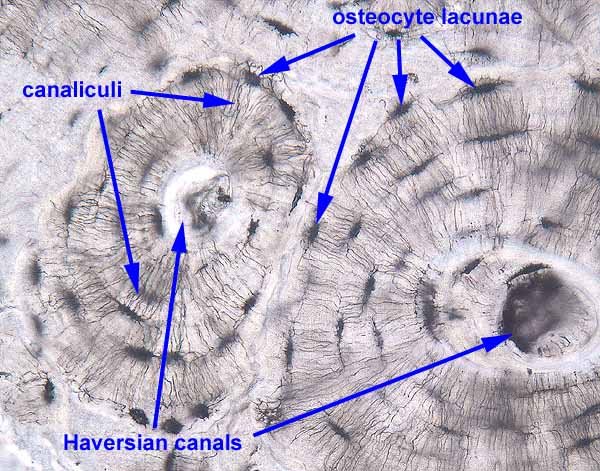
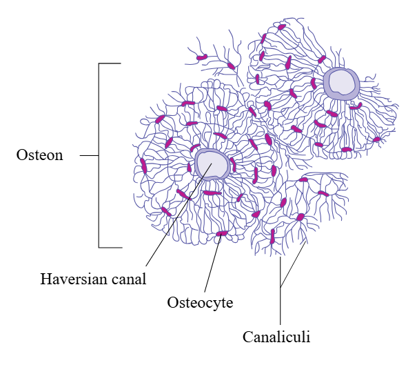
Lacunae
they hold the osteocytes
each one only contains one osteocyte
[Compact Bone]
Canaliculi
narrow crevices
this is what the osteocytes extend into
they penetrate the lamellae and connect the lacunae to central canal
Spongy Bone
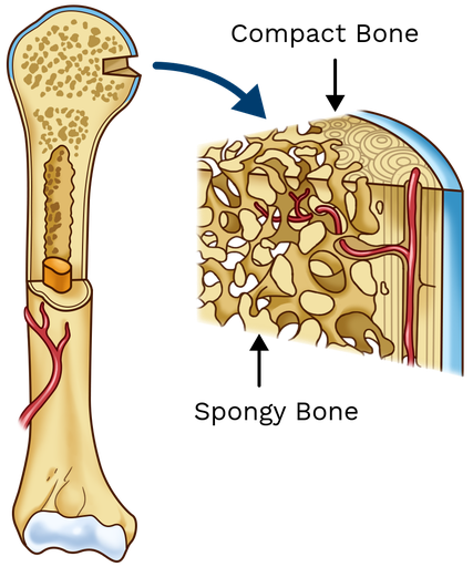
contains osteocytes housed in lacunae, but they’re not arranged in concentric circles
lacunae and osteocytes are found in lattice-like network of matrix called trabeculae
the trabeculae provide strength to the bone
the space between the network provide balance to the dense/heavy compact bone = lighter and easier to move
some spaces contain red marrow
[Blood/Nerve Supply]
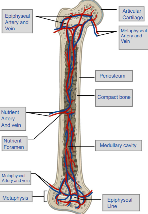
Nutrient Foramen
nutrient foramen is a tunnel that penetrates the diaphysis and provides access for blood vessels into the shaft of the bone
in order for bones to grow/be maintained, they require extensive blood supply
nutrient artery: transports oxygenated, nutrient-rich blood to the bone
nutrient vein: transports deoxygenated, waste0laden blood from the bone
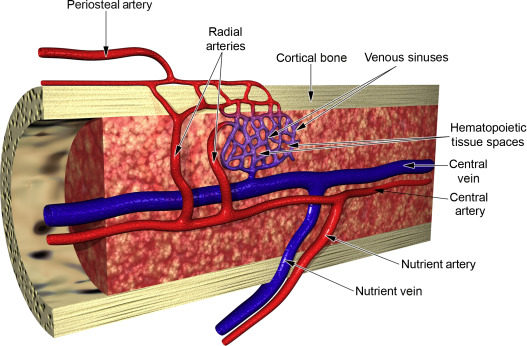
[Blood/Nerve Supply]
Metaphyseal Artery/Metaphyseal Vein
metaphyseal arteries deliver oxygenated blood and nutrients to support bone growth
metaphyseal veins remove deoxygenated blood and metabolic waste from the bone
they go from the metaphysis to the epiphysis
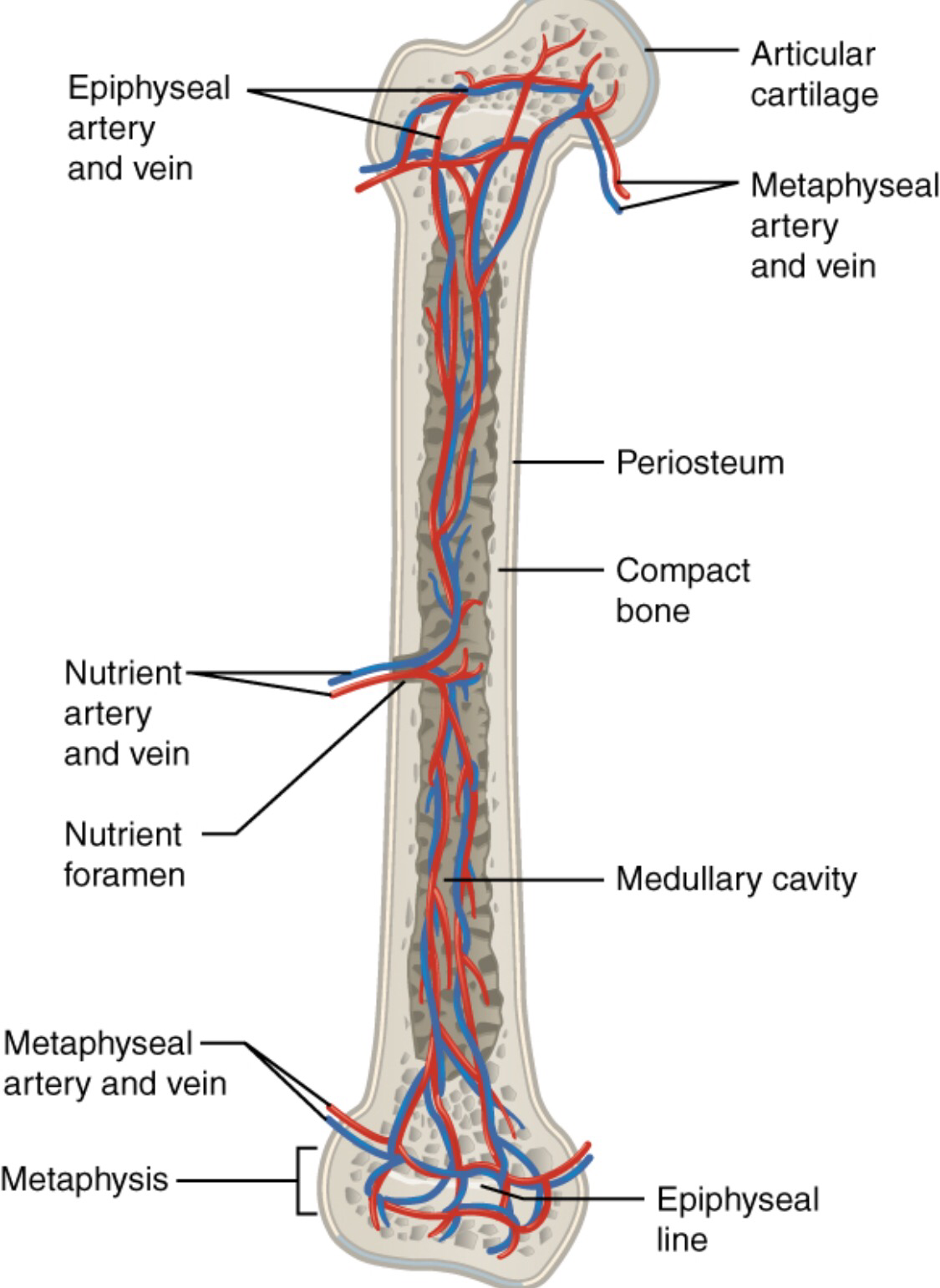
[Blood/Nerve Supply]
Nerves
nerves follow same paths into the bone
they concentrate in the more metabolically active regions of the bone
they sense pain and regulate blood supplies in bone growth
[Bone Formation/Development]
Cartilage Template
bone is a replacement tissue = uses model tissue (cartilage) to lay down mineral matrix
during fetal development, framework is laid down that determines where bones will form
flexible, semi-solid matrix produced by chondroblasts
cartilage is avascular = no blood vessels
functions are carried on by diffusion through the matrix
damaged cartilage doesn’t repair itself as easily as tissues
[Bone Formation/Development]
Intramembranous Ossification
formation of bones without a cartilage model
typical in flat bones, mandible, clavicles, and patella
begins around 8 weeks after fertilization
compact bone and spongy bone develops directly from sheets of connective tissue
[Bone Formation/Development]
Steps of Intramembranous Ossification
mesenchymal cells gather together and differentiate into specialized cells. some become osteogenic cells —> osteoblasts (appear in cluster called ossification center)
osteoblasts secrete osteoid (uncalcified matrix), which calcifies (hardens) in a few days with mineral salts = entraps osteoblasts inside. osteoblasts now become osteocytes.
osteoid (bone matrix) secreted makes trabecular matrix. the osteoblasts on the surface of spongy bone become the periosteum (protective layer of compact bone outside)
at birth, skull and clavicles are not fully ossified + sutures aren’t closed = deforms during passage through birth canal. last bones to ossify are flat bones of the face (reach adult size at end of growth spurt)
[Bone Formation/Development]
Endochondral Ossification
formation of bones using hyaline cartilage model
bone develops by replacing hyaline cartilage
around 6 weeks after fertilization, some mesenchymal cells differentiate into chondrocytes that form cartilaginous skeletal precursor
after, perichondrium appears
[Bone Formation/Development]
Steps of Endochondral Ossification
cavitation of hyaline shaft
chondrocytes calcify; impermeable matrix causes chondrocytes to die from lack of nutrients = deteriorates; blood vessels grow around edges of cartilage = medullary cavity
invasion of internal cavities
blood vessels invade spaces = enlarges and brings osteoblasts (this area is called the primary ossification center); the periosteal bud (fibroblasts, lymph vessels, nerve fibers, red marrow) migrate; osteoblasts secrete osteoid = spongy bone
formation of medullary cavity
primary ossification center enlarges; osteoclasts break down spongy bone + opens up medullary cavity; osseous tissue of outer shaft becomes thicker = compact bone
formation of epiphyses
secondary ossification center appears, cartilage calcifies/deteriorates = cavities for periosteal bud to enter; epiphyses are filled with spongy bone; cartilage remains
[Bone Formation/Development]
Longitudinal Growth: How Bones Grow in Length
1. Reserve Zone —> ANCHOR
Cells: Small, inactive chondrocytes.
Action: Anchors the growth plate to the epiphysis bone.
2. Proliferative Zone —> MULTIPLY & STACK
Cells: Chondrocytes doing mitosis.
Action: Divide rapidly and stack into tall columns to make new cells.
3. Hypertrophic Zone —> GROW & DIE
Cells: Older, larger chondrocytes.
Action: Cells swell up (hypertrophy), die, and the matrix breaks down.
4. Calcified Matrix Zone —> CALCIFY & INVADE
Cells: Mostly dead cells.
Action: Matrix hardens (calcifies); capillaries and osteoblasts invade from the shaft.
5. Ossification Zone —> BONE FORMATION
Cells: Osteoblasts.
Action: Osteoblasts turn leftover cartilage fragments (spicules) into spongy bone.
6. Early Twenties —> STOP
Action: Mitosis stops, cartilage turns entirely to bone, leaves an epiphyseal line.
[Bone Growth/Development]
Appositional Growth: How Bones Grow in Diameter
osteoprogenitor cells beneath the periosteum differentiate into osteoblasts + form new osteons on external bone surface
as bone is being added on the outside, osteoclasts remove/recycling lamellae in the inner surface = medullary cavity gradually enlarges as the bone increases in diameter
this process is called modeling
[Bone Growth/Development]
Bone Modeling
the process in which matrix is resorbed on one surface of a bone and deposited on another
takes place during bone growth
in adult life, bone undergoes remodeling: resorption of old/damaged bone takes place on same surface where osteoblasts lay new bone (injury, exercise, etc. leads to this)
[Fractures: Bone Repair]
What is a Fracture
a fracture is a broken bone
will heal regardless of if it’s reset to its original anatomical position
if it isn’t reset correctly, healing process will keep bone in deformed position
[Fractures: Bone Repair]
How to Classify a Fracture
type of reduction
when a broken bone is manipulated and set into natural position without surgery, it’s called a closed reduction
when it requires surgery to expose and reset: open reduction
bone location relative to skin
simple (closed): fracture where the skin remains intact
compound (open): fracture where at least one end of the broken bone tears through the skin = risk of infection
orientation of the break
transverse: break occurs perpendicular (90 degrees) to the axis of the bone
oblique: occurs at an angle that isn’t 90 degrees
spiral: bone segments are pulled apart as result of twisting motion
position of bone ends after the fracture
non-displaced: bone ends retain their position
displaced: bone end is out of normal alignment
[Fractures: Bone Repair]
Type of Fractures
comminuted: bone fragments into many pieces
compression/impacted: bone is crushed from upward/downward forces
depressed: broken bone is pressed inward (skull)
spiral: raged break as a result of excessive twisting of bone
greenstick: bone breaks incompletely (young patients)
colle’s: distal part of radius breaks
pott’s: malleolus of tibia/fibula break
[Fractures: Bone Repair]
Bone Repair
hematoma formation
blood vessels in bone tear = hemorrhage
hematoma (large blood clot) forms. disruption of blood flow to bone = death of bone cells around fracture
fibrocartilage callus formation
capillaries grow into hematoma and phagocytic cells invade area
fibroblasts/osteoblasts migrate to fraction and secrete cartilage matrix
chondrocytes from endosteum create internal callus by secreting matrix between two ends of broken bone, while chondrocytes from periosteum form external callus of hyaline cartilage/bone = stabilizes the fracture
bony callus formation
osteoclasts resorb the dead bone; osteogenic cells become very active
cartilage is replaced by trabecular bone
internal/external calli unite, compact bone replaces spongy bone = complete healing
this process takes several weeks + slight swelling may remain after
[Exercise, Nutrition, and Hormones]
Exercise and Bone Tissue
lack of mechanical stress causes bones to lose mineral salts/collagen fibers = weakens
mechanical stress stimulates growth/strengthening of mineral salts/collagen fibers
people who exercise regularly have greate bone density
any type of exercise stimulates strengthening, but resistance training is the best
resistant training slows down eventual bone loss due to aging (prevents osteoporosis)
[Exercise, Nutrition, and Hormones]
Nutrition and Bone Tissue
calcium: critical component of bone, must be obtained by diet because the body can’t make it.
can’t be absorbed in the intestine without vitamin D, so that’s also critical to bone
vitamin K: supports bone mineralization
magnesium: only small amount in body but 60% of it is in the skeleton
fluoride: helps stabilize and strengthen bone mineral
omega-3 fatty acids: reduce inflammation in the body (can interfere with function of osteoblasts = new osseous tissue)
[Exercise, Nutrition, and Hormones]
Hormones and Bone Tissue
hormones are involved in controlling bone growth, maintaining bone once formed, and remodeling it
growth hormone: triggers chondrocyte proliferation = increasing length of bones. increases calcium retention = enhanced mineralization, osteoblastic activity = greater bone density
thyroxine: promotes osteoblastic activity and synthesis of bone matrix
estrogen/testosterone: promote osteoblastic activity + production of bone matrix. responsible for growth spurt.
calcitriol: active form of vitamin D, stimulates calcium absorption
Calcium Homeostasis
Why Calcium Matters
Most abundant mineral in the body.
6 Functions to memorize: Bones, teeth, heartbeat strength, blood clotting, muscle contraction, and nerve signals.
The Two Conditions
Hypocalcemia (Too Low): Brittle bones, poor clotting, skipped heartbeats, muscle/nerve failure.
Hypercalcemia (Too High): Underactive nervous system —> Lethargy, slow reflexes, constipation, confusion, coma.
How the Body Fixes It
1. If Blood Calcium is Too LOW —> PTH raises it
How it senses it: Receptors on the Parathyroid Gland notice calcium isn't binding to them.
Bones: Activates osteoclasts to erode bone and release calcium into blood.
Intestines: Triggers kidneys to make Calcitriol (Vitamin D), which absorbs calcium from food.
Kidneys: Calcitriol tells kidneys to reabsorb calcium back into the blood.
2. If Blood Calcium is Too HIGH —> Calcitonin lowers it
Where it comes from: C cells of the Thyroid Gland.
Bones: Stops osteoclasts (bone eaters) but lets osteoblasts (bone builders) keep working to store calcium.
Intestines: Absorbs less calcium from food.
Kidneys: Prevents kidneys from keeping calcium so you pee it out.
The Skeleton's Big Picture
The skeleton is the primary manager of calcium homeostasis.
It also balances other ions, not just calcium.
[Bone Disorders]
Pituitary Growth Failure (dwarfism)
results from inadequate production of growth hormone
leads to reduced epiphyseal cartilage = abnormally short bones
rate in US because children can be treated with synthetic growth hormone
[Bone Disorders]
Achondroplasia
results from abnormal hyaline cartilage development
individuals have short, stocky limbs but the torso/head are normal size
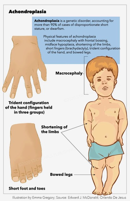
[Bone Disorders]
Marfan Syndrome
very tall with long, slender limbs
due to excessive cartilage formation at epiphyseal plates
can cause life-threatening cardiovascular problems
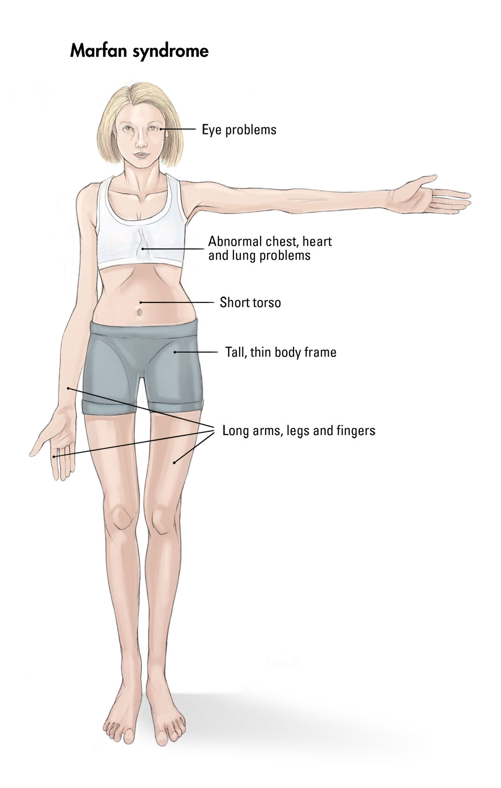
[Bone Disorders]
Gigantism
results from overproduction of growth hormone before puberty
puberty can be delayed
most common cause is pituitary tumor which can be treated with surgery, radiation, or drugs
[Bone Disorders]
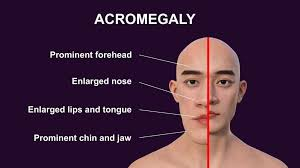
Acromegaly
results from too much growth hormone after epiphyseal plates close
bones don’t grow longer but instead thicker
bones of face, hands, jaw
[Bone Disorders]
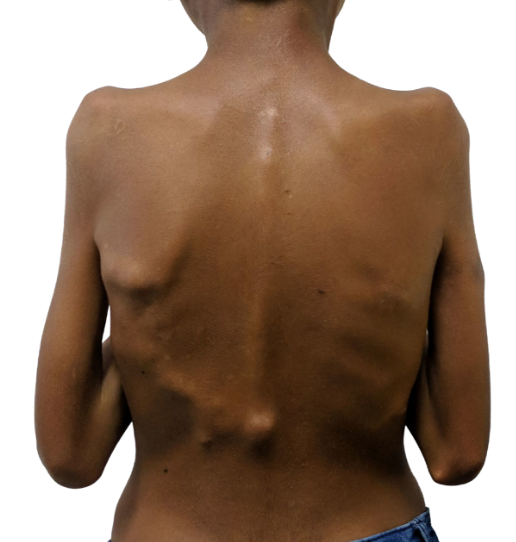
Fibrodysplasia Ossificans Progressiva (FOP)
rare gene mutation that causes deposition of bone around skeletal muscles and soft tissues
no effective treatment
painful/debilitating
patients rarely live into the 40’s
[Bone Disorders]
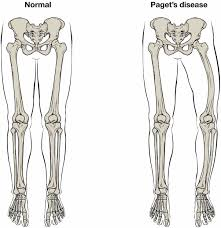
Paget’s Disease
overactive osteoclasts cause pores and weakening of long bones = bending/bowling
osteoblasts try to compensate for overactive osteoclasts
weak/brittle, prone to fractures
[Bone Disorders]
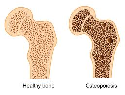
Osteoporosis
decrease in bone mass when bone resorption exceeds formation
common with aging
women lost bone mass more quickly than men starting at 50
50 is also start of menopause (ovaries reduce production of estrogen = less osteoblastic activity for production of bone matrix)
[Bone Disorders]
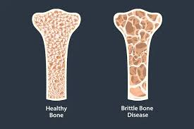
Osteogenesis Imperfecta (OI)
AKA brittle bone disease
genetic disease where bones don’t form properly = fragile/break easily
present from birth and stays throughout life
impacts body’s production of collagen
[Quiz]
Which are functions of skeletal system (4 answers)
Support , protection and movement
Thermoregulation
Vitamin D production
Mineral homeostasis
Blood cell production
Triglyceride storage
Communication
Support , protection and movement
Mineral homeostasis
Blood cell production
Triglyceride storage
[Quiz]
Match bones to shapes
long bones: ulna, tibia, radius, metacarpals
short bones: carpals/tarsals
flat bones; sternum, scapula, and cranium
irregular bones: vertebrae
seasmoid bones: patella
[Quiz]
Calcium plays critical roles in: (5 answers)
smooth muscle contraction
cardiac muscle contraction
blood clotting
endocrine function
bone remodeling
skeletal muscle contraction
smooth muscle contraction
cardiac muscle contraction
blood clotting
bone remodeling
skeletal muscle contraction
[Quiz]
What are extracellular fluid filled extensions of lacunae called?
canaliculi
[Quiz]
How are osteons in compact bone tissue aligned and how does the osteon arrangement affect bone strength?
Parallel to the length of the diaphysis; the shaft resists bending even when stresses are applied in certain directions.
[Quiz]
Match bone disorders to description
dislocation: Occurs when a bone slips out of place, tearing the ligaments that attach the bone at the joint
torn cartilage: Can result from a sharp blow to a joint or a severe twisting of a joint
bursitis: Painful inflammation of bursa, a fluid-filled sac that helps reduce friction in joints.
bunions: Painful swellings of the bursae in the first joints of big toes.
arthritis: Inflammation of a joint, resulting from an injury, natural wear and tear, or autoimmune disease.
[Quiz]
The cells active in fracture repair during the production of the hard (spongy bone) callus are called______
Osteoblasts
Images
Images
Images
osteons
central canal
concentric lamellae
canaliculi
lacuna
osteocytes
interstitial lamellae
multiple osteons